. 2022 Dec 16;3(4):101727.
doi: 10.1016/j.xpro.2022.101727. Epub 2022 Sep 27.

Preparation of a murine oral palate wound healing model

Affiliations

Preparation of a murine oral palate wound healing model

Trevor R Leonardo et al. STAR Protoc. .

Abstract

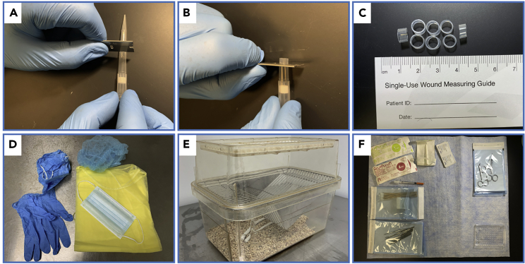
The oral cavity is a highly regenerative epithelial tissue that results in minimal scarring after injury. This protocol describes the preparation of a mouse palate wound model. The protocol includes steps to place an excisional wound on the mouse palate, followed by harvesting of wound tissue and bone decalcification. We detail how to overcome the technical challenge of limited anatomical space, avoid damaging the nasal cavity, manage bleeding, and collect tissue for downstream genomic or immunohistochemical analysis.

Keywords: Health sciences; Model organisms; Tissue engineering.


Conflict of interest statement

Declaration of interests The authors declare no competing interests.

Figures

None
Graphical abstract
Figure 1
Figure 1
Preparation for surgery (A) Image showing where to make the first cut on the pipette tip with a razor blade. (B) Image showing where to make the second cut. (C) Representative oral spacers after cutting pipette tips with the razor blade. (D) Personal protective equipment including: bonnet, gown, gloves, surgical mask, and foot covers. (E) Recovery cage. (F) Example surgical area with tools placed in easy to reach areas.
Figure 2
Figure 2
Palatal wound placement (A) Placement of forceps between incisors to hold the mouth open. (B) Wedging of spacer into place using forceps and buccal pouch as support. (C) Use of forceps to adjust spacer within buccal pouch. (D) A properly placed oral spacer. (E) 1 mm biopsy punch placement, with thumb and index finger of other hand supporting mouse head. (F) A properly placed 1 mm wound along the medial line and below first rugae. (G) Use forceps to physically remove excised tissue. (H) Appearance of palate after wound tissue is removed. (I) Use of cotton tipped applicator to apply pressure to the wound to achieve hemostasis.
Figure 3
Figure 3
Palate removal (A) Curved scissor placement to separate upper and lower mandible. (B) Initial incision inferior to palate with dissector scissors. (C) Lateral incision parallel and lateral to teeth. (D) Final incision using dissector scissors as this cuts through the incisors. (E) Removal of palate using forceps. (F) Fully removed palate after 1× PBS rinse.

References

    1. Byrd K.M., Piehl N.C., Patel J.H., Huh W.J., Sequeira I., Lough K.J., Wagner B.L., Marangoni P., Watt F.M., Klein O.D., et al. Heterogeneity within stratified epithelial stem cell populations maintains the oral mucosa in response to physiological stress. Cell Stem Cell. 2019;25:814–829.e6. doi: 10.1016/j.stem.2019.11.005. - DOI - PMC - PubMed
    1. Graves D.T., Nooh N., Gillen T., Davey M., Patel S., Cottrell D., Amar S. IL-1 plays a critical role in oral, but not dermal, wound healing. J. Immunol. 2001;167:5316–5320. doi: 10.4049/jimmunol.167.9.5316. - DOI - PubMed

Publication types

LinkOut - more resources