. 2022 Sep 27;40(13):111402.
doi: 10.1016/j.celrep.2022.111402.

Ventral hippocampus-lateral septum circuitry promotes foraging-related memory

Affiliations

Ventral hippocampus-lateral septum circuitry promotes foraging-related memory

Léa Décarie-Spain et al. Cell Rep. .

Abstract

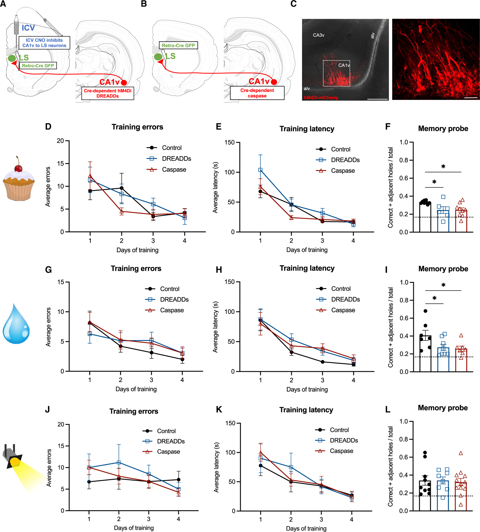
Remembering the location of a food or water source is essential for survival. Here, we reveal that spatial memory for food location is reflected in ventral hippocampus (HPCv) neuron activity and is impaired by HPCv lesion. HPCv mediation of foraging-related memory involves communication to the lateral septum (LS), as either reversible or chronic disconnection of HPCv-to-LS signaling impairs spatial memory retention for food or water location. This neural pathway selectively encodes appetitive spatial memory, as HPCv-LS disconnection does not affect spatial memory for escape location in a negative reinforcement procedure, food intake, or social and olfactory-based appetitive learning. Neural pathway tracing and functional mapping analyses reveal that LS neurons recruited during the appetitive spatial memory procedure are primarily GABAergic neurons that project to the lateral hypothalamus. Collective results emphasize that the neural substrates controlling spatial memory are outcome specific based on reinforcer modality.

Keywords: CP: Neuroscience; feeding; foraging; lateral hypothalamus; motivation; prefrontal cortex; reward.


Conflict of interest statement

Declaration of interests The authors declare no competing interests.

Figures

Figure 1.
Figure 1.. Bilateral HPCv lesions impair spatial memory for food location
(A) Spatial foraging task apparatus. (B) Diagram of stereotaxic viral infusion (AAV-hSYN-GCaMP7s) and placement of optic fiber in the HPCv for bulk photometry recordings. (C) Representative histology of GCaMP7s expression in HPCv neurons. Scale bar, 100 μm. (D) Variation in HPCv bulk calcium-dependent activity following a correct or incorrect investigation during a spatial food-seeking task (time × investigation type, p < 0.05). (E) Delta Z score 5 s following a correct versus incorrect investigation during a spatial food-seeking task (p < 0.05). (F) Simple linear regression of delta Z score 5 s following investigation relative to distance of investigated hole from correct escape hole (p < 0.01). (G) Representative HPCv lesion histology with NeuN immunohistochemistry. Scale bar, 500 μm. (H) Left: representative navigation paths of a control animal preferentially investigating correct (filled red) and adjacent holes (outlined orange) during spatial foraging memory probe. Right: representative navigation path of HPCv lesioned animal during spatial foraging memory probe. (I and J) Bilateral HPCv lesions did not impair learning of the spatial foraging task compared with controls, as measured by errors before locating correct hole during task training (I) and latency to locate correct hole during task training (J). (K) Bilateral HPCv lesions impaired retention of the spatial foraging task, as measured by the ratio of investigation of correct plus adjacent holes over total investigated during the first minute of the task (p < 0.05). Dotted line indicates chance performance level (0.1667). For graphs (D), (E), and (F), n = 6 (within-subjects). For graphs (I), (J), and (K), lesion n = 11, control n = 18. All values expressed as mean ± SEM.
Figure 2.
Figure 2.. Reversible and chronic CA1v-to-LS neural disconnection impairs spatial memory for food and water location but not for escape location
(A) Diagram of dual viral approach using a Cre-dependent inhibitory DREADDs (AAV-hSyn-DIO-hM4D(GI)-mCherry) approach to reversibly disconnect CA1v-to-LS neural pathway (with CNO injected 1 h before memory probe). (B) Diagram of dual viral approach using a Cre-dependent caspase (AAV1-Flex-taCasp3-TEVp) approach to chronically disconnect CA1v-to-LS neural pathway. (C) Representative injection site in CA1v demonstrating LS-projecting neurons infected with inhibitory DREADDs, which simultaneously drives expression of a fluorescent mCherry transgene. Scale bars, 500 μm (left) and 100 μm (right). (D and E) Neither reversible (DREADDs) nor chronic (caspase) disconnection of the CA1v-to-LS pathway impaired learning of the spatial food-seeking task compared with controls, as measured by errors before correct hole during task training (D) and latency to correct hole during task training (E). (F) Both reversible and chronic disconnection of the CA1v-to-LS pathway impaired retention of the food location as measured by the ratio of investigation of correct plus adjacent holes over total investigations during entire 2 min of the task (p < 0.05). (G–I) Likewise, reversible (DREADDs) and chronic (caspase) disconnection of the CA1v-to-LS pathway did not impair learning of the spatial water-seeking task compared with controls, as measured by errors before correct hole during task training (G) and latency to correct hole during task training (H), but impaired memory retention of the water location during the probe (I). (J and K) In contrast, disconnection of the CA1v-to-LS pathway either reversibly (DREADDs) or chronically (caspase) did not impair performance on the spatial escape task. There were no differences in learning as measured by errors before correct hole during task training (J) and latency to correct hole during task training (K). (L) Unlike the spatial foraging task, retention of the spatial escape task was not impaired by reversible or chronic disconnection of the CA1v-to-LS pathway. For graphs (E), (F), and (G) (CA1v-to-LS disconnect cohort 1), DREADDs n = 6, caspase n = 10, control n = 8. For graphs (H), (I), and (J) (CA1v-to-LS disconnect cohort 3), DREADDs n = 8, caspase n = 8, control n = 7). For graphs (K). (L), and (M) (CA1v-to-LS disconnect cohort 2), DREADDs n = 8, caspase n = 12, control n = 10. Dotted line indicates chance performance level (0.1667). All values expressed as mean ± SEM.
Figure 3.
Figure 3.. CA1v to mPFC projections do not contribute to appetitive spatial memory
(A) Diagram of dual viral approach to identify collateral targets of the CA1v-to-LS neural pathway. (B) Left: representative CA1v injection site from collateral identification approach. Scale bar, 500 μm. Middle: representative LS injection site from collateral identification approach in relation to the caudal (LSc) and rostral (LSr) subregions, the corpus callosum body (ccb), and lateral ventricle (VLa). Scale bar, 500 μm. Right: representative image collateral axons of the CA1v-to-LS pathway located in the prelimbic (PL) and infralimbic (ILA) areas of the mPFC. Scale bar, 50 μm. (C) Diagram of dual viral approach using Cre-dependent inhibitory DREADDs (AAV-hSyn-DIO-hM4D(GI)-mCherry) to acutely disconnect the CA1v-to-mPFC neural pathway (with CNO or aCSF injected 1 h before memory probe). (D) Representative injection site in CA1v demonstrating mPFC-projecting neurons infected with inhibitory DREADDs, which simultaneously drives expression of a fluorescent mCherry transgene. Scale bars, 100 μm. (E and F) Disconnection of the CA1v-to-mPFC neural pathway did not influence learning of the spatial foraging task compared with controls, as measured by errors before correct hole during training (E) and latency to correct hole during training (F). (G) Disconnection of the CA1v-to-mPFC pathway did not influence retention of the spatial foraging task in the memory probe. For graphs (E), (F), and (G), aCSF n = 6, CNO n = 8. Dotted line indicates chance performance level (0.1667). All values expressed as mean ± SEM.
Figure 4.
Figure 4.. LS neurons activated during food-seeking memory probe are predominantly GABAergic
(A–C) c-Fos and ChAT colocalization was observed in ~2% of LS neurons (A; scale bar, 500 μm), c-Fos and GAD1 colocalization was observed in ~88% of LS neurons (B; scale bar, 500 μm), and c-Fos and VGlut2 colocalization was observed in ~10% of LS neurons (C; scale bar, 500 μm) following the spatial memory probe test (for food reinforcement). (D) Percentage of c-Fos+ LS neurons expressing GABAergic, cholinergic and glutamatergic markers. Quantification from DREADDs vehicle group, n = 3.
Figure 5.
Figure 5.. Identification of second-order neural projections downstream of CA1v-to-LS projections
(A) Diagram of dual viral approach to identify brain regions that are second-order (2°) targets of the CA1v-to-LS neural pathway. (B) Representative LS injection site from second-order identification approach in relation to the caudal (LSc) and rostral (LSr) subregions, the corpus callosum body (ccb), and lateral ventricle (VLa). Scale bar, 100 μm. (C) Representative image of second-order fibers of the CA1v-to-LS pathway within the juxtadorsomedial (jd), juxtaventromedial dorsal (jvd), juxtaventromedial ventral (jvv), and subfornical anterior (sfa) subregions of the LHA. Scale bar, 200 μm. (D) Summary of the projection targets of LS neurons that receive input from CA1v (for expanded information see Table S1). The outputs of the right side of LS neurons receiving CA1v input are represented at the macroscale (gray matter region resolution) on a partial flatmap representation of the rat forebrain, adapted from Hahn et al. (2021). Connection weights are represented by block colors for each region following an ordinal scale ranging from weakest (0 = very weak or absent) to strongest (6 = strong), as there were no 7 (very strong) values. The lower panels represent one side of the brain with the part represented in the upper diagram outlined in magenta. (E–G) The COIN approach for further confirmation of second-order tracing strategy is depicted with FG injection site in the LHA (E; scale bar, 500 μm), representative AAV1-GFP injection site in the CA1v (F; scale bar, 100 μm), and FG-GFP appositions in the LS (G; scale bar, 100 μm). (H) Representative histology of c-Fos expression in the medial LHA (LHAjd and LHAjv) following i.c.v. infusion of aCSF. Scale bar, 200 μm. (I) Representative histology of c-Fos expression in the medial LHA (LHAjd and LHAjv) following i.c.v. infusion of CNO to inhibit LS-projecting CA1v neurons, Scale bar, 200 μm. (J) Counts of c-Fos-positive cells in the medial LHA (LHAjd and LHAjv) are reduced with i.c.v. administration of CNO (p < 0.05). aCSF n = 4 and CNO n = 4. Values expressed as mean ± SEM.

Similar articles

Cited by

References

    1. Aqrabawi AJ, and Kim JC (2018). Topographic organization of hippocampal inputs to the anterior olfactory nucleus. Front. Neuroanat 12, 12. - PMC - PubMed
    1. Aqrabawi AJ, Browne CJ, Dargaei Z, Garand D, Khademullah CS, Woodin MA, and Kim JC (2016). Top-down modulation of olfactory-guided behaviours by the anterior olfactory nucleus pars medialis and ventral hippocampus. Nat. Commun 7, 13721. - PMC - PubMed
    1. Arszovszki A, Borhegyi Z, and Klausberger T (2014). Three axonal projection routes of individual pyramidal cells in the ventral CA1 hippocampus. Front. Neuroanat 8, 53. - PMC - PubMed
    1. Barnes CA (1979). Memory deficits associated with senescence: a neurophysiological and behavioral study in the rat. J. Comp. Physiol. Psychol 93, 74–104. - PubMed
    1. Barson JR, Morganstern I, and Leibowitz SF (2013). Complementary roles of orexin and melanin-concentrating hormone in feeding behavior. Int. J. Endocrinol, 983964. - PMC - PubMed

Publication types

LinkOut - more resources