Observational Study
. 2022 Sep;10(9):e005323.
doi: 10.1136/jitc-2022-005323.

Single-cell transcriptomics identifies pathogenic T-helper 17.1 cells and pro-inflammatory monocytes in immune checkpoint inhibitor-related pneumonitis

Affiliations
Observational Study

Single-cell transcriptomics identifies pathogenic T-helper 17.1 cells and pro-inflammatory monocytes in immune checkpoint inhibitor-related pneumonitis

Amelie Franken et al. J Immunother Cancer. 2022 Sep.

Abstract

Background: Immune checkpoint inhibitor (ICI)-related pneumonitis is the most frequent fatal immune-related adverse event associated with programmed cell death protein-1/programmed death ligand-1 blockade. The pathophysiology however remains largely unknown, owing to limited and contradictory findings in existing literature pointing at either T-helper 1 or T-helper 17-mediated autoimmunity. In this study, we aimed to gain novel insights into the mechanisms of ICI-related pneumonitis, thereby identifying potential therapeutic targets.

Methods: In this prospective observational study, single-cell RNA and T-cell receptor sequencing was performed on bronchoalveolar lavage fluid of 11 patients with ICI-related pneumonitis and 6 demographically-matched patients with cancer without ICI-related pneumonitis. Single-cell transcriptomic immunophenotyping and cell fate mapping coupled to T-cell receptor repertoire analyses were performed.

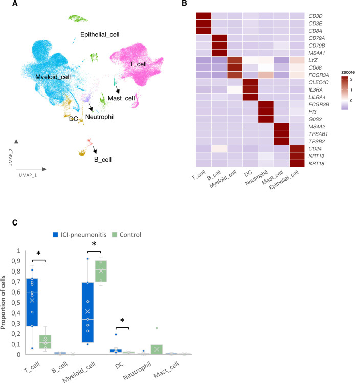
Results: We observed enrichment of both CD4+ and CD8+ T cells in ICI-pneumonitis bronchoalveolar lavage fluid. The CD4+ T-cell compartment showed an increase of pathogenic T-helper 17.1 cells, characterized by high co-expression of TBX21 (encoding T-bet) and RORC (ROR-γ), IFN-G (IFN-γ), IL-17A, CSF2 (GM-CSF), and cytotoxicity genes. Type 1 regulatory T cells and naïve-like CD4+ T cells were also enriched. Within the CD8+ T-cell compartment, mainly effector memory T cells were increased. Correspondingly, myeloid cells in ICI-pneumonitis bronchoalveolar lavage fluid were relatively depleted of anti-inflammatory resident alveolar macrophages while pro-inflammatory 'M1-like' monocytes (expressing TNF, IL-1B, IL-6, IL-23A, and GM-CSF receptor CSF2RA, CSF2RB) were enriched compared with control samples. Importantly, a feedforward loop, in which GM-CSF production by pathogenic T-helper 17.1 cells promotes tissue inflammation and IL-23 production by pro-inflammatory monocytes and vice versa, has been well characterized in multiple autoimmune disorders but has never been identified in ICI-related pneumonitis.

Conclusions: Using single-cell transcriptomics, we identified accumulation of pathogenic T-helper 17.1 cells in ICI-pneumonitis bronchoalveolar lavage fluid-a phenotype explaining previous divergent findings on T-helper 1 versus T-helper 17 involvement in ICI-pneumonitis-,putatively engaging in detrimental crosstalk with pro-inflammatory 'M1-like' monocytes. This finding yields several novel potential therapeutic targets for the treatment of ICI-pneumonitis. Most notably repurposing anti-IL-23 merits further research as a potential efficacious and safe treatment for ICI-pneumonitis.

Keywords: Autoimmunity; Computational Biology; Immunotherapy; Lung Neoplasms; Programmed Cell Death 1 Receptor.


Conflict of interest statement

Competing interests: None declared.

Figures

Figure 1
Figure 1
Cell composition of ICI-pneumonitis and control patients’ bronchoalveolar lavage fluid (BALF). (A) UMAP plot of 141,056 cells present in ICI-pneumonitis and control BALF. (B) Heatmap of canonical marker gene expression used for main cell type annotation. (C) Comparison of relative cell type abundance, revealed accumulation of T cells and dendritic cells in ICI-pneumonitis BALF, while other myeloid cells (monocytes/macrophages) were relatively depleted. Wilcoxon rank-sum test was used; significance is shown as *p<0.05. DC, dendritic cell; ICI, immune checkpoint inhibitor; UMAP, Uniform Manifold Approximation and Projection.
Figure 2
Figure 2
T-cell phenotypes and relative abundance in ICI-pneumonitis and control bronchoalveolar lavage fluid. (A) Uniform Manifold Approximation and Projection plot of 65,293 T cells, (B) annotated according to canonical marker gene expression of CD4+ (left panel) and CD8+ T cells, MAIT, and NK-cells (right panel). (C) A comparison of relative T-cell subtype abundance, revealed accumulation of CD4+ T-helper 17.1 cells with a pathogenic phenotype (TH17.1_TBX21) in ICI-pneumonitis, as well as enrichment of CD4+ regulatory type 1 T cells (TR1), CD4+ naïve-like T cells (TN), and CD8+ effector memory (TEM) T cells. Wilcoxon rank-sum test was used; significance is shown as *p<0.05. (D) Volcano plot showing differentially expressed genes in T cells comparing ICI-pneumonitis and control T cells. P values were obtained by the model-based analysis of single-cell transcriptomics (MAST) test and Bonferroni-corrected (see online supplemental table S1 for all differentially expressed genes). (E) Differential gene set enrichment analysis (DGSEA) on differentially expressed genes for ICI-pneumonitis versus control T cells using the R package hypeR. Only significant genes (adjusted p value<0.05) and genes with a log-fold change higher than 0.5 or lower than −0.5 were used (see online supplemental tables S2 and S3 for all differentially expressed gene sets in ICI-pneumonitis and control T cells, respectively). CD4_N, CD4+ naïve-like T cells; CD4_EM, CD4+ effector memory T cells; CD4_TR1, CD4+ regulatory type 1 T cells; CD4_Th17.1_RORC, CD4+ T-helper 17.1 lymphocytes with predominant non-pathogenic features; CD4_Th17.1_TBX21, CD4+ T-helper 17.1 lymphocytes with predominant (pathogenic) T-helper 1-like features; CD4_FH, CD4+ follicular helper T-cells; CD4_REG, CD4+ regulatory T cells; CD8_N, CD8+ naïve-like T cells; CD8_EM, CD8+ effector memory T cells; CD8_RM, CD8+ resident memory T cells; CD8_EMRA, CD8+ recently activated effector memory T cells; CD8_EX, CD8+ exhausted T cells; CD8_gd, CD8+ gamma delta T cells; DGEA, differential gene expression analysis; FDR, false discovery rate; ICI, immune checkpoint inhibitor; MAIT, mucosal associated invariant T cells; NK, natural killer; NK_cyto, cytotoxic NK-cells; NK_infla, inflammatory NK-cells.
Figure 3
Figure 3
Cell fate mapping and T-cell receptor repertoire analysis of CD4+ T cells in ICI-pneumonitis and control bronchoalveolar lavage fluid. (A) Uniform Manifold Approximation and Projection plot of 11,570 CD4+ T cells with cell fate trajectories and (B) latent time as calculated by the CellRank algorithm, showing TEM were connected to the TH17.1_RORC cluster, which branched into either TH17.1_TBX21 or TR1 as terminal states, or formed a terminal state itself. (C) Continuous gene expression profiling along the TH17.1_TBX21 trajectory. (D) Barplot of relative cell abundance in ICI-pneumonitis and control bronchoalveolar lavage fluid (BALF) along the TH17.1_TBX21 trajectory. (E) Line graph of the Gini coefficient for ICI-pneumonitis and control BALF T-cell receptor repertoire along the TH17.1_TBX21 trajectory, as calculated by the DescTools algorithm. CD4_N, CD4+ naïve-like T cells; CD4_EM, CD4+ effector memory T cells; CD4_TR1, CD4+ regulatory type 1 T cells; CD4_Th17.1_RORC, CD4+ T-helper 17.1 lymphocytes with predominant non-pathogenic features; CD4_Th17.1_TBX21, CD4+ T-helper 17.1 lymphocytes with predominant (pathogenic) T-helper 1-like features; CD4_FH, CD4+ follicular helper T cells; ICI, immune checkpoint inhibitor.
Figure 4
Figure 4
Macrophage and monocyte phenotypes and relative abundance in ICI-pneumonitis and control bronchoalveolar lavage fluid. (A) Uniform Manifold Approximation and Projection plot of 63,330 macrophages and monocytes, (B) annotated according to canonical marker gene expression. (C) Heatmap of functional gene expression patterns in macrophages and monocytes, showing pro-inflammatory/anti-inflammatory and antigen presentation-related gene expression patterns. (D) Comparison of relative cell subtype abundance, showing a relative enrichment of monocyte-derived macrophages (Mac_Md) and IL-1Bhigh pro-inflammatory monocytes (Monocyte_IL-1B) and a relative depletion of alveolar resident macrophages (Mac_Alveolar) in ICI-pneumonitis bronchoalveolar lavage fluid. Wilcoxon rank-sum test was used; significance is shown as *p<0.05. (E) Volcano plot showing differentially expressed genes in monocytes/macrophages comparing ICI-pneumonitis and control samples. P values were obtained by the model-based analysis of single-cell transcriptomics (MAST) test and Bonferroni-corrected (see online supplemental table S4 for all differentially expressed genes). (F) Differential gene set enrichment analysis (DGSEA) on differentially expressed genes for ICI-pneumonitis versus control monocytes/macrophages using the R package hypeR. Only significant genes (adjusted p value<0.05) and genes with a log-fold change higher than 0.5 or lower than −0.5 were used (see online supplemental table S5 and S6 for all differentially expressed gene sets in ICI-pneumonitis and control monocytes/macrophages, respectively). DGEA, differential gene expression analysis; FDR, false discovery rate; ICI, immune checkpoint inhibitor; IL, interleukin; Macrophage_Md_LGMN monocyte-derived macrophage.
Figure 5
Figure 5
Dendritic cell phenotypes and relative abundance in ICI-pneumonitis and control bronchoalveolar lavage fluid. (A) Uniform Manifold Approximation and Projection plot of 2642 dendritic cells, (B) annotated according to canonical marker gene expression. (C) A comparison of relative dendritic cell subtype abundance showed no statistically significant differences between ICI-pneumonitis and control bronchoalveolar lavage fluid. Wilcoxon rank-sum test was used. cDC1, classical type I dendritic cell; cDC2, classical type II dendritic cell; ICI, immune checkpoint inhibitor; migDC, migratory dendritic cell; pDC, plasmacytoid dendritic cell.

References

    1. Waldman AD, Fritz JM, Lenardo MJ. A guide to cancer immunotherapy: from T cell basic science to clinical practice. Nat Rev Immunol 2020;20:651–68. 10.1038/s41577-020-0306-5 - DOI - PMC - PubMed
    1. Haanen JBAG, Carbonnel F, Robert C, et al. . Management of toxicities from immunotherapy: ESMO clinical practice guidelines for diagnosis, treatment and follow-up. Ann Oncol 2017;28:iv119–42. 10.1093/annonc/mdx225 - DOI - PubMed
    1. Suresh K, Voong KR, Shankar B, et al. . Pneumonitis in non-small cell lung cancer patients receiving immune checkpoint immunotherapy: incidence and risk factors. J Thorac Oncol 2018;13:1930–9. 10.1016/j.jtho.2018.08.2035 - DOI - PubMed
    1. Wang DY, Salem J-E, Cohen JV, et al. . Fatal toxic effects associated with immune checkpoint inhibitors: a systematic review and meta-analysis. JAMA Oncol 2018;4:1721–8. 10.1001/jamaoncol.2018.3923 - DOI - PMC - PubMed
    1. Reynolds KL, Arora S, Elayavilli RK, et al. . Immune-Related adverse events associated with immune checkpoint inhibitors: a call to action for collecting and sharing clinical trial and real-world data. J Immunother Cancer 2021;9:e002896. 10.1136/jitc-2021-002896 - DOI - PMC - PubMed

Publication types

Substances