. 2022 Sep 28;13(1):5695.
doi: 10.1038/s41467-022-33315-8.

Activation of the human insulin receptor by non-insulin-related peptides

Affiliations

Activation of the human insulin receptor by non-insulin-related peptides

Nicholas S Kirk et al. Nat Commun. .

Abstract

The human insulin receptor signalling system plays a critical role in glucose homeostasis. Insulin binding brings about extensive conformational change in the receptor extracellular region that in turn effects trans-activation of the intracellular tyrosine kinase domains and downstream signalling. Of particular therapeutic interest is whether insulin receptor signalling can be replicated by molecules other than insulin. Here, we present single-particle cryoEM structures that show how a 33-mer polypeptide unrelated to insulin can cross-link two sites on the receptor surface and direct the receptor into a signalling-active conformation. The 33-mer polypeptide engages the receptor by two helical binding motifs that are each potentially mimicable by small molecules. The resultant conformation of the receptor is distinct from-but related to-those in extant three-dimensional structures of the insulin-complexed receptor. Our findings thus illuminate unexplored pathways for controlling the signalling of the insulin receptor as well as opportunities for development of insulin mimetics.


Conflict of interest statement

Y.W., H.H., J.F.E., F.A.M., F.M.-O., Q.C., V.V.K., and D.G.B. are employees of Eli Lilly and Company. During employment, employees are minor stockholders in Eli Lilly and Company. A.L.A. is an employee of Advanced Testing Laboratory, a company that provides scientific services to Eli Lilly and Company. M.C.L, N.S.K., and M.B.M. received funding from Eli Lilly and Company to undertake the work described in the manuscript.

Figures

Fig. 1
Fig. 1. IR-binding peptides and their biochemical properties.
a [Site-2]-[Site-1] fusion peptides S519, IM459 and IM172 and their isolated [Site 2]- and [Site 1] components. Non-native amino acids and modifications are as follows:- a: 2-aminoisobutyric acid; p: N-acetylproline; y: O-methyltyrosine; #: N-terminal (4-aminomethyl)phenylacetylation; @: N-terminal phenylacetylation; *: C-terminal amidation. b Receptor inhibition (ki) and auto-phosphorylation (EC50) constants (see Supplementary Fig. 2b–f). BHI = biosynthetic human insulin (control). Errors are SEM; n = 3 independent replicates. Source data are provided as a Source Data file. c The [Site-1]-[Site-2] fusion peptide S961. Individual residues colored green and mauve within panels a and c are those that are part of the defining motifs of the [Site 2]- and [Site 1] peptides, respectively (see Supplementary Fig. 1); residues involved in disulfide bridge formation are colored in yellow, with the corresponding bridge shown as yellow square bracket.
Fig. 2
Fig. 2. CryoEM structure of the IM459-bound hIR-Aecto.
a NuPAGE 4-12% Bis-Tris gel (non-reducing conditions) of pooled fractions evidencing the presence of the IM459 peptide within the purified complex. This gel experiment was conducted n = 1 times. b Focus-refined cryoEM map showing the fit of IR domains L1, CR, L2, FnIII-1′, and L2′ and the residual density attributable to IM459; density corresponding to domain FnIII-1 is asterisked. c,d Map as in b, showing respective orthogonal views of the rigid-body fit of the co-complex of S519C16 and the hIR L1-CR module (PDB entry 5J3H). e-g Comparison of the respective half-structures of the IM459-bound hIR-Aecto, the apo hIR ectodomain (PDB entry 4ZXB), and the four-insulin-bound hIR ectodomain (PDB entry 6SOF), displayed with a common alignment of their respective domains FnIII-1′. In e, the location of domains FnIII-2′ and FnIII-3′ are indicative only. See also Supplementary Fig. 3. In panels b-g, receptor domain colors match those in the primary structure domain layout provided in Supplementary Fig. 2a.
Fig. 3
Fig. 3. CryoEM structure of the IM172N22-complexed IR-Aecto + insulin.
a Overview of the cryoEM map associated with the receptor “head” region. Asterisks (*) indicate the two copies of IM172N22 (light magenta and green respectively). Receptor domain colors are as follows: orange, L1-CR-L2-[FnIII-1] module; cyan: L2′-[FnIII-1]′ module and αCT’ segment; pink: insulin A chain; dark blue: insulin B chain. b Atomic model of IM172N22 (carbon atoms green) bound to IR-Aecto domain FnIII-1′ (carbon atoms light cyan). c Atomic model of IM172N22 (carbon atoms light magenta) bound to IR-Aecto domain FnIII-1 (carbon atoms light orange). In panels b and c, oxygen atoms are in red, nitrogen atoms in blue and sulfur atoms in yellow. d Relative affinity ratio (Ki(mutant)/Ki(control)) and relative phosphorylation inhibition ratio (IC50(mutant)/IC50(control)) of mutant peptides. Mutations indicated are with respect to the control peptide (= IM172N22 devoid of N-terminal phenylacetylation). Error bars reflect standard error of the mean of each ratio, based on n = 3 independent technical replicates and with aberrant measurements omitted as indicated in the associated Source Data file. #: ratio >4000.
Fig. 4
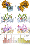
Fig. 4. IM459N21 solution structure and comparison of the modes of binding of IM172N22 and insulin.
a Correspondence of the backbone conformation of the hIR-bound IM172N22 peptide (green) with that of IM459N21 peptide (grey) determined in solution (for clarity, only six of the NMR-based IM459N21 models are shown). The orange bonds depict the IM172N22 cysteine residues and their associated disulfide bond; the white bonds depict those of IM459N21. Selected IM172N22 residues are labelled. b Overlay of the structure of IM172N22 (green) bound to domain FnIII-1′ (grey) with that of insulin (A chain pink, B chain dark blue) bound to the same domain (PDB 6SOF). The orange bonds depict the disulfide linkages within IM172N22 and within insulin. c, Correspondence of key [FnIII-1′]-engaging residues of IM172N22 with those of insulin. Chain termini are labelled and colors are as in panel b. d Proximity (asterisked) of the N terminus of a Site 1 peptide with that of the C terminus of a Site 2 peptide within the map of the IM459-bound hIR ectodomain. Peptides models are derived respectively from PDB 5J3H (S519C16 bound to the hIR L1-CR module) and from the IM172N22-bound IRΔβ-zip structure presented in this manuscript. The IM172N22 peptide is in green, the S519C16 peptide is in light magenta, receptor domain colors are as in the primary structure domain layout provided in Supplementary Fig. 2a.

Similar articles

Cited by

References

    1. Saltiel AR, Kahn CR. Insulin signalling and the regulation of glucose and lipid metabolism. Nature. 2001;414:799–806. doi: 10.1038/414799a. - DOI - PubMed
    1. Scapin G, et al. Structure of the insulin receptor–insulin complex by single-particle cryo-EM analysis. Nature. 2018;556:122–125. doi: 10.1038/nature26153. - DOI - PMC - PubMed
    1. Weis F, et al. The signalling conformation of the insulin receptor ectodomain. Nat. Commun. 2018;9:4420. doi: 10.1038/s41467-018-06826-6. - DOI - PMC - PubMed
    1. Uchikawa E, Choi E, Shang G, Yu H, Bai X-C. Activation mechanism of the insulin receptor revealed by cryo-EM structure of the fully liganded receptor-ligand complex. eLife. 2019;8:e48630. doi: 10.7554/eLife.48630. - DOI - PMC - PubMed
    1. Gutmann T, et al. Cryo-EM structure of the complete and ligand-saturated insulin receptor ectodomain. J. Cell Biol. 2020;219:e201907210. doi: 10.1083/jcb.201907210. - DOI - PMC - PubMed

Publication types