. 2022 Sep 28;10(1):158.
doi: 10.1186/s40168-022-01353-5.

Selected commensals educate the intestinal vascular and immune system for immunocompetence

Affiliations

Selected commensals educate the intestinal vascular and immune system for immunocompetence

Rossana Romero et al. Microbiome. .

Abstract

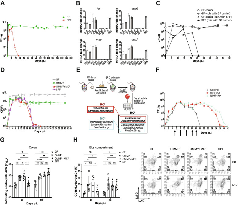
Background: The intestinal microbiota fundamentally guides the development of a normal intestinal physiology, the education, and functioning of the mucosal immune system. The Citrobacter rodentium-carrier model in germ-free (GF) mice is suitable to study the influence of selected microbes on an otherwise blunted immune response in the absence of intestinal commensals.

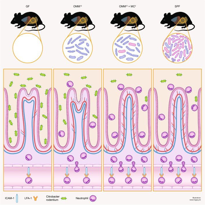
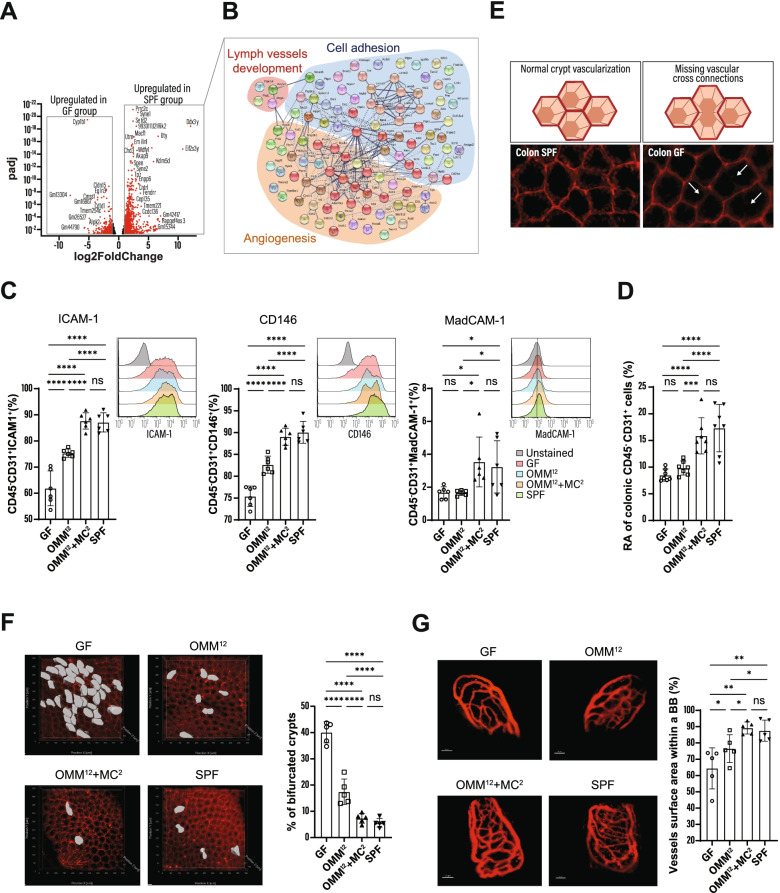
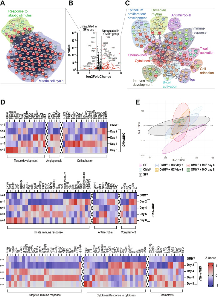
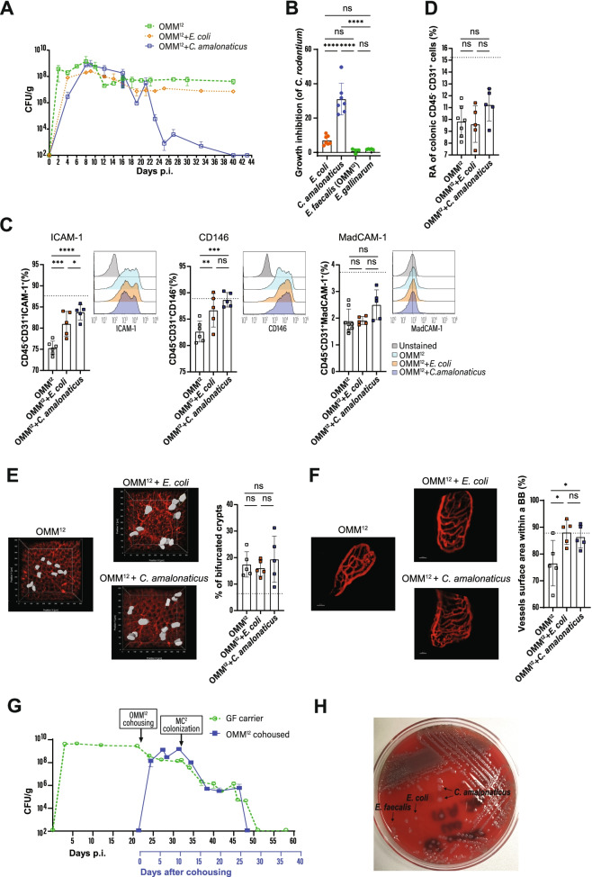
Results: Here, we describe that colonization of adult carrier mice with 14 selected commensal microbes (OMM12 + MC2) was sufficient to reestablish the host immune response to enteric pathogens; this conversion was facilitated by maturation and activation of the intestinal blood vessel system and the step- and timewise stimulation of innate and adaptive immunity. While the immature colon of C. rodentium-infected GF mice did not allow sufficient extravasation of neutrophils into the gut lumen, colonization with OMM12 + MC2 commensals initiated the expansion and activation of the visceral vascular system enabling granulocyte transmigration into the gut lumen for effective pathogen elimination.

Conclusions: Consortium modeling revealed that the addition of two facultative anaerobes to the OMM12 community was essential to further progress the intestinal development. Moreover, this study demonstrates the therapeutic value of a defined consortium to promote intestinal maturation and immunity even in adult organisms. Video Abstract.

Keywords: Asymptomatic infection; Blood vessel development; C. rodentium; Colonization resistance; Commensal imprinting; Endothelial cells; Enteric pathogen; Genome-guided microbiota; Intestinal maturation; Microbial consortia; Neutrophils; Oligo-Mouse-Microbiota.


Conflict of interest statement

The authors declare that they have no competing interests.

Figures

Fig. 1
Fig. 1
Commensals induce C. rodentium elimination by enhancing colonic neutrophil recruitment. A Course of C. rodentium infection in GF and SPF. B Kinetic of virulence gene expression in C. rodentium. C CFUs of C. rodentium in GF or SPF mice cohoused with C. rodentium-carrier mice. D Fecal CFUs in indicated mice after infection with C. rodentium. E Schema for identification of facultative anaerobic bacteria from the fecal microbiota of SPF mice. F C. rodentium-infected SPF mice were treated i.p. with neutrophil-depleting antibodies at indicated time points (arrows), and fecal CFUs were quantified. G Absolute cell number (ACN) of infiltrating neutrophils into the colon of mice at days 8 and 10 p.i. H Neutrophil frequencies in the colonic intraepithelial compartment at days 8 and 10 p.i. Animals were orally infected with (109 CFU/ml) C. rodentium. Data shown are mean ± SD of 2–3 independent experiments; each symbol represents the individual value for one mouse (D and E). One-way ANOVA was used: ****p < 0.0001; **p ≤ 0.01; *p ≤ 0.05; ns, not significant
Fig. 2
Fig. 2
Influence of commensals on maturation of the vascular and immune system. Sketch shows crypts, blood vessels, C. rodentium and neutrophils under different colonization conditions. GF mice have an immature colon and when infected with C. rodentium, they become lifelong carriers with high pathogen titers and low numbers of neutrophils in the colon. Colonization of GF animals with the OMM12 microbiota prior to C. rodentium infection leads to partial maturation of the colonic blood vessels and crypts. Furthermore, OMM12 colonization activates endothelial cells allowing neutrophils to extravasate into the lamina propria and lumen of the colon, with partial elimination of C. rodentium. Addition of MC2 to the OMM12 consortium further enhances tissue maturation of the colon allowing clearance of C. rodentium comparable to SPF mice
Fig. 3
Fig. 3
Commensals activate endothelial cells and promote intestinal angiogenesis. A Volcano plot showing fold change of gene expression in colonic endothelial cells of GF and SPF mice. Significant differences in gene expression between the two groups (log2-fold change ≥ 1; p-adj < 0.05) are marked in red. B STRING analysis for significantly upregulated transcripts of colonic endothelial cells from SPF mice. C Expression of ICAM1+, CD146+, and MadCAM1+ on colonic endothelial cells in indicated mice. D Relative abundance (RA) of colonic endothelial cells in the LP of indicated mice. E Schematic and microscopic representation of vascular cross connections in the colon. F Representative images and quantification of whole-mount staining indicated in mice. Partial vascularized crypts were quantified by insertion of contour surface modules (gray areas). G Intravital, multiphoton microscopy of small intestinal blood capillaries after i.v. injection of Qtracker™ 655. Vessel areas were quantified with trace image processing for morphometric measurements within a bounding box (BB). Scale bar: 30 μm. Results are representative of 4–5 experiments. Shown are representative data of at least two independent experiments with means ± SD; each symbol represents the individual value for one mouse (C-I). One-way ANOVA was used: ****p < 0.0001; **p ≤ 0.01; *p ≤ 0.05; ns, not significant
Fig. 4
Fig. 4
Commensal bacteria initiate gene expression of vascular and immune networks. AC Volcano plot and STRING analysis showing fold change of gene expression in whole colon of GF and OMM12 mice (B). STRING analysis for significantly upregulated transcripts in the colon of GF (A) and OMM12 mice (C). D Heat map of differentially expressed gene clusters in OMM12 and OMM12 + MC2 at indicated time points after MC2 addition, log2-fold change ≥ 1; p-adj < 0.05. n = 4 biological replicates per group. E Principal component analysis of colonic gene expression in differently colonized mice. The axes correspond to principal component 1 (x-axis) and 2 (y-axis)
Fig. 5
Fig. 5
Impact of E. coli and C. amalonaticus on intestinal maturation and colonization resistance. A Course of C. rodentium infection in indicated mice. B In vitro growth inhibition of C. rodentium in the presence of indicated bacteria. Data are shown as fold change compared to C. rodentium O/N culture control. C Expression of ICAM1+, CD146+, and MadCAM1+ on colonic endothelial cells. D Relative abundance (RA) of colonic LP CD45 CD31+ endothelial cells in colonized mice. E Representative quantification of vascularization in stained whole-mount tissues in indicated mice. Partial vascularized crypts were quantified by insertion of contour surface modules (gray areas). F Intravital, multiphoton microscopy of small intestinal blood capillaries after i.v. injection of Qtracker™ 655. Vessel areas were quantified with trace image processing to make morphometric measurements within a bounding box (BB). Scale bar: 30 μm. Results are representative of 4–5 experiments. G C. rodentium carriers were cohoused with OMM12 on day 22 p.i. All mice were gavaged 10 days later with MC2 bacteria. CFUs of C. rodentium were determined in feces. H Fecal colonies of E.coli, E.faecalis (from OMM12) and C.amalonaticus on Columbia blood agar universal plate from mice of experiment (G) after pathogen elimination. Animals were orally infected with (109 CFU/ml) C. rodentium. Data shown are mean ± SD of 2–3 independent experiments; each symbol represents the value for one mouse (DF). Dotted line represents the reference range for OMM12 + MC2 mice (DF). One-way ANOVA was used: ****p < 0.0001; **p ≤ 0.01; *p ≤ 0.05; ns, not significant

Similar articles

Cited by

References

    1. Round JL, Mazmanian SK. The gut microbiota shapes intestinal immune responses during health and disease. Nat Rev Immunol. 2009;9:313–323. doi: 10.1038/nri2515. - DOI - PMC - PubMed
    1. Derrien M, Alvarez AS, de Vos WM. The gut microbiota in the first decade of life. Trends Microbiol. 2019;27:997–1010. doi: 10.1016/j.tim.2019.08.001. - DOI - PubMed
    1. Perez PF, Doré J, Leclerc M, Levenez F, Benyacoub J, Serrant P, et al. Bacterial imprinting of the neonatal immune system: lessons from maternal cells? Pediatrics. 2007;119:e724–e732. doi: 10.1542/peds.2006-1649. - DOI - PubMed
    1. Al Nabhani Z, Dulauroy S, Marques R, Cousu C, Al Bounny S, Déjardin F, et al. A weaning reaction to microbiota is required for resistance to immunopathologies in the adult. Immunity. 2019;50:1276–1288.e5. doi: 10.1016/j.immuni.2019.02.014. - DOI - PubMed
    1. Kennedy EA, King KY, Baldridge MT. Mouse microbiota models: comparing germ-free mice and antibiotics treatment as tools for modifying gut bacteria. Front Physiol. 2018;0:1534. doi: 10.3389/fphys.2018.01534. - DOI - PMC - PubMed

Publication types