

A miniaturized and integrated dual-channel fluorescence module for multiplex real-time PCR in the portable nucleic acid detection system
- PMID: 36172017
- PMCID: PMC9510591
- DOI: 10.3389/fbioe.2022.996456
A miniaturized and integrated dual-channel fluorescence module for multiplex real-time PCR in the portable nucleic acid detection system
Abstract
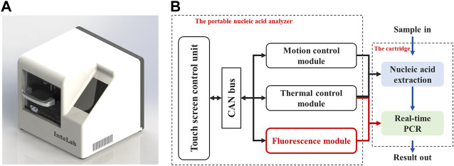
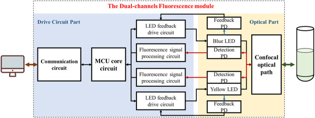
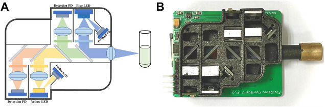
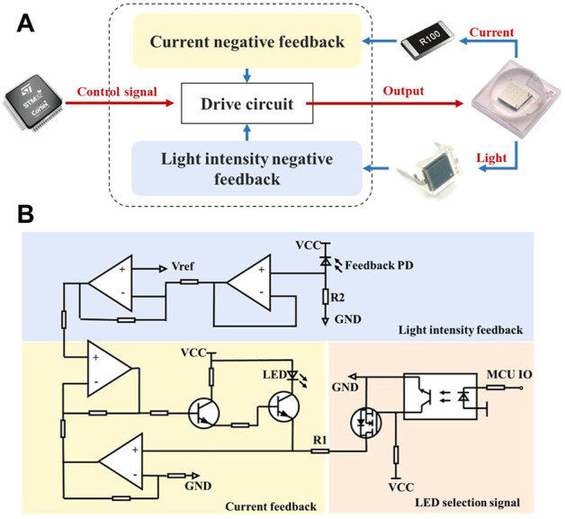
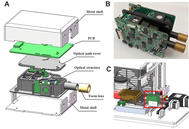
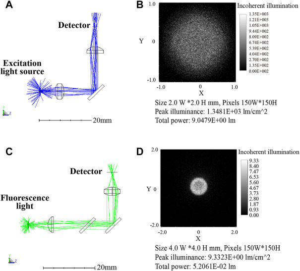
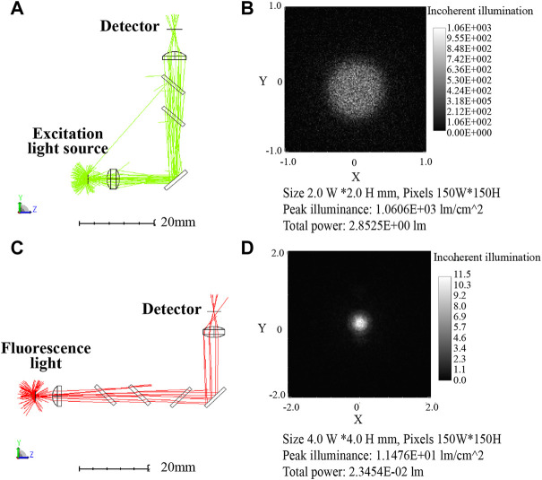
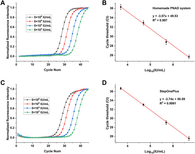
A portable nucleic acid detection (PNAD) system based on real-time polymerase chain reaction (real-time PCR) has been developed for point-of-care testing (POCT) of infectious disease pathogens. In order to achieve "sample-in, result-out" while keeping the system compact, the hardware system integrates optical, thermal and motion control modules in a limited space for nucleic acid extraction, purification, amplification and detection. Among these hardware modules, the fluorescence module is one of the most important modules, because its performance directly affects the accuracy and sensitivity of the testing results. In this paper, a miniaturized, high-sensitivity and integrated dual-channel fluorescence module have been proposed for the homemade PNAD system. Based on the principle of confocal optical path, two group of excitation-emission optical paths of different wavelengths are integrated in a small space. In terms of circuitry, a current-light dual negative feedback light emitting diode (LED) drive circuit is applied to improve the stability of the excited light source. All optical and electronic components are integrated in a metal box of 55 mm × 45 mm × 15 mm, that helps miniaturize the detection system. Two different modules have been assembled to fit various fluorescent dyes or probes with the set of excitation and emission as follow: module 1#: 470 nm/525 nm, 570 nm/630 nm; module 2#: 520 nm/570 nm, 630 nm/690 nm. Finally, hepatitis B virus (HBV) concentration gradient detection and multiplex detection of different gene targets of SARS-CoV-2 are carried out on the PNAD system equipped with these two fluorescence modules for evaluating their performances. Compared with the commercial real-time PCR instrument, our fluorescence module has good stability and detection sensitivity.
Keywords: LED drive circuit; confocal optical path; fluorescence detection; point-of-care testing; real-time PCR.
Copyright © 2022 Fang, Wang, Su, Liu, Chen, Chen, Jin and He.
Conflict of interest statement
The authors declare that the research was conducted in the absence of any commercial or financial relationships that could be construed as a potential conflict of interest.
Figures
Similar articles
-
A Highly Integrated and Diminutive Fluorescence Detector for Point-of-Care Testing: Dual Negative Feedback Light-Emitting Diode (LED) Drive and Photoelectric Processing Circuits Design and Implementation.Biosensors (Basel). 2022 Sep 16;12(9):764. doi: 10.3390/bios12090764. Biosensors (Basel). 2022. PMID: 36140149 Free PMC article.
-
Fast and Accurate Control Strategy for Portable Nucleic Acid Detection (PNAD) System Based on Magnetic Nanoparticles.J Biomed Nanotechnol. 2021 Mar 1;17(3):407-415. doi: 10.1166/jbn.2021.3028. J Biomed Nanotechnol. 2021. PMID: 33875075
-
[Development of a portable micro-liquid chromatograph].Se Pu. 2021 Sep;39(9):1030-1037. doi: 10.3724/SP.J.1123.2021.06029. Se Pu. 2021. PMID: 34486843 Free PMC article. Chinese.
-
[Research progress on analysis of human papillomavirus by microchip capillary electrophoresis].Se Pu. 2020 Oct 8;38(10):1179-1188. doi: 10.3724/SP.J.1123.2020.05016. Se Pu. 2020. PMID: 34213114 Review. Chinese.
-
Portable nucleic acid thermocyclers.Chem Soc Rev. 2013 Nov 21;42(22):8769-98. doi: 10.1039/c3cs60144g. Chem Soc Rev. 2013. PMID: 24030680 Review.
Cited by
-
Miniaturized electrophoresis: An integrated microfluidic cartridge with functionalized hydrogel-assisted LAMP for sample-to-answer analysis of nucleic acid.Biomicrofluidics. 2024 Dec 4;18(6):064104. doi: 10.1063/5.0211812. eCollection 2024 Dec. Biomicrofluidics. 2024. PMID: 39649103
-
FRETting about CRISPR-Cas Assays: Dual-Channel Reporting Lowers Detection Limits and Times-to-Result.ACS Sens. 2024 Jul 26;9(7):3616-3624. doi: 10.1021/acssensors.4c00652. Epub 2024 Jul 8. ACS Sens. 2024. PMID: 38978209 Free PMC article.
-
BRET-based biosensors for SARS-CoV-2 oligonucleotide detection.Front Bioeng Biotechnol. 2024 Jun 3;12:1353479. doi: 10.3389/fbioe.2024.1353479. eCollection 2024. Front Bioeng Biotechnol. 2024. PMID: 38887615 Free PMC article.
References
-
- Abdallah Z., Rumeau A., Fernandez A., Cibiel G., Llopis O. (2014). Nonlinear equivalent-circuit modeling of a fast photodiode. IEEE Phot. Technol. Lett. 26 (18), 1840–1842. 10.1109/lpt.2014.2337352 - DOI
LinkOut - more resources
Full Text Sources
Miscellaneous