Review
. 2022 Sep 12:10:985692.
doi: 10.3389/fbioe.2022.985692. eCollection 2022.

Application of medical imaging methods and artificial intelligence in tissue engineering and organ-on-a-chip

Affiliations
Review

Application of medical imaging methods and artificial intelligence in tissue engineering and organ-on-a-chip

Wanying Gao et al. Front Bioeng Biotechnol. .

Abstract

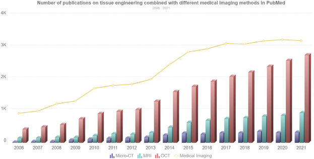
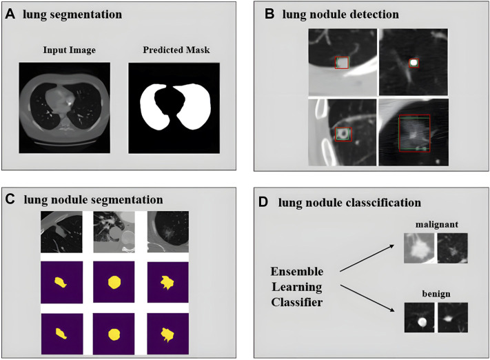
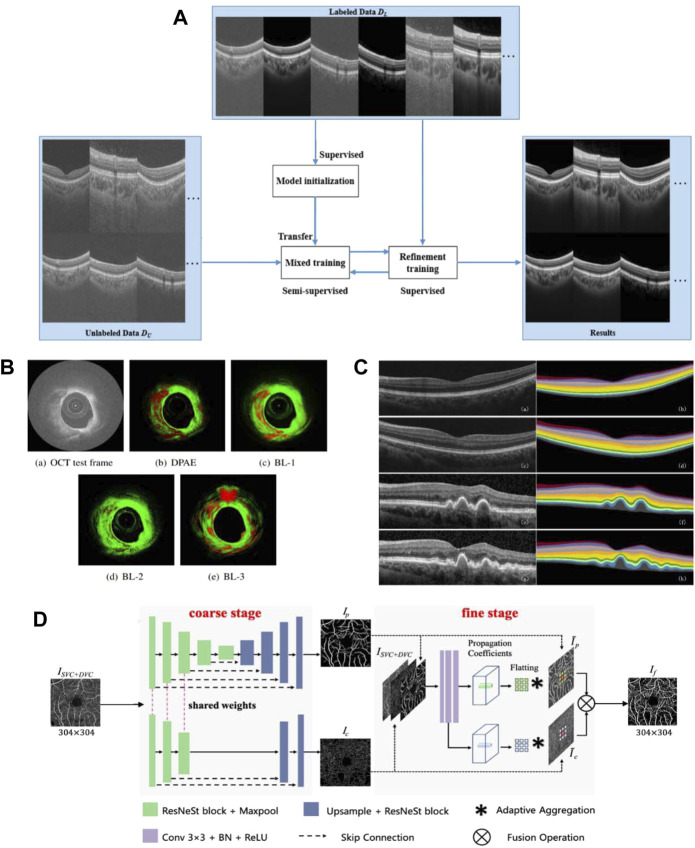
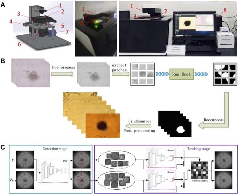
Organ-on-a-chip (OOC) is a new type of biochip technology. Various types of OOC systems have been developed rapidly in the past decade and found important applications in drug screening and precision medicine. However, due to the complexity in the structure of both the chip-body itself and the engineered-tissue inside, the imaging and analysis of OOC have still been a big challenge for biomedical researchers. Considering that medical imaging is moving towards higher spatial and temporal resolution and has more applications in tissue engineering, this paper aims to review medical imaging methods, including CT, micro-CT, MRI, small animal MRI, and OCT, and introduces the application of 3D printing in tissue engineering and OOC in which medical imaging plays an important role. The achievements of medical imaging assisted tissue engineering are reviewed, and the potential applications of medical imaging in organoids and OOC are discussed. Moreover, artificial intelligence - especially deep learning - has demonstrated its excellence in the analysis of medical imaging; we will also present the application of artificial intelligence in the image analysis of 3D tissues, especially for organoids developed in novel OOC systems.

Keywords: artificial intelligence; deep learning; medical imaging; organ-on-a-chip; tissue engineering.


Conflict of interest statement

Authors Xijing Zhang and Jianmin Yuan were employed by United Imaging Group. The remaining authors declare that the research was conducted in the absence of any commercial or financial relationships that could be construed as a potential conflict of interest.

Figures

FIGURE 1
FIGURE 1
The representative chips of Organ-on-a-chip. (A) Heart on a chip (adapted and modified from Marsano et al., 2016). (B) a Glomerulus Chip (adapted and modified from Musah et al., 2018). (C) Lung chip (adapted and modified from Huh et al., 2010). (D) Intestinal chip (adapted and modified from Kim et al., 2012).
FIGURE 2
FIGURE 2
Four different types of organoids constructed by researchers. (A) Intestinal organoids (adapted and modified from Gjorevski et al., 2016). (B) Skin organoids (adapted and modified from Lee et al., 2021). (C) Organoid Models of Human Liver Cancers (adapted and modified from Nuciforo et al., 2018). (D) Human blood vessel organoids (adapted and modified from Wimmer et al., 2019).
FIGURE 3
FIGURE 3
A typical process of 3D bioprinting includes 6 steps: 3D modeling, bioink selection, bioprinting, post-processing and application. (adapted and modified from Murthy et al., 2014; Vijayavenkataraman et al., 2018; Lee et al., 2021).
FIGURE 4
FIGURE 4
Number of publications on tissue engineering combined with different medical imaging methods in PubMed. The line chart represents the overall trend of the number of searches for medical imaging keywords, and the bar chart represents the number of searches for micro-CT, MRI, and OCT from 2006 to 2021.
FIGURE 5
FIGURE 5
Application of Different Imaging Methods in Tissue Engineering. (A) Noninvasive MRI images of labeled and unlabeled stent-grafts in mice, a, b) RARE T2-weighted images of labeled (a) and unlabeled. (b) seed scaffolds after implantation. Boxes represents the location of the graft and K represents kidneys. c, d) Corresponding T2 maps of a, b) (adapted and modified from Harrington et al., 2011). (B) micro-CT scanning of collagen-based scaffold (adapted and modified from Bartoš et al., 2018). (C) OCT imaging contrasting the effects of pulsatile stimulation on tissue-engineered vascular grafts culture, (a–f) are images with arterial stimulation, (g–l) are images without arterial stimulation (adapted and modified from Chen et al., 2017).
FIGURE 6
FIGURE 6
Research on artificial tissue with potential application in medical imaging. (A) Related research on artificial microvascular system. (a)Microvascular Networks Using Laser Patterns in Polyethylene Glycol Hydrogels. (b) 3D printed heart perfusion model (adapted and modified from Fleischer et al., 2020). (B) Brightfield and fluorescence images of brTEBV. (a) Brightfield image of brTEBV with a branch angle of 45° considering MC adhesion, where the dashed circles mark the inlet, side, main regions. (b) Fluorescence images of green-labeled MCs in the brTEBV region (adapted and modified from Lee et al., 2021).
FIGURE 7
FIGURE 7
Deep Learning Image Processing and Analysis Using Brain MRI as an Example. (A) Image reconstruction of brain MRI (adapted and modified from Lundervold et al., 2019). (B) Image denoising of brain MRI (adapted and modified from Lehtinen et al., 2018). (C) Smallest brain metastasis detected by artificial intelligence method marked with red bounding box (adapted and modified from Zhang et al., 2020). (D) Brain Tumor Segmentation Using UNet++ (adapted and modified from Zhou et al., 2020). (E) Feature images extracted by Parkinson’s diagnostic network (adapted and modified from Sivaranjini et al., 2020).
FIGURE 8
FIGURE 8
Some network frameworks applied in MRI image processing and analysis. (A) Spatial connectivity-aware network including LSTM blocks, exploiting sagittal information from adjacent slices. (adapted and modified from Li et al., 2019). (B) The Faster R-CNN network structure has two branches, the bounding box regression network and the classification network. The region proposal network is used to recommend bounding boxes that may have targets. (adapted and modified from Ren et al., 2015). (C) U-net is often used as a basic network. The blue boxes represent feature maps with different number of channels, the white boxes represent the copied feature maps, and the arrows represent operations such as convolution, pooling, etc. (adapted and modified from Ronneberger et al., 2015). (D) The UNet++ network obtained by improving U-Net, the downward arrow indicates downsampling, the upward arrow indicates down adoption, and the dot arrow indicates skip connections between feature maps. (adapted and modified from Zhou et al., 2019).
FIGURE 9
FIGURE 9
Achievements related to the realization of pulmonary nodule CAD system. (A) Input image and predicted mask for lung segmentation (adapted and modified from Tan et al., 2020). (B) Lung nodule detection results using deep learning. The green rectangle box represents ground truth and the red rectangle box represents the detection results (adapted and modified from Cao et al., 2019). (C) Segmentation results of large nodules, the first row is the original image, the second row is the radiologist’s manual annotation results, and the third row is the result of the network prediction (adapted and modified from Shi et al., 2020). (D) Classification of lung nodules into malignant and benign using an ensemble learning classifier (adapted and modified from Zhang et al., 2019).
FIGURE 10
FIGURE 10
Related achievements of AI processing OCT images. (A) structure of the proposed semi-supervised system (adapted and modified from Wang et al., 2021). (B) The result of plaque detection, the red area represents the detected plaque, and the green area represents the normal tissue (adapted and modified from Roy et al., 2016). (C) Retinal 10-layer segmentation prediction results, The left is the original image, the right is the segmentation result (adapted and modified from Li et al., 2019). (D) Architecture of the proposed OCTA-Net network (adapted and modified from Ma et al., 2021).
FIGURE 11
FIGURE 11
Relevant results of artificial intelligence combined with organoids. (A–B) System and process for edge detection of tumor spheres (adapted and modified from Chen et al., 2021b). (A) The SMART system for automated imaging and analysis. 1) Condenser with light source; 2) sample plate; 3) motorized x,y stage; 4) motorized Z-axis module; 5) objective wheel; 6) filter wheel; 7) CCD; 8) computer to control SMART system with developed software interface. (B) The process of tumor sphere edge detection. (C) Pipeline for organoids tracking (adapted and modified from Bian et al., 2021).

Similar articles

Cited by

References

    1. Abdullah K. A., Reed W. (2018). 3D printing in medical imaging and healthcare services. J. Med. Radiat. Sci. 65 (3), 237–239. 10.1002/jmrs.292 - DOI - PMC - PubMed
    1. Alkadi R., Taher F., El-baz A., Werghi N. (2018). A deep learning-based approach for the detection and localization of prostate cancer in T2 magnetic resonance images. J. Digit. Imaging 32 (5), 793–807. 10.1007/s10278-018-0160-1 - DOI - PMC - PubMed
    1. Amoroso N., La Rocca M., Bellantuono L., Diacono D., Fanizzi A., Lella E., et al. (2019). Deep learning and multiplex networks for accurate modeling of brain age. Front. Aging Neurosci. 11, 115. 10.3389/fnagi.2019.00115 - DOI - PMC - PubMed
    1. Andermatt S., Pezold S., Cattin P. (2016). “Multi-dimensional gated recurrent units for the segmentation of biomedical 3D-data,” in 2nd International Workshop on Deep Learning in Medical Image Analysis (DLMIA)/1st International Workshop on Large-Scale Annotation of Biomedical Data and Expert Label Synthesis (LABELS) (Athens, GREECE.
    1. Artegiani B., Clevers H. (2018). Use and application of 3D-organoid technology. Hum. Mol. Genet. 27 (R2), R99–R107. 10.1093/hmg/ddy187 - DOI - PubMed