

Functional network alterations in young brain tumor patients with radiotherapy-induced memory impairments and vascular injury
- PMID: 36172034
- PMCID: PMC9511024
- DOI: 10.3389/fneur.2022.921984
Functional network alterations in young brain tumor patients with radiotherapy-induced memory impairments and vascular injury
Abstract
Background: Cognitive impairment and cerebral microbleeds (CMBs) are long-term side-effects of cranial radiation therapy (RT). Previously we showed that memory function is disrupted in young patients and that the rate of cognitive decline correlates with CMB development. However, vascular injury alone cannot explain RT-induced cognitive decline. Here we use resting-state functional MRI (rsfMRI) to further investigate the complex mechanisms underlying memory impairment after RT.
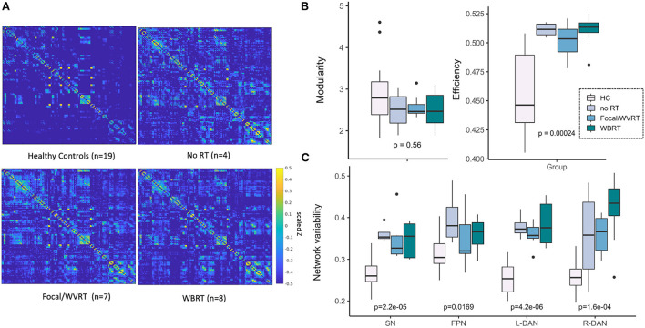
Methods: Nineteen young patients previously treated with or without focal or whole-brain RT for a brain tumor underwent cognitive testing followed by 7T rsfMRI and susceptibility-weighted imaging for CMB detection. Global brain modularity and efficiency, and rsfMRI signal variability within the dorsal attention, salience, and frontoparietal networks were computed. We evaluated whether MR metrics could distinguish age- and sex-matched controls (N = 19) from patients and differentiate patients based on RT exposure and aggressiveness. We also related MR metrics with memory performance, CMB burden, and risk factors for cognitive decline after RT.
Results: Compared to controls, patients exhibited widespread hyperconnectivity, similar modularity, and significantly increased efficiency (p < 0.001) and network variability (p < 0.001). The most abnormal values were detected in patients treated with high dose whole-brain RT, having supratentorial tumors, and who did not undergo RT but had hydrocephalus. MR metrics and memory performance were correlated (R = 0.34-0.53), though MR metrics were more strongly related to risk factors for cognitive worsening and CMB burden with evidence of functional recovery.
Conclusions: MR metrics describing brain connectivity and variability represent promising candidate imaging biomarkers for monitoring of long-term cognitive side-effects after RT.
Keywords: 7T MRI; brain connectivity; brain tumors; fMRI; memory; radiation therapy; vascular injury.
Copyright © 2022 Morrison, Walter, Mueller, Felton, Jakary, Stoller, Molinaro, Braunstein, Hess and Lupo.
Conflict of interest statement
The authors declare that the research was conducted in the absence of any commercial or financial relationships that could be construed as a potential conflict of interest.
Figures


Similar articles
-
Rate of radiation-induced microbleed formation on 7T MRI relates to cognitive impairment in young patients treated with radiation therapy for a brain tumor.Radiother Oncol. 2021 Jan;154:145-153. doi: 10.1016/j.radonc.2020.09.028. Epub 2020 Sep 20. Radiother Oncol. 2021. PMID: 32966846 Free PMC article.
-
Relationship between 7T MR-angiography features of vascular injury and cognitive decline in young brain tumor patients treated with radiation therapy.J Neurooncol. 2021 May;153(1):143-152. doi: 10.1007/s11060-021-03753-3. Epub 2021 Apr 24. J Neurooncol. 2021. PMID: 33893923 Free PMC article.
-
Risk factors of radiotherapy-induced cerebral microbleeds and serial analysis of their size compared with white matter changes: A 7T MRI study in 113 adult patients with brain tumors.J Magn Reson Imaging. 2019 Sep;50(3):868-877. doi: 10.1002/jmri.26651. Epub 2019 Jan 20. J Magn Reson Imaging. 2019. PMID: 30663150 Free PMC article.
-
Cerebral Microbleeds Are Associated With Increased Brain Iron and Cognitive Impairment in Patients With Cerebral Small Vessel Disease: A Quantitative Susceptibility Mapping Study.J Magn Reson Imaging. 2022 Sep;56(3):904-914. doi: 10.1002/jmri.28092. Epub 2022 Jan 31. J Magn Reson Imaging. 2022. PMID: 35099829
-
Longitudinal assessment of chemotherapy-induced changes in brain and cognitive functioning: A systematic review.Neurosci Biobehav Rev. 2018 Sep;92:304-317. doi: 10.1016/j.neubiorev.2018.05.019. Epub 2018 May 20. Neurosci Biobehav Rev. 2018. PMID: 29791867
Cited by
-
Longitudinal changes of regional spontaneous brain activity in nasopharyngeal carcinoma patients receiving chemoradiotherapy.Front Neurosci. 2025 Jul 25;19:1607727. doi: 10.3389/fnins.2025.1607727. eCollection 2025. Front Neurosci. 2025. PMID: 40787243 Free PMC article.
-
Depression and Anxiety After Radiation-Induced Brain Injury: A Review of Current Research Progress.Curr Oncol. 2025 Jul 26;32(8):419. doi: 10.3390/curroncol32080419. Curr Oncol. 2025. PMID: 40862788 Free PMC article. Review.
References
-
- Morrison MA, Mueller S, Felton E, Jakary A, Stoller S, Avadiappan S, et al. . Rate of radiation-induced microbleed formation on 7T MRI relates to cognitive impairment in young patients treated with radiation therapy for a brain tumor. Radiother Oncol. 154:145–53. (2020) 10.1016/j.radonc.2020.09.028 - DOI - PMC - PubMed