

Differences in brain activations between micro- and macro-expressions based on electroencephalography
- PMID: 36172039
- PMCID: PMC9511965
- DOI: 10.3389/fnins.2022.903448
Differences in brain activations between micro- and macro-expressions based on electroencephalography
Abstract
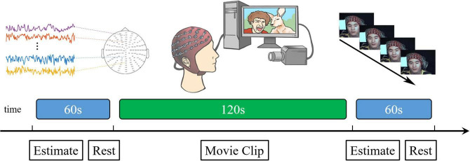
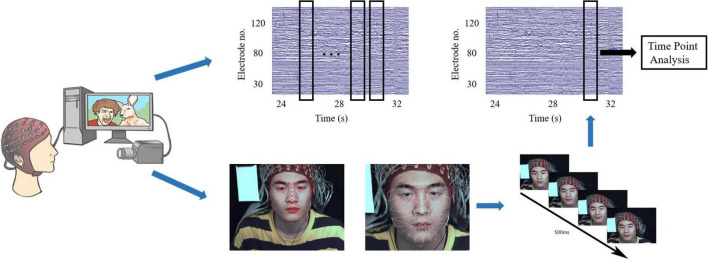
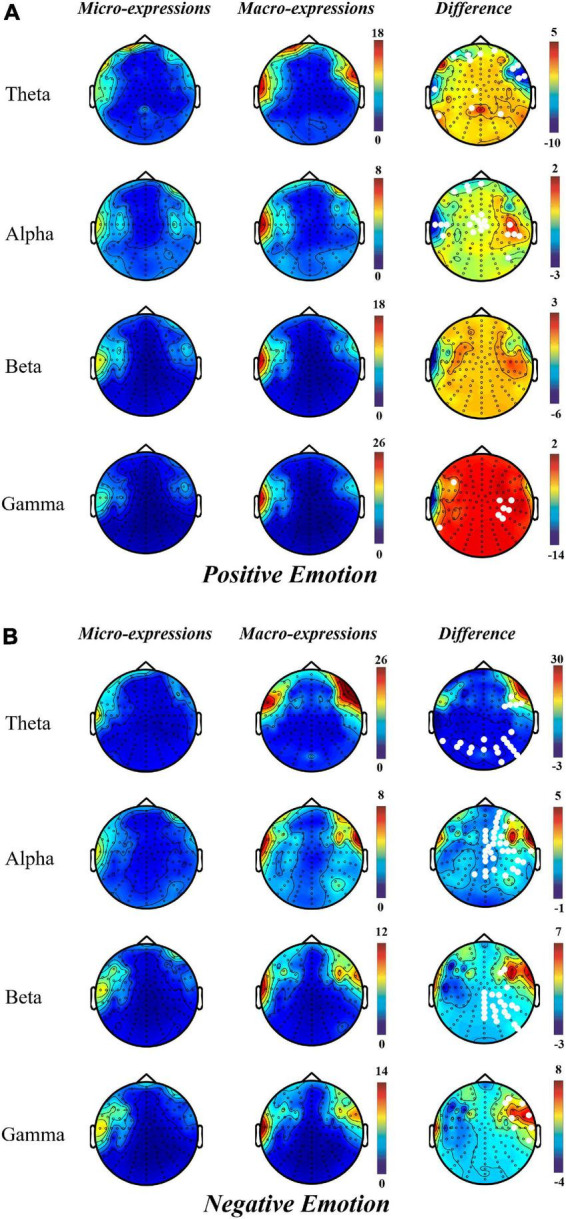
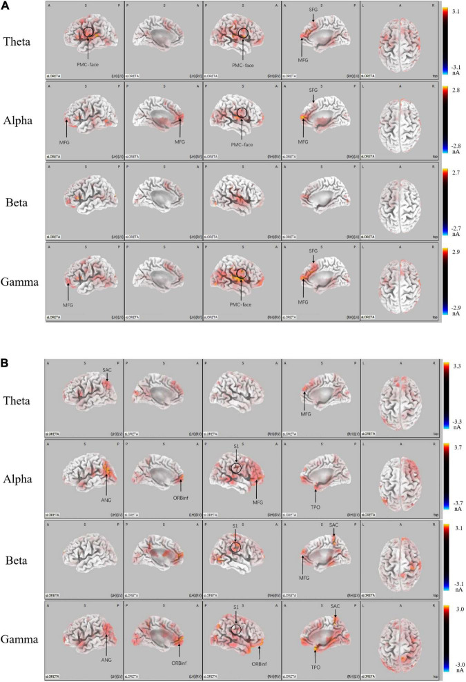
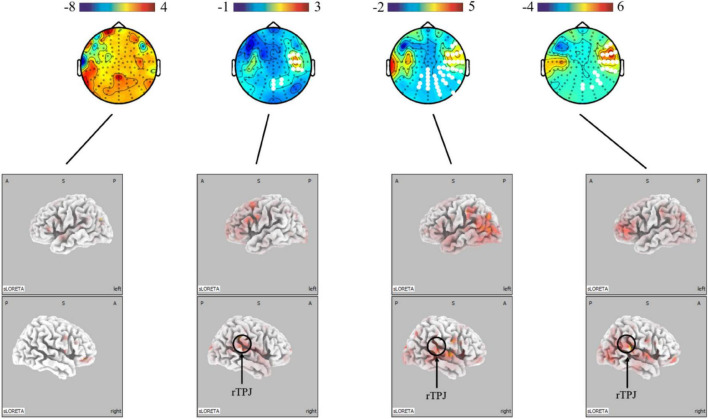
Micro-expressions can reflect an individual's subjective emotions and true mental state and are widely used in the fields of mental health, justice, law enforcement, intelligence, and security. However, the current approach based on image and expert assessment-based micro-expression recognition technology has limitations such as limited application scenarios and time consumption. Therefore, to overcome these limitations, this study is the first to explore the brain mechanisms of micro-expressions and their differences from macro-expressions from a neuroscientific perspective. This can be a foundation for micro-expression recognition based on EEG signals. We designed a real-time supervision and emotional expression suppression (SEES) experimental paradigm to synchronously collect facial expressions and electroencephalograms. Electroencephalogram signals were analyzed at the scalp and source levels to determine the temporal and spatial neural patterns of micro- and macro-expressions. We found that micro-expressions were more strongly activated in the premotor cortex, supplementary motor cortex, and middle frontal gyrus in frontal regions under positive emotions than macro-expressions. Under negative emotions, micro-expressions were more weakly activated in the somatosensory cortex and corneal gyrus regions than macro-expressions. The activation of the right temporoparietal junction (rTPJ) was stronger in micro-expressions under positive than negative emotions. The reason for this difference is that the pathways of facial control are different; the production of micro-expressions under positive emotion is dependent on the control of the face, while micro-expressions under negative emotions are more dependent on the intensity of the emotion.
Keywords: electroencephalography (EEG); emotion; expression inhibition; macro-expressions; micro-expressions.
Copyright © 2022 Zhao, Liu, Chen, Wang, Chen, Wang and Liu.
Conflict of interest statement
The authors declare that the research was conducted in the absence of any commercial or financial relationships that could be construed as a potential conflict of interest.
Figures
Similar articles
-
Responses of functional brain networks in micro-expressions: An EEG study.Front Psychol. 2022 Oct 28;13:996905. doi: 10.3389/fpsyg.2022.996905. eCollection 2022. Front Psychol. 2022. PMID: 36389479 Free PMC article.
-
Micro-Expression Recognition Based on Nodal Efficiency in the EEG Functional Networks.IEEE Trans Neural Syst Rehabil Eng. 2024;32:887-894. doi: 10.1109/TNSRE.2023.3347601. Epub 2024 Feb 26. IEEE Trans Neural Syst Rehabil Eng. 2024. PMID: 38190663
-
Affection of facial artifacts caused by micro-expressions on electroencephalography signals.Front Neurosci. 2022 Nov 24;16:1048199. doi: 10.3389/fnins.2022.1048199. eCollection 2022. Front Neurosci. 2022. PMID: 36507351 Free PMC article.
-
Emotion recognition in EEG signals using deep learning methods: A review.Comput Biol Med. 2023 Oct;165:107450. doi: 10.1016/j.compbiomed.2023.107450. Epub 2023 Sep 9. Comput Biol Med. 2023. PMID: 37708717 Review.
-
Perceiving emotional expressions in others: Activation likelihood estimation meta-analyses of explicit evaluation, passive perception and incidental perception of emotions.Neurosci Biobehav Rev. 2016 Dec;71:810-828. doi: 10.1016/j.neubiorev.2016.10.020. Epub 2016 Nov 9. Neurosci Biobehav Rev. 2016. PMID: 27836460 Review.
Cited by
-
EEG-Based Micro-Expression Recognition: Flexible Brain Network Reconfiguration Supporting Micro-Expressions Under Positive Emotion.Psychol Res Behav Manag. 2025 Apr 2;18:781-796. doi: 10.2147/PRBM.S506311. eCollection 2025. Psychol Res Behav Manag. 2025. PMID: 40191181 Free PMC article.
References
-
- Adolphs R. (2002). Neural systems for recognizing emotion. Curr. Opin. Neurobiol. 12 169–177. - PubMed
-
- Arrington C. M., Carr T. H., Mayer A. R., Rao S. M. (2000). Neural mechanisms of visual attention: Object-based selection of a region in space. J. Cogn. Neurosci. 12 106–117. - PubMed
-
- Asthana A., Zafeiriou S., Cheng S., Pantic M. (2013). “Robust discriminative response map fitting with constrained local models,” in Proceedings of the 2013 IEEE conference on computer vision and pattern recognition, Portland, OR.
LinkOut - more resources
Full Text Sources