. 2022 Sep 12:13:1011065.
doi: 10.3389/fphar.2022.1011065. eCollection 2022.

Proteomics unite traditional toxicological assessment methods to evaluate the toxicity of iron oxide nanoparticles

Affiliations

Proteomics unite traditional toxicological assessment methods to evaluate the toxicity of iron oxide nanoparticles

Junyuan Han et al. Front Pharmacol. .

Abstract

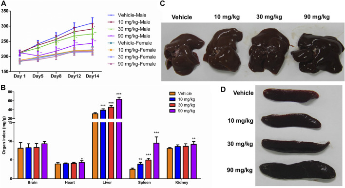
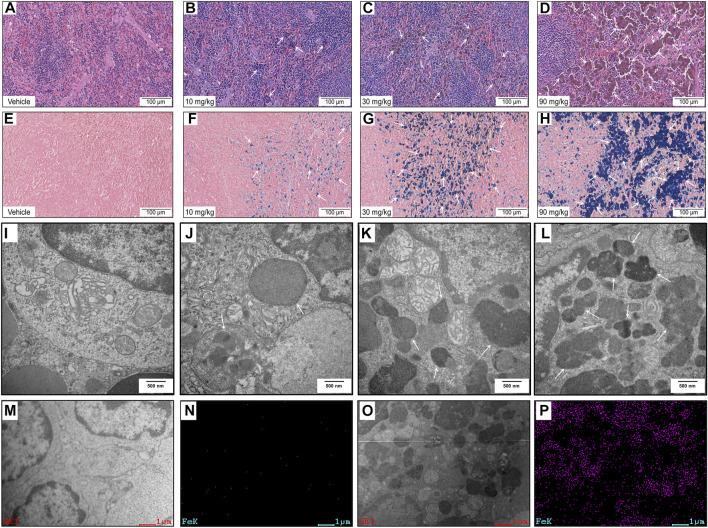
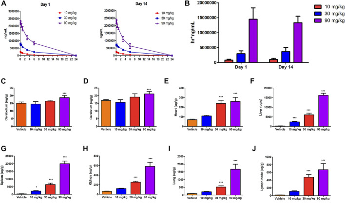
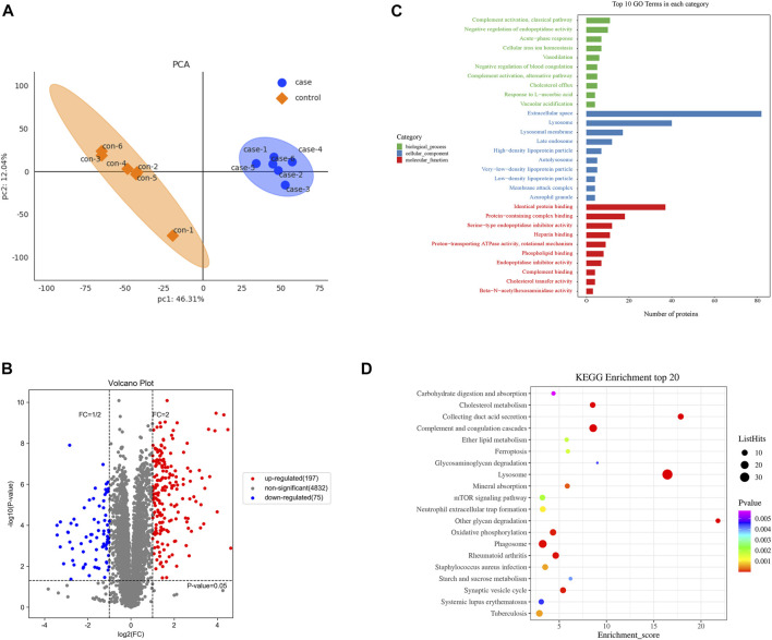
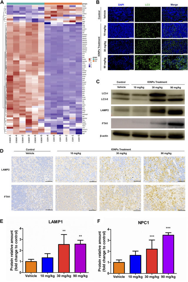
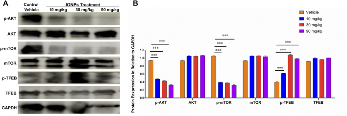
Iron oxide nanoparticles (IONPs) are the first generation of nanomaterials approved by the Food and Drug Administration for use as imaging agents and for the treatment of iron deficiency in chronic kidney disease. However, several IONPs-based imaging agents have been withdrawn because of toxic effects and the poor understanding of the underlying mechanisms. This study aimed to evaluate IONPs toxicity and to elucidate the underlying mechanism after intravenous administration in rats. Seven-week-old rats were intravenously administered IONPs at doses of 0, 10, 30, and 90 mg/kg body weight for 14 consecutive days. Toxicity and molecular perturbations were evaluated using traditional toxicological assessment methods and proteomics approaches, respectively. The administration of 90 mg/kg IONPs induced mild toxic effects, including abnormal clinical signs, lower body weight gain, changes in serum biochemical and hematological parameters, and increased organ coefficients in the spleen, liver, heart, and kidneys. Toxicokinetics, tissue distribution, histopathological, and transmission electron microscopy analyses revealed that the spleen was the primary organ for IONPs elimination from the systemic circulation and that the macrophage lysosomes were the main organelles of IONPs accumulation after intravenous administration. We identified 197 upregulated and 75 downregulated proteins in the spleen following IONPs administration by proteomics. Mechanically, the AKT/mTOR/TFEB signaling pathway facilitated autophagy and lysosomal activation in splenic macrophages. This is the first study to elucidate the mechanism of IONPs toxicity by combining proteomics with traditional methods for toxicity assessment.

Keywords: AKT/mTOR/TFEB signaling pathway; autophagy; iron oxide nanoparticles; lysosome; proteomics; toxicity.


Conflict of interest statement

The authors declare that the research was conducted in the absence of any commercial or financial relationships that could be construed as a potential conflict of interest.

Figures

FIGURE 1
FIGURE 1
General physiological findings and organ coefficients after IONPs administration: (A) Changes in the body weight of male and female rats treated with iron oxide nanoparticles (IONPs) for 14 days. Results are expressed as means ± standard deviation (n = 11). (B) Organ coefficients for the brain, heart, liver, spleen, and kidneys in rats administered indicated doses of IONPs relative to those administered the vehicle. Results are expressed as means ± standard deviation (n = 10). (C,D) Representative images of the liver and spleen following the administration of vehicle or IONPs for 14 days. *p < 0.05, **p < 0.01, ***p < 0.001 versus vehicle.
FIGURE 2
FIGURE 2
Histopathology and ultrastructural alterations in the spleen: (A–H) Representative images of specimens stained with hematoxylin and eosin (A–D) and Perls’ Prussian blue (E–H) after treatment with vehicle or IONPs for 14 days. (I–L) Representative transmission electron microscopy (TEM) images of spleen specimens following treatment with vehicle or IONPs for 14 days. (M–P) TEM-energy-dispersive spectrometry (EDS) analysis of the spleen after treatment with vehicle or 90 mg/kg IONPs for 14 days; (M,O) and (N,P) show the TEM and corresponding iron element distribution in the EDS images of the spleens after treatment with vehicle and IONPs, respectively. The EDS images show the element distribution of iron in the entire TEM image.
FIGURE 3
FIGURE 3
Toxicokinetics and tissue distribution after IONPs administration: (A) Mean serum concentration–time curves of IONPs after the first (day 1) and last doses (day 14). (B) The area under the concentration–time curve on days 1 and 14 of IONPs administration at different doses (C–J) Organ-specific distribution of IONPs after administration for 14 consecutive days. All results are expressed as means ± standard deviation (n = 6). *p < 0.05, **p < 0.01, ***p < 0.001 vs. vehicle.
FIGURE 4
FIGURE 4
Proteomics analysis of differentially expressed proteins in the spleen after IONPs administration: (A) Principal Component Analysis of case (90 mg/kg IONPs) and control (vehicle) samples. (B) Volcano plots showing the number of differentially abundant proteins and the distribution of significance and fold change between the vehicle and 90 mg/kg IONPs groups. The gray dots are proteins with nonsignificant difference in expression, and the red and blue dots indicate significantly upregulated and downregulated proteins, respectively. (C) The Gene Ontology (GO) enrichment analysis graph showing top 10 terms enriched by p values in each of the ontological categories of biological process, cellular component, and molecular function in the 90 mg/kg IONPs group. (D) The Kyoto Encyclopedia of Genes and Genomes pathways enriched by DEPs identified between the 90 mg/kg IONPs and vehicle groups. The ListHits indicates the number of differentially abundant proteins enriched in specific pathways.
FIGURE 5
FIGURE 5
IONPs administration leads to lysosomal activation and autophagy in splenic macrophages: (A) Clustering heatmap of differentially expressed proteins associated with lysosomal activation (B) Immunofluorescence staining showing the expression of light chain 3 (LC3) in spleen specimens collected from rats treated with vehicle or different doses of IONPs. Nuclei are stained with DAPI. Scale bars: 50 μm. (C) The protein levels of LC3, FTH1, and LAMP2 were analyzed using western blotting, and β-actin was used as internal reference (D) Immunochemical staining showing the expression of ferritin heavy chain 1 (FTH1) and lysosome-associated membrane protein (LAMP) 2 in spleen specimens of rats treated with vehicle or different doses of IONPs. (E) and (F) The verification of LAMP1 and NPC1 abundance using parallel reaction monitoring. Data are shown as means ± standard deviation (n = 6). **p < 0.01, ***p < 0.001 vs. vehicle.
FIGURE 6
FIGURE 6
The AKT/mTOR/TFEB signaling pathway facilitates in IONPs-induced autophagy: (A) Representative western blotting image for p-ATK, AKT, p-mTOR, mTOR, p-TFEB, and TFEB in spleen samples of rats after IONPs administration. The protein expression levels of p-ATK, AKT, p-mTOR, mTOR, p-TFEB, and TFEB were semiquantitatively determined by measuring the integrated optical density of bands based on the western blot images. GAPDH was used as an internal reference. (B) Corresponding graph showing relative changes in protein expression levels. ***p < 0.001 vs. vehicle.

References

    1. Allard-Vannier E., Cohen-Jonathan S., Gautier J., Herve-Aubert K., Munnier E., Souce M., et al. (2012). Pegylated magnetic nanocarriers for doxorubicin delivery: A quantitative determination of stealthiness in vitro and in vivo . Eur. J. Pharm. Biopharm. 81 (3), 498–505. 10.1016/j.ejpb.2012.04.002 - DOI - PubMed
    1. Anderson S. D., Gwenin V. V., Gwenin C. D. (2019). Magnetic functionalized nanoparticles for biomedical, drug delivery and imaging applications. Nanoscale Res. Lett. 14 (1), 188. 10.1186/s11671-019-3019-6 - DOI - PMC - PubMed
    1. Andrews N. C. (2000). Iron homeostasis: Insights from genetics and animal models. Nat. Rev. Genet. 1 (3), 208–217. 10.1038/35042073 - DOI - PubMed
    1. Askri D., Cunin V., Beal D., Berthier S., Chovelon B., Arnaud J., et al. (2019a). Investigating the toxic effects induced by iron oxide nanoparticles on neuroblastoma cell line: An integrative study combining cytotoxic, genotoxic and proteomic tools. Nanotoxicology 13 (8), 1021–1040. 10.1080/17435390.2019.1621399 - DOI - PubMed
    1. Askri D., Cunin V., Ouni S., Beal D., Rachidi W., Sakly M., et al. (2019b). Effects of iron oxide nanoparticles (gamma-Fe2O3) on liver, lung and brain proteomes following sub-acute intranasal exposure: A new toxicological assessment in rat model using iTRAQ-based quantitative proteomics. Int. J. Mol. Sci. 20 (20), E5186. 10.3390/ijms20205186 - DOI - PMC - PubMed