. 2022 Sep 21:15:2003-2012.
doi: 10.2147/CCID.S379609. eCollection 2022.

The Microbiome in Comedonal Contents of Inflammatory Acne Vulgaris is Composed of an Overgrowth of Cutibacterium Spp. and Other Cutaneous Microorganisms

Affiliations

The Microbiome in Comedonal Contents of Inflammatory Acne Vulgaris is Composed of an Overgrowth of Cutibacterium Spp. and Other Cutaneous Microorganisms

Narifumi Akaza et al. Clin Cosmet Investig Dermatol. .

Abstract

Background: Acne vulgaris (acne) and cutaneous resident microorganisms are considered to be closely related. However, the bacterial and fungal microbiota in the comedonal contents of inflammatory acne lesions have not yet been investigated in detail.

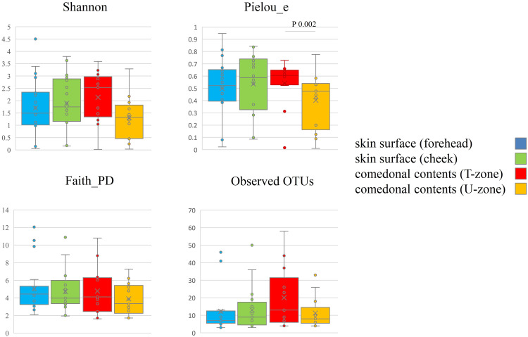
Purpose: To clarify the relationship between cutaneous microorganisms and acne, we examined the microbiome in the comedonal contents of inflammatory acne and on the facial skin of patients with acne using 16s rRNA and ITS gene sequencing with a next-generation sequencer (NGS).

Patients and methods: Twenty-two untreated Japanese acne outpatients were examined. The comedonal contents of inflammatory acne lesions on the face were collected using a comedo extractor. Skin surface samples from facial skin were collected using the swab method.

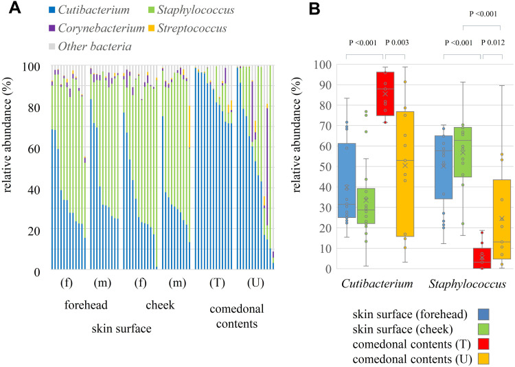
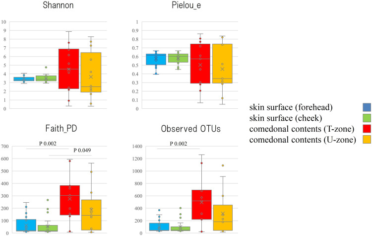
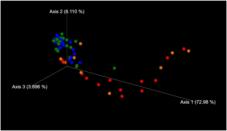
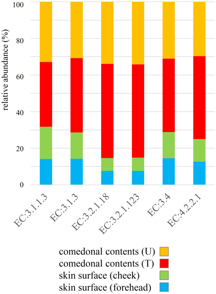
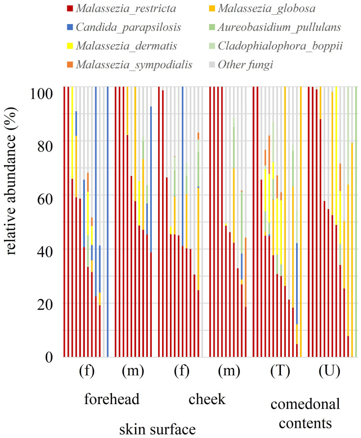
Results: The results obtained revealed that the predominant bacteria in the comedonal contents of inflammatory acne were Cutibacterium spp. (more prominent in areas with large amounts of sebum), while those on the skin surface were Staphylococcus spp. Malassezia spp., particularly Malassezia restricta, were the predominant fungi in both the comedonal contents of inflammatory acne and on the skin surface. The bacterial microbiome in comedonal contents exhibited stronger metabolic activity, including the production of enzymes related to acne, than that on the skin surface.

Conclusion: These results indicate that acne is an inflammatory disease involving the overgrowth of Cutibacterium acnes and other cutaneous resident microorganisms, including Malassezia spp.

Keywords: Cutibacterium; Malassezia; Staphylococcus; acne vulgaris; microbiome; microbiota; next generation sequencing.


Conflict of interest statement

The authors report no conflicts of interest in this work.

Figures

Figure 1
Figure 1
Genus classification of 16s rRNA clones in comedonal contents of inflammatory acne and on the skin surface of acne patients. (A) Individual distribution of the main bacterial genus arranged in descending order of Cutibacterium spp.; (B) boxplots of Cutibacterium and Staphylococcus spp. are shown. f, Female; m, male; T, T-zone; U, U-zone. Only bacteria with an average detection rate of 1% or higher are listed, while those with an average detection rate of less than 1% are classified as others. The Mann–Whitney U-test was employed for a statistical analysis of the skin surface of the forehead and cheek, the comedonal contents of the T-zone and U-zone, the skin surface of the forehead and the comedonal contents of T-zone, and the skin surface of cheek and the comedonal contents of the U-zone. Significant differences were observed between Cutibacterium and Staphylococcus spp. in the comedonal contents of the T-zone and on the skin surface of the forehead (P < 0.05). Significant differences were also noted between Cutibacterium and Staphylococcus spp. in the comedonal contents of the T- and U-zones (P < 0.05). No significant differences were found in any bacteria on the skin surface (forehead and cheek) or in comedonal contents (T- and U-zones) between females and males.
Figure 2
Figure 2
Alpha diversity of 16s rRNA clones in comedonal contents of inflammatory acne and on the skin surface of acne patients. The Mann–Whitney U-test was employed for a statistical analysis of the skin surface of the forehead and cheek, the comedonal contents of the T-zone and U-zone, the skin surface of the forehead and the comedonal contents of the T-zone, and the skin surface of the cheek and the comedonal contents of the U-zone. In the analysis using Faith’s phylogenetic diversity and the observed OTUs, diversities were higher in the comedonal contents of the T- and U-zones than on the skin surface of the forehead and cheek (P < 0.05); however, no significant differences were observed by the Shannon index or Pielou’s evenness index. No significant differences were found in either index on the skin surface (forehead and cheek) or in comedonal contents (T- and U-zones) between females and males.
Figure 3
Figure 3
Beta diversity (weighted UniFrac values) of 16s rRNA clones in comedonal contents of inflammatory acne and on the skin surface of acne patients. Blue, skin surface (forehead); green, skin surface (cheek); red, comedonal contents of the T-zone; orange, comedonal contents of the U-zone. Significant differences were observed between the bacterial microbiome of the comedonal contents and that on the skin surface by PERMANOVA. P value; the skin surface of the forehead versus the comedonal contents of the T-zone, <0.001; the skin surface of the cheek versus the comedonal contents of the U-zone, 0.006.
Figure 4
Figure 4
Functional prediction of 16s rRNA clones in comedonal contents of inflammatory acne and on the skin surface of acne patients. Data are arranged in descending order of comedonal contents. f, Female; m, male; T, T-zone; U, U-zone. The median value of each group was used as data, and ratios are shown in the graph. Enzymes related to acne; lipase, EC 3.1.1.3; phosphatase, EC 3.1.3; neuraminidase, EC 3.2.1.18; endoglycoceramidase, EC 3.2.1.123; protease, EC 3.4; hyaluronate lyase, EC 4.2.2.1.
Figure 5
Figure 5
Species classifications of ITS clones in comedonal contents of inflammatory acne and on the skin surface of acne patients. The individual distribution of the main fungal species arranged in descending order of M. restricta is shown. f, Female; m, male; T, T-zone; U, U-zone. Only fungi with an average detection rate of 1% or higher are listed, and fungi with an average detection rate of less than 1% are classified as others. The Mann–Whitney U-test was employed for a statistical analysis of the skin surface of the forehead and cheek, the comedonal contents of the T-zone and U-zone, the skin surface of the forehead and the comedonal contents of the T-zone, and the skin surface of the cheek and the comedonal contents of the U-zone. No significant differences were observed between comedonal contents and the skin surface. Furthermore, no significant differences were noted in any fungus on the skin surface (forehead and cheek) or in comedonal contents (T- and U-zones) between females and males.
Figure 6
Figure 6
Alpha diversity of ITS clones in comedonal contents of inflammatory acne and on the skin surface of acne patients. f, Female; m, male; T, T-zone; U, U-zone. The Mann–Whitney U-test was employed for a statistical analysis of the skin surface of the forehead and cheek, the comedonal contents of the T-zone and U-zone, the skin surface of the forehead and the comedonal contents of the T-zone, and the skin surface of the cheek and the comedonal contents of the U-zone. In an analysis using Pielou’s evenness index, diversities in comedonal contents were higher in the T-zone than in the U-zone, while no significant differences were observed by Shannon’s index, Pielou’s evenness index, or the observed OTUs (P < 0.05).
Figure 7
Figure 7
Beta diversity (weighted UniFrac values) of ITS clones in comedonal contents of inflammatory acne and on the skin surface of acne patients. Blue, skin surface (forehead); green, skin surface (cheek); red, comedonal contents of the T-zone; orange, comedonal contents of the U-zone. There were no significant differences between the fungal microbiome of the comedonal contents and that on the skin surface by PERMANOVA (P > 0.05).

References

    1. Xu H, Li H. Acne, the skin microbiome, and antibiotic treatment. Am J Clin Dermatol. 2019;20:335–344. doi:10.1007/s40257-018-00417-3 - DOI - PMC - PubMed
    1. Akaza N, Akamatsu H, Kishi M, et al. Effects of Propionibacterium acnes on various mRNA expression levels in normal human epidermal keratinocytes in vitro. J Dermatol. 2009;36:213–223. doi:10.1111/j.1346-8138.2009.00626.x - DOI - PubMed
    1. Ingham E, Holland KT, Gowland G, Cunliffe WJ. Partial purification and characterization of lipase (EC 3.1.1.3) from Propionibacterium acnes. J Gen Microbiol. 1981;124:393–401. doi:10.1099/00221287-124-2-393 - DOI - PubMed
    1. Jarrousse V, Castex-Rizzi N, Khammari A, Charveron M, Dreno B. Modulation of integrins and filaggrin expression by Propionibacterium acnes extracts on keratinocytes. Arch Dermatol Res. 2007;299:441–447. doi:10.1007/s00403-007-0774-5 - DOI - PubMed
    1. Iinuma K, Sato T, Akimoto N, et al. Involvement of Propionibacterium acnes in the augmentation of lipogenesis in hamster sebaceous glands in vivo and in vitro. J Invest Dermatol. 2009;129:2113–2119. doi:10.1038/jid.2009.46 - DOI - PubMed