Clinicopathological features and individualized treatment of kidney involvement in B-cell lymphoproliferative disorder
- PMID: 36172352
- PMCID: PMC9510618
- DOI: 10.3389/fimmu.2022.903315
Clinicopathological features and individualized treatment of kidney involvement in B-cell lymphoproliferative disorder
Abstract
Background: Due to the various clinical and pathological manifestations of kidney involvement in lymphoproliferative disorder (LPD), the whole spectrum of kidney disease in LPD is still unclear, and data on kidney prognosis is scarce.
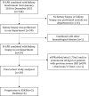
Methods: We retrospectively reviewed the renal pathology profiles from January 2010 to December 2021, and 28 patients with B-cell LPD combined with intact renal biopsy data were included.
Results: There were 20 men and eight women aging 41 to 79 years at the time of renal biopsy (median age 62 years). According to hematological diagnosis, patients were classified into four groups: chronic lymphocytic leukemia (CLL) (group1, n=7), Waldenström macroglobulinemia/lymphoplasmacytic lymphoma (WM/LPL) (group 2, n=8; WM, n=6; LPL, n=2), Other non-Hodgkin's lymphomas (NHL) (group3, n=7; diffuse large B-cell lymphoma (DLBCL), n=2; mucosa-associated lymphoid tissue (MALT) lymphoma, n=4; Low grade B-cell lymphoma, n=1), and monoclonal gammopathy of undetermined significance/monoclonal gammopathy of renal significance (MGUS/MGRS) (group 4, n=6). Median serum creatinine (Scr) level was 129 (range,59-956) umol/L. Eight patients (29%) were presented with acute kidney injury (AKI), and five patients (18%) required hemodialysis upon admission. Twenty-three patients (82%) presented with proteinuria (median protein excretion, 2.14 g/d), 11(39%) of whom had the nephrotic syndrome. Interstitial malignant infiltration was the most frequent renal lesion (n=6). Eight patients underwent immunohistochemistry of renal tissues, of which three patients (CLL, n=1; LPL, n=1; WM, n=1) had confirmed lymphoma infiltrates, and the infiltrating cells in the remaining five patients (CLL, n=1; MALT lymphoma, n=2; MGUS, n=2) were considered unrelated to lymphoma. The most common glomerular diseases were renal amyloidosis (n=4) and membranous nephropathy (n=4). Only 20 patients were treated, 13 of whom were treated with rituximab separately or in combination. The median follow-up time was 11 months. Of these, six had achieved hematological response, complete response in five cases. Eight had achieved renal response. At the end-of-study visit, four patients died and two progressed to end stage kidney disease (ESKD).
Conclusion: In conclusion, the clinicopathological spectrum of renal involvement in BLPD is diverse. Renal biopsy and immunohistochemistry are required for early diagnosis and prognostic assessment.
Keywords: Waldenström macroglobulinemia/lymphoplasmacytoid lymphoma; chronic lymphocytic leukemia; kidney involvement; lymphoproliferative disorders; monoclonal gammopathy of undetermined significance; non-Hodgkin’s lymphomas.
Copyright © 2022 Nie, Sun, Zhang, Yuan, Mao, Wang, Li, Duan, Xing and Zhang.
Conflict of interest statement
The authors declare that the research was conducted in the absence of any commercial or financial relationships that could be construed as a potential conflict of interest.
Figures


Similar articles
-
Kidney Involvement of Patients with Waldenström Macroglobulinemia and Other IgM-Producing B Cell Lymphoproliferative Disorders.Clin J Am Soc Nephrol. 2018 Jul 6;13(7):1037-1046. doi: 10.2215/CJN.13041117. Epub 2018 May 30. Clin J Am Soc Nephrol. 2018. PMID: 29848505 Free PMC article.
-
Clinicopathological spectrum of renal parenchymal involvement in B-cell lymphoproliferative disorders.Kidney Int. 2019 Jul;96(1):94-103. doi: 10.1016/j.kint.2019.01.027. Epub 2019 Mar 15. Kidney Int. 2019. PMID: 30987838
-
Kidney diseases associated with monoclonal immunoglobulin M-secreting B-cell lymphoproliferative disorders: a case series of 35 patients.Am J Kidney Dis. 2015 Nov;66(5):756-67. doi: 10.1053/j.ajkd.2015.03.035. Epub 2015 May 16. Am J Kidney Dis. 2015. PMID: 25987261
-
Rituximab: a review of its use in non-Hodgkin's lymphoma and chronic lymphocytic leukaemia.Drugs. 2003;63(8):803-43. doi: 10.2165/00003495-200363080-00005. Drugs. 2003. PMID: 12662126 Review.
-
Diagnostic framing of IgM monoclonal gammopathy: Focus on Waldenström macroglobulinemia.Hematol Oncol. 2019 Apr;37(2):117-128. doi: 10.1002/hon.2539. Epub 2018 Sep 7. Hematol Oncol. 2019. PMID: 30192023 Review.
Cited by
-
Case report: Acute kidney injury as the initial manifestation of chronic lymphocytic leukemia/small lymphocytic lymphoma.Front Med (Lausanne). 2023 Oct 20;10:1279005. doi: 10.3389/fmed.2023.1279005. eCollection 2023. Front Med (Lausanne). 2023. PMID: 37928472 Free PMC article.
-
The rare phenomenon of glomerulonephritis presentation in extra-nodal marginal zone lymphoma: summary of two cases and review of the literature.Ann Hematol. 2025 Feb;104(2):1127-1138. doi: 10.1007/s00277-025-06245-w. Epub 2025 Feb 7. Ann Hematol. 2025. PMID: 39918599 Free PMC article. Review.
-
Modified Endothelial Activation and Stress Index: A New Predictor for Survival Outcomes in Classical Hodgkin Lymphoma Treated with Doxorubicin-Bleomycin-Vinblastine-Dacarbazine-Based Therapy.Diagnostics (Basel). 2025 Jan 14;15(2):185. doi: 10.3390/diagnostics15020185. Diagnostics (Basel). 2025. PMID: 39857069 Free PMC article.
-
Case Report: Membranous nephropathy associated with mantle cell lymphoma: a rare case.Front Med (Lausanne). 2025 Aug 15;12:1627010. doi: 10.3389/fmed.2025.1627010. eCollection 2025. Front Med (Lausanne). 2025. PMID: 40893891 Free PMC article.
References
-
- Leung N, Bridoux F, Batuman V, Chaidos A, Cockwell P, D'Agati VD, et al. . The evaluation of monoclonal gammopathy of renal significance: a consensus report of the international kidney and monoclonal gammopathy research group. Nat Rev Nephrol (2019) 15:45–59. doi: 10.1038/s41581-018-0077-4 - DOI - PMC - PubMed
Publication types
MeSH terms
Substances
LinkOut - more resources
Full Text Sources

