

Case Reports
doi: 10.1016/j.case.2022.06.003.
eCollection 2022 Sep.
Percutaneous Intramyocardial Septal Radiofrequency Ablation Relieving Residual Left Ventricular Outflow Tract Obstruction Following Alcohol Septal Ablation
Affiliations
- PMID: 36172476
- PMCID: PMC9510671
- DOI: 10.1016/j.case.2022.06.003
Item in Clipboard
Case Reports
Percutaneous Intramyocardial Septal Radiofrequency Ablation Relieving Residual Left Ventricular Outflow Tract Obstruction Following Alcohol Septal Ablation
CASE (Phila).
.
No abstract available
Keywords: Hypertrophic obstructive cardiomyopathy (HOCM); Liwen procedure; Percutaneous intramyocardial septal radiofrequency ablation (PIMSRA); Residual left ventricular outflow tract obstruction (LVOTO).
Figures


(A) Pre-PIMSRA echocardiography showed thickened IVS on the parasternal long-axis view. (B) The LVOT-PGmax at Valsalva maneuver was 76 mm Hg. (C) The LVOT-PGmax under maximal treadmill exercise tolerance was 158 mm Hg. (D) The pre-PIMSRA CCTA showed large septal branches. (E) The CMR demonstrated nearly transmural (certainly >75%) late gadolinium enhancement with an endocardial base on the right ventricular side of the septum. The location is distal to the mitral-septal contact location during systolic anterior motion. (F) The MCE demonstrated abundant ventricular septal blood supply on the apical 4-chamber view.

The process of PIMSRA. (A) Lower the color signal scale to ensure the guideline avoids the coronary artery and the right ventricular cavity. (B) The needle was inserted along the guideline to the central part of IVS (arrows indicate the needle artifact). The posterior (zones 3(C) and 4(D)) and anterior (zones 1(E) and 2(F)) septum were ablated successively from the basal part to the middle part (arrows indicate the needle artifact).
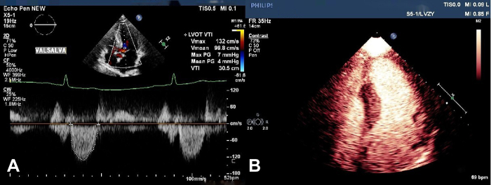
(A) Post-PIMSRA echocardiography shows the LVOT-PGmax at Valsalva was 7 mm Hg. (B) Post-PIMSRA MCE shows the ablated filling defect in the middle and basal septum on the apical 4-chamber view.
References
-
- Maron M.S., Olivotto I., Betocchi S., Casey S.A., Lesser J.R., Losi M.A., et al. Effect of left ventricular outflow tract obstruction on clinical outcome in hypertrophic cardiomyopathy. N Engl J Med. 2003;348:295–303. - PubMed
-
- Liebregts M., Vriesendorp P.A., Ten B.J. Alcohol septal ablation for obstructive hypertrophic cardiomyopathy: a word of endorsement. J Am Coll Cardiol. 2017;70:481–488. - PubMed
-
- Fifer M.A., Sigwart U. Controversies in cardiovascular medicine. Hypertrophic obstructive cardiomyopathy: alcohol septal ablation. Eur Heart J. 2011;32:1059–1064. - PubMed
Publication types
LinkOut - more resources
Full Text Sources
