. 2022 Oct 1;15(10):dmm049707.
doi: 10.1242/dmm.049707. Epub 2022 Oct 24.

An adherent-invasive Escherichia coli-colonized mouse model to evaluate microbiota-targeting strategies in Crohn's disease

Affiliations

An adherent-invasive Escherichia coli-colonized mouse model to evaluate microbiota-targeting strategies in Crohn's disease

Adeline Sivignon et al. Dis Model Mech. .

Abstract

Adherent-invasive Escherichia coli (AIEC) were investigated for their involvement in the induction/chronicity of intestinal inflammation in Crohn's disease (CD). AIEC gut establishment is favoured by overexpression of the glycoprotein CEACAM6 in the ileal epithelium. We generated a transgenic mouse model, named 'Vill-hCC6', in which the human CEACAM6 gene was under the control of the villin promoter, conditioning expression in the small intestine. We demonstrated that CEACAM6 is strongly expressed in the small intestine mucosa and is correlated with numerous glycosylations displayed at the brush border of enterocytes. Ex vivo, the AIEC-enterocyte interaction was enhanced by CEACAM6 expression and necessitated the presence of the bacterial adhesive factor FimH. Finally, AIEC bacteria preferentially persisted in a FimH-dependent manner in the ileal mucosa of Vill-hCC6 mice compared to wild-type mice. This preclinical model opens new perspectives in the mechanistic study of the AIEC pathobiont and represents a valuable tool to evaluate the efficacy of new strategies to eliminate AIEC implanted in the ileal mucosa, such as phages, inhibitory and/or anti-virulence molecules, or CRISPR-based strategies targeting virulence or fitness factors of AIEC bacteria.

Keywords: AIEC; CEACAM6; Crohn's disease; Ileal colonization; Mouse model.


Conflict of interest statement

Competing interests The authors declare no competing or financial interests.

Figures

Fig. 1.
Fig. 1.
Generation of a Vill-hCC6 knock-in mouse model harbouring the human CEACAM6 gene under the control of the villin promoter at the Hprt locus. The solid lines represent intronic sequences. In the targeting vector, hatched black and red boxes represent murine and human Hprt exons, respectively. Neomycin and loxP sites are represented by a grey arrow and blue triangles, respectively, and the red box depicts hGHpolyA. In recombined E14Tg2a (E14) embryo-derived stem (ES) cells, the Hprt gene was reconstituted by introduction of the human promoter (pink box) and exons 1 and 2. Orange and green boxes represent human CEACAM6 cDNA and the villin promoter, respectively. Recombined ES cell clones were injected into blastocysts to generate chimeric animals. Heterozygous females were obtained following the breeding of the chimeras with wild-type (WT) females. Yellow boxes with red crosses and hatched black boxes represent the knock-in allele and the WT allele, respectively.
Fig. 2.
Fig. 2.
Global strategy for the generation of hemizygous male mice. Yellow boxes with red crosses and hatched black boxes represent the knock-in allele and the WT allele, respectively. The asterisks represent the X chromosome carrying the human CEACAM6 transgene under the control of the villin promoter.
Fig. 3.
Fig. 3.
Mucosal CEACAM6 expression in the intestine of Vill-hCC6 mice. (A) Segments of the gastrointestinal tract from Vill-hCC6 mice were opened and washed with cold PBS, and mucosa were collected by scraping with a glass slide. Total RNA was extracted and used for mRNA analysis. The graph shows the RT-qPCR analysis for mRNA expression levels of CEACAM6 relative to Rps26 (26S), n=3 mice. Data are presented as mean±s.e.m., *P<0.05 (Kruskal–Wallis test). (B) Total proteins were extracted from mucosa collected from the gastrointestinal tract of WT or Vill-hCC6 mice. CEACAM6 accumulation was assessed by western blotting using a monoclonal antibody raised against CEACAM6. Quantification of CEACAM6 expression relative to GAPDH was performed for Vill-hCC6 mice by assessing band intensities (n=4 mice) using the Image Lab software. Data are presented as mean±s.e.m., *P<0.05 (Kruskal–Wallis test). (C) CEACAM6 immunohistochemical staining of intestinal sections of WT or Vill-hCC6 mice. Slides were scanned and assessed with an AxioScan Z1 using Zen 2.3 Pro software.
Fig. 4.
Fig. 4.
Analysis of glycosylation at the brush border of enterocytes isolated from intestinal mucosa of Vill-hCC6 or WT mice. Sections (3 cm) of distal ileum or colon were collected from WT or Vill-hCC6 mice, and isolated enterocytes were prepared as described in the Materials and Methods section. (A) Enterocytes of WT or Vill-hCC6 mice were incubated for 1 h with FITC-labelled concanavalin A at 50 µg/ml in PBS and visualized using phase-contrast microscopy and fluorescence (400×, immersion oil, Axio Observer, Zeiss). (B) The graph shows average fluorescence intensities (intensity/µm2) at the brush border of ileal or colonic enterocytes from WT or Vill-hCC6 mice (25-30 enterocytes/mouse; three mice/group). Values represent mean±s.e.m., ****P<0.0001 (Kruskal–Wallis test).
Fig. 5.
Fig. 5.
Assessment of the ability of the adherent-invasive Escherichia coli (AIEC) LF82 bacterial strain to adhere to the brush border of ileal enterocytes of Vill-hCC6. Sections (3 cm) of distal ileum were collected from WT or Vill-hCC6 mice, and enterocytes were prepared as described in the Materials and Methods section. Enterocytes were infected for 2 h with bacteria (∼10 bacteria/enterocyte) in cell culture medium without antibiotics then were washed in PBS to eliminate nonadherent bacteria. (A) Adhesion of GFP-expressing AIEC LF82 bacteria to the brush border of an ileal enterocyte from a Vill-hCC6 mouse (phase-contrast/fluorescence microscopy, 400×, immersion oil, Axio Observer, Zeiss). (B) Adhesion index of GFP-expressing AIEC LF82 bacteria to ileal enterocytes, according to the mouse genotype (total enterocytes counted: 197 for WT mice, 236 for Vill-hCC6 mice). (C) Adhesion index to ileal enterocytes from Vill-hCC6 mice of a nonpiliated LF82-ΔfimH mutant or LF82 WT in the absence or presence of 2% d-mannose (total enterocytes counted: 245 for LF82 WT, 125 for LF82-ΔfimH and 192 for LF82+d-mannose). Adhesion index was expressed as the mean number of bacteria adhering to the brush border of one enterocyte±s.e.m. Three independent experiments were performed. ****P<0.0001 (B, Mann–Whitney test; C, Kruskal–Wallis test).
Fig. 6.
Fig. 6.
Ability of the AIEC LF82 WT strain or of the nonpiliated LF82-ΔfimH mutant to colonize Vill-hCC6 mice. Vill-hCC6 mice were treated with streptomycin for 2 days (day −3) and then received water (day −1) for 24 h. Mice were orally administered (day 0) 109 colony-forming units (CFU) of AIEC LF82 or LF82-ΔfimH bacteria and were killed at 10 days postinfection (dpi). (A) Quantification of AIEC bacteria in the faeces at 3, 5, 7 and 10 dpi (CFU/g of faeces). (B) Quantification of AIEC bacteria associated with the ileal mucosa at 10 dpi (CFU/g of tissue). (C) Disease activity index (DAI) score assessment at 3, 7 and 10 dpi. n=6 per group. Values represent median with interquartile range, *P<0.05, **P<0.01 (Mann–Whitney test).
Fig. 7.
Fig. 7.
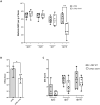
AIEC gut colonization after an oral challenge with a mix of four AIEC strains (CEA614S, CEA618U, CEA212U and CEA304S) in Vill-hCC6 or WT mice. Vill-hCC6 or WT mice were treated with streptomycin for 2 days (day −3) and then received water (day −1) for 24 h. Mice were orally administered (day 0) 109 CFU of AIEC bacterial mix (CEA614S, CEA618U, CEA212U and CEA304S) and were killed at day 21 after administration. (A) Quantification of AIEC bacteria in the faeces at 1, 7, 14 and 21 dpi (CFU/g of faeces). (B) Quantification of AIEC bacteria associated with the intestinal mucosa at 21 dpi (CFU/g of tissue). WT mice, n=6; Vill-hCC6 mice, n=7. Values represent median with interquartile range, *P<0.05 (Mann–Whitney test).

References

    1. Agus, A., Massier, S., Darfeuille-Michaud, A., Billard, E. and Barnich, N. (2014). Understanding host-adherent-invasive Escherichia coli interaction in Crohn's disease: opening up new therapeutic strategies. Biomed. Res. Int. 2014, 567929. 10.1155/2014/567929 - DOI - PMC - PubMed
    1. Barnich, N., Carvalho, F. A., Glasser, A.-L., Darcha, C., Jantscheff, P., Allez, M., Peeters, H., Bommelaer, G., Desreumaux, P., Colombel, J.-F.et al. (2007). CEACAM6 acts as a receptor for adherent-invasive E. coli, supporting ileal mucosa colonization in Crohn disease. J. Clin. Invest. 117, 1566-1574. 10.1172/JCI30504 - DOI - PMC - PubMed
    1. Boudeau, J., Barnich, N. and Darfeuille-Michaud, A. (2001). Type 1 pili-mediated adherence of Escherichia coli strain LF82 isolated from Crohn's disease is involved in bacterial invasion of intestinal epithelial cells. Mol. Microbiol. 39, 1272-1284. 10.1111/j.1365-2958.2001.02315.x - DOI - PubMed
    1. Bretin, A., Lucas, C., Larabi, A., Dalmasso, G., Billard, E., Barnich, N., Bonnet, R. and Nguyen, H. T. T. (2018). AIEC infection triggers modification of gut microbiota composition in genetically predisposed mice, contributing to intestinal inflammation. Sci. Rep. 8, 12301. 10.1038/s41598-018-30055-y - DOI - PMC - PubMed
    1. Buisson, A., Vazeille, E., Fumery, M., Pariente, B., Nancey, S., Seksik, P., Peyrin-Biroulet, L., Allez, M., Ballet, N., Filippi, J.et al. (2021). Faster and less invasive tools to identify patients with ileal colonization by adherent-invasive E. coli in Crohn's disease. United European Gastroenterol. J. 9, 1007-1018. 10.1002/ueg2.12161 - DOI - PMC - PubMed

Publication types