. 2023 Dec;36(6):638-650.
doi: 10.1177/19714009221130490. Epub 2022 Sep 29.

Clinical applications of arterial spin labeling of the intracranial compartment in vascular anomalies-A case-based review

Affiliations

Clinical applications of arterial spin labeling of the intracranial compartment in vascular anomalies-A case-based review

Amara Ahmed et al. Neuroradiol J. 2023 Dec.

Abstract

Arterial spin labeling (ASL) is a magnetic resonance perfusion technique that allows for quantification of cerebral blood flow (CBF) without the use of contrast or radiation. Several applications of ASL have been described in diagnosis of strokes and stroke mimics, intracranial tumors, and other conditions. Various vascular anomalies exhibit specific CBF patterns that correlate with different signal intensities on ASL. In this case-based review, we demonstrate the utility of ASL in diagnosis and surveillance of vascular anomalies in the intracranial compartment.

Keywords: Arterial spin labeling; CEA; arteriovenous malformations; cerebral blood flow; neuroimaging; vascular anomalies.


Conflict of interest statement

Declaration of conflicting interestsThe author(s) declared no potential conflicts of interest with respect to the research, authorship, and/or publication of this article.

Figures

Figure 1.
Figure 1.
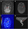
A 79-year-old male presenting with intermittent headaches. Given his history of renal failure, he could not receive contrast for neuroimaging. (A) Focal round increased signal in the right sylvian fissure (blue arrow) from blood pooling in the right MCA aneurysm sac on 3D pCASL. Also, note the asymmetric increased signal in the right occipital lobe (yellow arrow). Time of flight (TOF) MRA (B) with 3D reconstruction (C) and T2 weighted MRI (D) demonstrate a trilobed aneurysm at the right MCA bifurcation (red arrows). Conventional angiography images show pre (E) and post (F) aneurysm coiling and embolization.
Figure 2.
Figure 2.
Left ICA aneurysm. A 58-year-old male presenting with headaches. Focal increased ASL signal is seen in the region of the left suprasellar cistern (A), (C) corresponding to a large posterior projecting aneurysm sac (red arrows) off the left supraclinoid ICA (B), (D). 3D pCASL in grayscale (top left) and color (bottom left) ASL signal in the aneurysm sac demonstrates a lack of complete thrombosis of the sac, despite intermediate signal changes seen on TOF MRA (B).
Figure 3.
Figure 3.
Grayscale 3D pCASL image (A) demonstrates an easily detectable focus of increased signal corresponding to the aneurysm (red arrow). The increase in signal occurs due to the pooling of spins within the aneurysm. In addition, asymmetric increased ASL signal within the right occipital lobe was demonstrated (yellow arrow). Incidentally, the same patient had a right fetal PCA (blue arrow), as seen anatomically on TOF MRA (B) and 3D reconstructions (C), (D). This case shows the importance of ASL imaging in patients with contrast contraindication.
Figure 4.
Figure 4.
Vascular variant-dolichoectasia of vertebrobasilar system. A 46-year-old male with poorly controlled hypertension. No history of connective tissue disorder. Grayscale and color 3D pCASL (top images) Intravascular ASL signal in basilar artery depicts dilated/ectatic caliber (red arrows) of the vessel (A and B). Post gadolinium contrast T1 axial (C) and coronal (D) sequences confirm vertebrobasilar arterial anatomy correlating to the location of ASL abnormality.
Figure 5.
Figure 5.
A. Dural arteriovenous fistula (dAVF). A 69-year-old male with left facial droop and weakness. A) Axial T2W demonstrates a prominent left parietal venous flow void (white arrow, top left). B) Axial SWI shows linear susceptibility artifact corresponding to the prominent left parietal flow void (blue arrow, top right). C and D) MRA MIP and MRA show an early draining venous structure in the left temporoparietal region (white arrows) contiguous with the flow void. 5A continued – E) High flow dural arteriovenous fistula of the left temporal/occipital region receiving contributions from the posterior division of middle meningeal, left occipital, and ascending pharyngeal branches of the left ECA as well as the vidian branch of the left ICA and early venous drainage into the vein of Labbe and superior sagittal sinus (white and yellow arrows). F) Successful embolization of the left ECA branches, including posterior division of the middle meningeal, left occipital, and ascending pharyngeal arteries using glue n-BCA with complete stasis at these levels. 5B2D pASL images demonstrate asymmetrically increased signal within the early draining vein compatible with shunting (blue arrows).
Figure 6.
Figure 6.
Dural arteriovenous fistula (dAVF). A 77-year-old female with upper facial pain, headache, and nausea. Grayscale and color map 3D pCASL images (A and B) show increased signal in the left parasagittal anterior cranial fossa corresponding to a prominent vessel on the MR Axial T1 post-contrast image (C) suggestive of arteriovenous fistula (red arrows). Digital subtraction angiography image (D) shows a dAVF in the left anterior cranial fossa (blue arrow). Supply from the ethmoidal branch of the internal maxillary artery and venous drainage with ectasia through a cortical vein.
Figure 7.
Figure 7.
Arteriovenous Malformation (AVM), Spetzler-Martin grade 3. 50-year-old male with nausea, vomiting, and headache. MR axial T1 post-contrast image (A), digital subtraction angiography (B), and 3D reconstructed images (D) demonstrate right cerebellar AVM (red arrows). Grayscale 3D pCASL image (C) shows serpiginous areas of increased signal within the right cerebellar hemisphere corresponding to the AVM on anatomic images.
Figure 8.
Figure 8.
Arteriovenous malformation (AVM), Spetzler-Martin grade 1. 25-year-old male with headache. Color map and grayscale 3D pCASL images (A and B, respectively) demonstrate a focal area of increased signal in the left temporal region corresponding to the subtle vascular malformation (red arrows) on MRA image (D) and MR axial T1 post-contrast image (E). Digital subtraction angiography image (C) shows a left temporal AVM. The focal area of increased signal on ASL is obvious and easy to detect. However, it is nonspecific. In this case, it could be an aneurysm or vascular malformation Correlation with anatomic images is essential for the distinction.
Figure 9.
Figure 9.
Arteriovenous Malformation (AVM). 53-year-old with hemorrhage in the left occipital junction. MR DWI, axial T2 weighted image, and MRA image (A, C, and D, respectively) show small nidus of dysplastic arteries in the left occipital region with poorly defined margins (red arrows), consistent with residual AVM corresponding to the serpiginous bright signal on color map 2D pASL image (B).
Figure 10.
Figure 10.
Arteriovenous Malformation (AVM). 53-year-old with hemorrhage in the left occipital junction. A, B) Digital subtraction angiography images show the AVM (red arrows) supplied by the left posterior inferior temporal branch of the left PCA and temporal occipital branches of the left MCA with C, D) venous drainage via a dilated cortical vein (yellow arrows) which drains into the superior sagittal sinus.
Figure 11.
Figure 11.
A 67-year-old Korean female with end-stage renal disease, hypertension, diabetes mellitus, and prior pontine infarct presented to the hospital from the dialysis center with sudden onset left facial droop, slurred speech, and bilateral upper extremity weakness for initial concern of acute stroke. Her serum potassium level was 7.5 mmol/L, creatinine was 11 mg/dL, and blood urea nitrogen was 93 mg/dL in the setting of a missed dialysis session. (A)Axial diffusion-weighted acquisition demonstrated diffuse cytotoxic changes in the putamina and inferior right caudate (red arrows). There is low signal (blue arrow) on apparent diffusion coefficient map (B). On axial FLAIR acquisition (C), there is an associated increased signal in the region of restricted diffusion (yellow arrow) with additional peripheral hyperintense signal extending along the putamen (green arrow), indicative of vasogenic edema. 3D pASL (D) shows a corresponding symmetric increased signal from an accumulation of labeled blood in the putamen (yellow arrows), confirming toxic etiology, that is, uremic encephalopathy in this given clinical setting.
Figure 12.
Figure 12.
A 73-year-old female with hypertension, hyperlipidemia, and elective carotid endarterectomy (CEA) one week before right carotid artery stenosis presented to the emergency department after witnessed seizures, left-sided gaze deviation, and recent history of daily right-sided headaches. A–C) 2D pASL shows asymmetric increased signal in the right cerebral hemisphere denoting cerebral hyperperfusion (yellow arrows). D–F) Axial FLAIR images demonstrate signal abnormality in the regions within the right cerebral hemisphere corresponding to the areas of increased ASL signal.
Figure 13.
Figure 13.
A 52-year old male with past medical history of hypertension, hyperlipidemia, transient ischemic attack status post CEA presented with left-sided numbness. He was found to have occlusion of right posterior cerebral artery and was transferred to our hospital for further care. A, B) 3D pseudocontinuous ASL showed high signal in the left sigmoid sinus (blue arrow) and right transverse sinus (yellow arrow). C) Contrast-enhanced CT showed compression of the brachiocephalic vein (red arrow) between the sternum and aortic arch. D) DSA in the venographic phase after vertebral artery injection showed relative hemostasis in left sigmoid sinus.

Similar articles

References

    1. Daftari Besheli L, Ahmed A, Hamam O, et al. Arterial Spin Labeling technique and clinical applications of the intracranial compartment in stroke and stroke mimics - A case-based review. The Neuroradiology J; 35: 197140092210988. - PMC - PubMed
    1. Weinstein JD, Hamam O, Urrutia VC, et al. Added value of arterial spin labeling in detecting posterior reversible encephalopathy syndrome as a stroke mimic on baseline neuroimaging: a single center experience. Front Neurol 2022; 13: 831218. DOI: 10.3389/FNEUR.2022.831218 - DOI - PMC - PubMed
    1. Yedavalli V, Tong E. The Potential Utility of Arterial Spin Labeling in Detecting and Localizing Posterior Circulation Occlusions in Every Day Practice: A Clinical Report of Selected Cases. J Clin Imaging Sci 2020; 10: 78. DOI: 10.25259/JCIS_118_2020 - DOI - PMC - PubMed
    1. Proisy M, Bruneau B, Rozel C, et al. Arterial spin labeling in clinical pediatric imaging. Diagn Interv Imaging 2016; 97(2): 151–158. DOI: 10.1016/J.DIII.2015.09.001 - DOI - PubMed
    1. Wang J, Licht DJ. Pediatric Perfusion MR Imaging Using Arterial Spin Labeling. Neuroimaging Clin North America 2006; 16(1): 149–167. DOI: 10.1016/J.NIC.2005.10.002 - DOI - PubMed

MeSH terms

LinkOut - more resources