. 2022 Nov 30;45(11):855-867.
doi: 10.14348/molcells.2022.0104. Epub 2022 Sep 28.

CBP-Mediated Acetylation of Importin α Mediates Calcium-Dependent Nucleocytoplasmic Transport of Selective Proteins in Drosophila Neurons

Affiliations

CBP-Mediated Acetylation of Importin α Mediates Calcium-Dependent Nucleocytoplasmic Transport of Selective Proteins in Drosophila Neurons

Jae Ho Cho et al. Mol Cells. .

Abstract

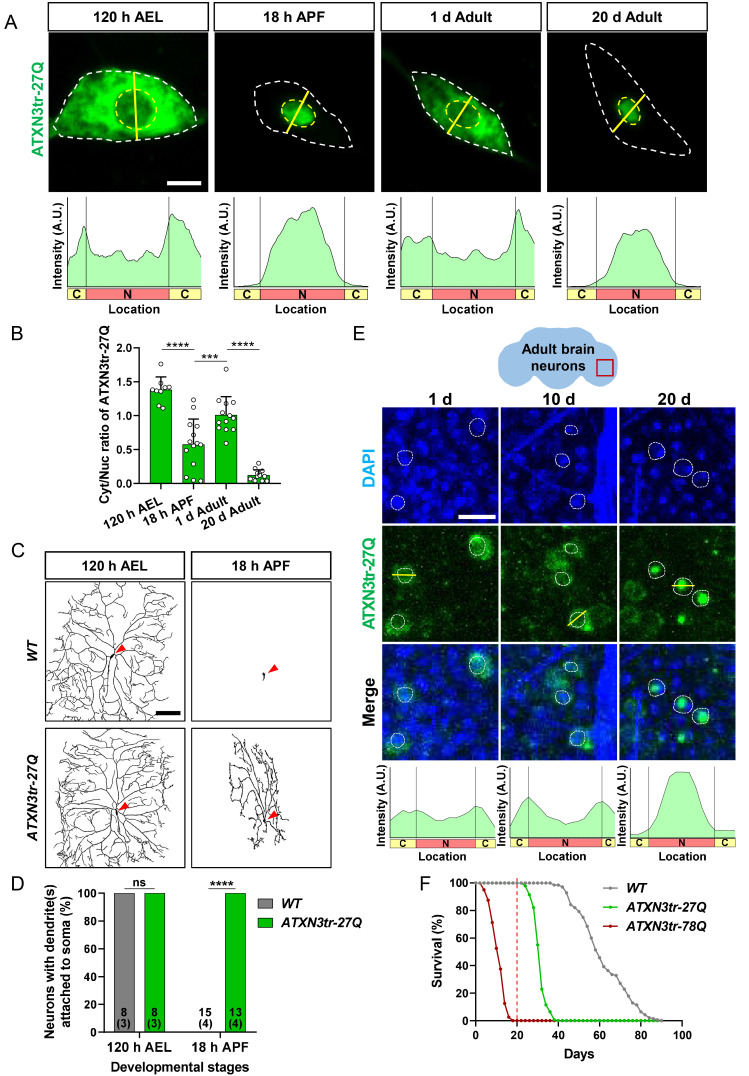
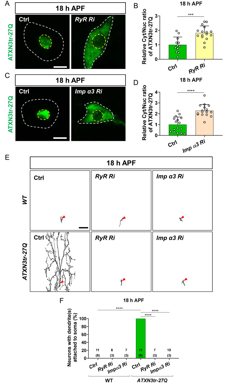
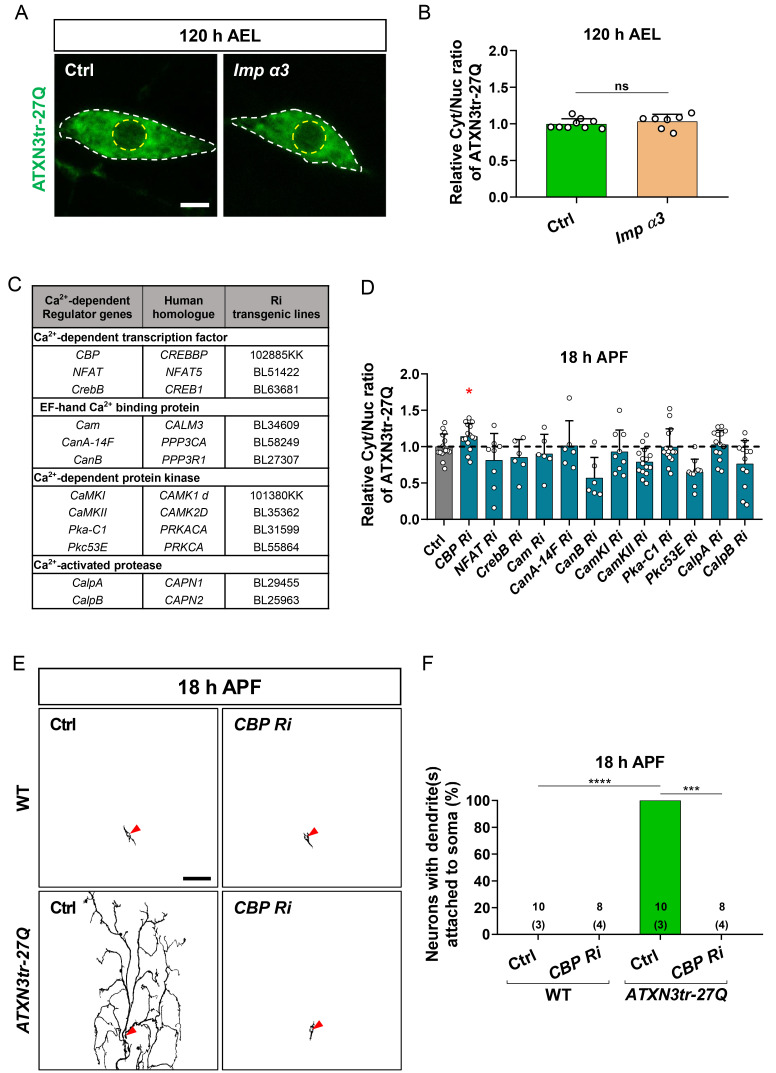
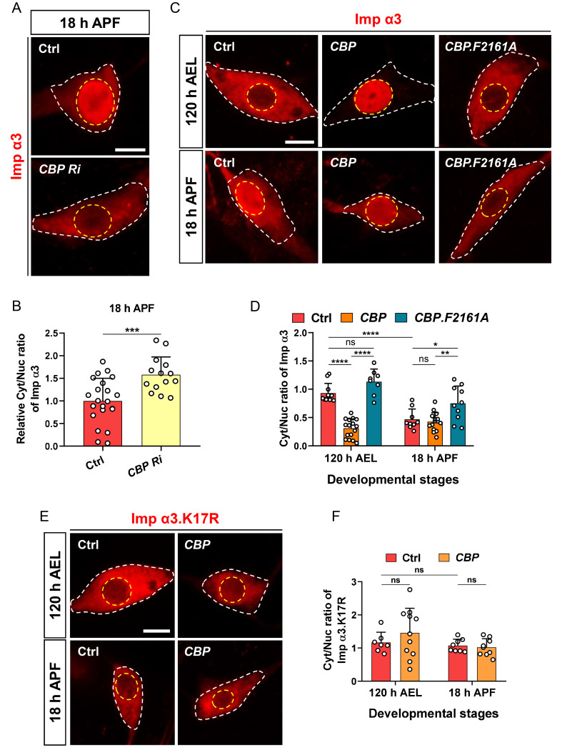
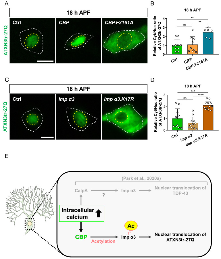
For proper function of proteins, their subcellular localization needs to be monitored and regulated in response to the changes in cellular demands. In this regard, dysregulation in the nucleocytoplasmic transport (NCT) of proteins is closely associated with the pathogenesis of various neurodegenerative diseases. However, it remains unclear whether there exists an intrinsic regulatory pathway(s) that controls NCT of proteins either in a commonly shared manner or in a target-selectively different manner. To dissect between these possibilities, in the current study, we investigated the molecular mechanism regulating NCT of truncated ataxin-3 (ATXN3) proteins of which genetic mutation leads to a type of polyglutamine (polyQ) diseases, in comparison with that of TDP-43. In Drosophila dendritic arborization (da) neurons, we observed dynamic changes in the subcellular localization of truncated ATXN3 proteins between the nucleus and the cytosol during development. Moreover, ectopic neuronal toxicity was induced by truncated ATXN3 proteins upon their nuclear accumulation. Consistent with a previous study showing intracellular calcium-dependent NCT of TDP-43, NCT of ATXN3 was also regulated by intracellular calcium level and involves Importin α3 (Imp α3). Interestingly, NCT of ATXN3, but not TDP-43, was primarily mediated by CBP. We further showed that acetyltransferase activity of CBP is important for NCT of ATXN3, which may acetylate Imp α3 to regulate NCT of ATXN3. These findings demonstrate that CBP-dependent acetylation of Imp α3 is crucial for intracellular calcium-dependent NCT of ATXN3 proteins, different from that of TDP-43, in Drosophila neurons.

Keywords: ATXN3; CBP; Importin α; acetylation; calcium; nucleocytoplasmic transport.


Conflict of interest statement

CONFLICT OF INTEREST

The authors have no potential conflicts of interest to disclose.

Figures

Fig. 1
Fig. 1. ATXN3tr-27Q undergoes dynamic nucleocytoplasmic translocation in a cellular context-dependent manner and induces neuronal toxicity upon nuclear accumulation in Drosophila neurons.
(A) Subcellular localization of overexpressed HA-ATXN3tr-27Q proteins in C4da neurons during development stages (120 h AEL, 18 h APF, 1-day adult, and 20-day adult) [+/+;ppk1a-GAL4>UAS-CD4-tdGFP/UAS-HA-ATXN3tr-27Q]. Outer and inner dashed lines indicate the borders of cell bodies and nuclei, respectively. Scale bar = 5 μm. The intensity profile of fluorescent signals representing ATXN3tr-27Q proteins across cell bodies along yellow lines are presented at the bottom. (B) Quantification of cytosol/nuclear (Cyt/Nuc) ratio of HA-ATXN3tr-27Q proteins during development stages. Values are presented as mean ± SD. ****P < 0.0001, ***P = 0.0005 by one-way ANOVA with Tukey post hoc test; n = 9 for 120 h AEL, n = 14 for 18 h APF, n = 13 for 1 d adult, n = 13 for 20 d adult. (C) Skeletonized dendrite images of C4da neurons of 120 h AEL and 18 h APF [WT, +/+;ppk1a-GAL4>UAS-CD4-tdGFP/+, HA-ATXN3tr-27Q, +/+; ppk1a-GAL4>UAS-CD4-tdGFP/UAS-HA-ATXN3tr-27Q]. Red-colored arrowheads indicate cell bodies of C4da neurons. Scale bar = 100 μm. (D) Penetrance of dendrite defects at 120 h AEL and 18 h APF. Values are presented as mean ± SD. ****P < 0.0001 by Fisher’s exact test; ns, not significant. The number of neurons for each genotype is shown in the figure, and the number of animals is shown below in parentheses. (E) Subcellular localization of overexpressed HA-ATXN3tr-27Q proteins in adult brain neurons along aging (1 day, 10 days, and 20 days adults) [+/+;elav-GAL4/UAS-HA-ATXN3tr-27Q]. Red-colored box indicates imaging area of adult brain neurons. (F) Comparison of survival rates over time between control flies and flies pan-neuronally expressing HA-ATXN3tr-27Q and HA-ATXN3tr-78Q [WT, +/+;elav-GAL4/+, ATXN3tr-27Q, +/+;elav-GAL4/UAS-HA-ATXN3tr-27Q, ATXN3tr-78Q, +/+;elav-GAL4/UAS-HA-ATXN3tr-78Q].
Fig. 2
Fig. 2. Nuclear accumulation of ATXN3tr-27Q inducing dendrite remodeling defects during metamorphosis is regulated by the intracellular calcium level and involves Imp α3 in neurons.
(A) Subcellular localization of overexpressed HA-ATXN3tr-27Q proteins in C4da neurons of control (Ctrl) or expressing RyR Ri at 18 h APF [Ctrl, +/+;ppk1a-GAL4/UAS-HA-ATXN3tr-27Q, RyR Ri, UAS-RyR RNAi/+;ppk1a-GAL4/UAS-HA-ATXN3tr-27Q]. Outer and inner dashed lines indicate borders of cell bodies and nuclei, respectively. Scale bar = 5 μm. (B) Quantification of Cyt/Nuc ratio of HA-ATXN3tr-27Q proteins in C4da neurons of Ctrl or expressing RyR Ri at 18 h APF. Values are presented as mean ± SD. ***P = 0.0003 by two-tailed t-test; n = 12 neurons for Ctrl, n = 16 neurons for for RyR Ri. (C) Subcellular localization of overexpressed HA-ATXN3tr-27Q in C4da neurons of Ctrl or expressing Imp α3 Ri at 18 h APF [Ctrl, +/+;ppk1a-GAL4/UAS-HA-ATXN3tr-27Q, Imp α3 Ri, UAS-Imp α3 RNAi/+;ppk1a-GAL4/UAS-HA-ATXN3tr-27Q]. Outer and inner dashed lines indicate borders of cell bodies and nuclei, respectively. Scale bar = 5 μm. (D) Quantification of Cyt/Nuc ratio of HA-ATXN3tr-27Q proteins in C4da neurons of Ctrl or expressing Imp α3 Ri at 18 h APF. Values are presented as mean ± SD. ****P < 0.0001 by two-tailed t-test; n = 19 neurons for Ctrl, n = 19 neuron for Imp α3 Ri. (E) Skeletonized dendrite images of C4da neurons of 18 h APF [Ctrl in WT, +/+;ppk1a-GAL4>UAS-CD4-tdGFP/+, RyR Ri in WT, UAS-RyR RNAi/+;ppk1a-GAL4>UAS-CD4-tdGFP/+, Imp α3 Ri in WT, UAS-Imp α3 RNAi/+;ppk1a-GAL4>UAS-CD4-tdGFP/+, Ctrl in ATXN3tr-27Q, +/+;ppk1a-GAL4>UAS-CD4-tdGFP/UAS-HA-ATXN3tr-27Q, RyR Ri in ATXN3tr-27Q, UAS-RyR RNAi/+;ppk1a-GAL4>UAS-CD4-tdGFP/UAS-HA-ATXN3tr-27Q, Imp α3 Ri in ATXN3tr-27Q, UAS-Imp α3 RNAi/+;ppk1a-GAL4>UAS-CD4-tdGFP/UAS-HA-ATXN3tr-27Q]. Scale bar = 50 μm. (F) Penetrance of dendrite defects at 18 h APF. Values are presented as mean ± SD. ****P < 0.0001 by Fisher’s exact test. The number of neurons for each genotype is shown in the figure, and the number of animals is shown below in parentheses.
Fig. 3
Fig. 3. Calcium-dependent NCT of ATXN3tr-27Q is mediated by CBP in neurons.
(A) Subcellular localization of overexpressed HA-ATXN3tr-27Q proteins in C4da neurons of control (Ctrl) or expressing Imp α3 at 120 h AEL [Ctrl, +/+;ppk1a-GAL4/UAS-HA-ATXN3tr-27Q, Imp α3, UAS-2xFlag-Imp α3/+;ppk1a-GAL4/UAS-HA-ATXN3tr-27Q]. Outer and inner dashed lines indicate borders of cell bodies and nuclei, respectively. Scale bar = 5 μm. (B) Quantification of Cyt/Nuc ratio of HA-ATXN3tr-27Q proteins in C4da neurons of Ctrl or expressing Imp α3 at 120 h AEL. Values are presented as mean ± SD. P > 0.05 (P = 0.4235) by two-tailed t-test; ns, not significant; n = 9 for Ctrl, n = 7 for Imp α3. (C) List of intracellular calcium-dependent regulators screened in this study. (D) Quantification of Cyt/Nuc ratio of HA-ATXN3tr-27Q proteins in C4da neurons of Ctrl or expressing denoted transgenes described in C. [Ctrl, ppk-Gal4/+;UAS-HA-ATXN3tr-27Q, CBP Ri, ppk-Gal4/UAS-CBP RNAi;UAS-HA-ATXN3tr-27Q, NFAT Ri, ppk-Gal4/+;UAS-HA-ATXN3tr-27Q/UAS-NFAT RNAi, CrebB Ri, ppk-Gal4/UAS-CrebB RNAi;UAS-HA-ATXN3tr-27Q/+, Cam Ri, ppk-Gal4/+;UAS-HA-ATXN3tr-27Q/UAS-Cam RNAi, CanA-14F Ri, ppk-Gal4/CanA-14F;UAS-HA-ATXN3tr-27Q/+, CanB Ri, ppk-Gal4/+;UAS-HA-ATXN3tr-27Q/UAS-CanB RNAi, CaMKI Ri, ppk-Gal4/UAS-CaMKI RNAi;UAS-HA-ATXN3tr-27Q/+, CaMKII Ri, ppk-Gal4/+;UAS-HA-ATXN3tr-27Q/UAS-CamKII RNAi, Pka-C1 Ri, ppk-Gal4/+;UAS-HA-ATXN3tr-27Q/UAS-Pka-C1 RNAi, Pkc53E Ri, ppk-Gal4/+;UAS-HA-ATXN3tr-27Q/UAS-Pkc53E RNAi, CalpA Ri, ppk-Gal4/+;UAS-HA-ATXN3tr-27Q/CalpA Ri, CalpB Ri, ppk-Gal4/+; UAS-HA-ATXN3tr-27Q/UAS-CalpB RNAi]. Values are presented as mean ± SD. *P = 0.0358 by two-tailed t-test; n = 15 for Ctrl, n = 15 for CBP Ri, n = 8 for NFAT Ri, n = 6 for CrebB Ri, n = 6 for Cam Ri, n = 6 for CanA-14F Ri, n = 6 for CanB Ri, n = 9 for CamKI Ri, n = 15 for CamKII Ri, n = 15 for Pka-C1 Ri, n = 10 for Pkc53E Ri, n= 15 for CalpA Ri, n = 12 for CalpB Ri. (E) Skeletonized dendrite images of C4da neurons of 18 h APF [Ctrl in WT, +/+;ppk1a-GAL4>UAS-CD4-tdGFP/+, CBP Ri in WT, UAS-CBP RNAi/+; ppk1a-GAL4>UAS-CD4-tdGFP/+, Ctrl in ATXN3tr-27Q, +/+;ppk1a-GAL4>UAS-CD4-tdGFP/UAS-HA-ATXN3tr-27Q, CBP Ri in ATXN3tr-27Q, UAS-CBP RNAi/+;ppk1a-GAL4>UAS-CD4-tdGFP/UAS-HA-ATXN3tr-27Q]. Scale bar = 50 μm. (F) Penetrance of dendrite defects at 18 h APF. Values are presented as mean ± SD. ****P < 0.0001 by Fisher’s exact test. The number of neurons for each genotype is shown in the figure, and the number of animals is shown below in parentheses.
Fig. 4
Fig. 4. CBP regulates NCT of Drosophila Imp α3 via acetyltransferase activity in neurons.
(A) Subcellular localization of overexpressed Flag-Imp α3 proteins in C4da neurons of control (Ctrl) or expressing CBP Ri at 18 h APF [Ctrl, UAS-2xFlag-Imp α3/+;ppk1a-Gal4/+, CBP Ri, UAS-2xFlag-Imp α3/UAS-CBP RNAi;ppk1a-Gal4/+]. Outer and inner dashed lines indicate borders of cell bodies and nuclei, respectively. Scale bar = 5 μm. (B) Quantification of Cyt/Nuc ratio of Flag-Imp α3 proteins in C4da neurons of Ctrl or expressing CBP Ri at 18 h APF. Values are presented as mean ± SD. ***P = 0.0009 by two-tailed t-test; n = 22 for Ctrl, n = 14 for CBP Ri. (C) Subcellular localization of overexpressed Flag-Imp α3 proteins in C4da neurons of Ctrl or expressing CBP or CBP.F2161A at 120 h AEL and 18 h APF [Ctrl, UAS-2xFlag-Imp α3/+;ppk1a-Gal4/+, CBP, UAS-2xFlag-Imp α3/+;ppk1a-Gal4/UAS-CBP.wt-V5, CBP.F2161A, UAS-2xFlag-Imp α3/UAS-CBP.F2161A-V5;ppk1a-Gal4/+]. Outer and inner dashed lines indicate borders of cell bodies and nuclei, respectively. Scale bar = 5 μm. (D) Quantification of Cyt/Nuc ratio of Flag-Imp α3 proteins in C4da neurons of Ctrl or expressing CBP or CBP.F2161A at 120 h AEL and 18 h APF. Values are presented as mean ± SD. P = 0.28, ****P < 0.0001, **P = 0.0025, *P = 0.0377 by two-way ANOVA with Tukey’s post hoc test; ns, not significant; n = 11 for Ctrl at 120 h AEL, n = 16 for CBP at 120 h AEL, n = 8 for CBP.F2161A at 120 h AEL, n = 9 for Ctrl at 18 h APF, n = 15 for CBP at 18 h APF, n = 10 for CBP.F2161A at 18 h APF. (E) Subcellular localization of overexpressed Flag-Imp α3.K17R proteins in C4da neurons of Ctrl or expressing CBP at 120 h AEL and 18 h APF [Ctrl, UAS-2xFlag-Imp α3.K17R/+;ppk1a-Gal4/+, CBP, UAS-2xFlag-Imp α3.K17R/+;ppk1a-Gal4/UAS-CBP.wt-V5]. Outer and inner dashed lines indicate borders of cell bodies and nuclei, respectively. Scale bar = 5 μm. (F) Quantification of Cyt/Nuc ratio of Flag-Imp α3.K17R proteins in C4da neurons of Ctrl or expressing CBP at 120 h AEL and 18 h APF. Values are presented as mean ± SD. P > 0.3 by two-way ANOVA with Tukey’s post hoc test; ns, not significant; n = 7 for Ctrl at 120 h AEL, n = 11 for CBP at 120 h AEL, n = 8 for Ctrl at 18 h APF, n = 9 for CBP at 18 h APF.
Fig. 5
Fig. 5. CBP-mediated acetylation of Imp α3 is crucial for NCT of ATXN3tr-27Q in neurons.
(A) Subcellular localization of overexpressed HA-ATXN3tr-27Q proteins in C4da neurons of control (Ctrl) or expressing CBP or CBP.F2161A at 18 h APF [Ctrl, ppk-Gal4/+; UAS-HA-ATXN3tr-27Q/+, CBP, ppk-gal4/+;UAS-HA-ATXN3tr-27Q/UAS-CBP.wt-V5, CBP.F2161A, ppk-Gal4/UAS-CBP.F2161A-V5;HA-ATXN3tr-27Q/+]. Outer and inner dashed lines indicate borders of cell bodies and nuclei, respectively. Scale bar = 5 μm. (B) Quantification of Cyt/Nuc ratio of HA-ATXN3tr-27Q proteins in C4da neurons of Ctrl or expressing CBP or CBP.F2161A at 18 h APF. Values are presented as mean ± SD. P = 0.9498, **P < 0.004 by one-way ANOVA with Tukey’s post hoc test; ns, not significant; n = 10 for Ctrl, n = 9 for CBP, n = 6 for CBP.F2161A. (C) Subcellular localization of overexpressed HA-ATXN3tr-27Q proteins in C4da neurons of Ctrl or expressing Flag-Imp α3 or Flag-Imp α3.K17R at 18 h APF [Ctrl, +/+;ppk1a-Gal4/UAS-HA-ATXN3tr-27Q, Imp α3, UAS-2xFlag-Imp α3/+;ppk1a-Gal4/HA-ATXN3tr-27Q, Imp α3.K17R, UAS-2xFlag-Imp α3.K17R/+;ppk1a-Gal4/UAS-HA-ATXN3tr-27Q]. Outer and inner dashed lines indicate borders of cell bodies and nuclei, respectively. Scale bar = 5 μm. (D) Quantification of Cyt/Nuc ratio of HA-ATXN3tr-27Q proteins in C4da neurons of Ctrl or expressing Flag-Imp α3 or Flag-Imp α3.K17R at 18 h APF. Values are presented as mean ± SD. P = 0.2972, ***P = 0.0002, ****P < 0.0001 by one-way ANOVA with Tukey’s post hoc test; ns, not significant; n = 10 for Ctrl, n = 11 for Imp α3, n = 12 for Imp α3.K17R. (E) A schematic illustration for the molecular mechanism regulating NCT of ATXN3tr-27Q proteins.

Similar articles

Cited by

References

    1. Bagur R., Hajnoczky G. Intracellular Ca2+ sensing: its role in calcium homeostasis and signaling. Mol. Cell. 2017;66:780–788. doi: 10.1016/j.molcel.2017.05.028. - DOI - PMC - PubMed
    1. Bannister A.J., Kouzarides T. The CBP co-activator is a histone acetyltransferase. Nature. 1996;384:641–643. doi: 10.1038/384641a0. - DOI - PubMed
    1. Bannister A.J., Miska E.A., Gorlich D., Kouzarides T. Acetylation of importin-alpha nuclear import factors by CBP/p300. Curr. Biol. 2000;10:467–470. doi: 10.1016/S0960-9822(00)00445-0. - DOI - PubMed
    1. Bauer N.C., Doetsch P.W., Corbett A.H. Mechanisms regulating protein localization. Traffic. 2015;16:1039–1061. doi: 10.1111/tra.12310. - DOI - PubMed
    1. Bichelmeier U., Schmidt T., Hubener J., Boy J., Ruttiger L., Habig K., Poths S., Bonin M., Knipper M., Schmidt W.J., et al. Nuclear localization of ataxin-3 is required for the manifestation of symptoms in SCA3: in vivo evidence. J. Neurosci. 2007;27:7418–7428. doi: 10.1523/JNEUROSCI.4540-06.2007. - DOI - PMC - PubMed

LinkOut - more resources