. 2022 Oct;31(10):e4433.
doi: 10.1002/pro.4433.

Effects of targeting signal mutations in a mitochondrial presequence on the spatial distribution of the conformational ensemble in the binding site of Tom20

Affiliations

Effects of targeting signal mutations in a mitochondrial presequence on the spatial distribution of the conformational ensemble in the binding site of Tom20

Xiling Han et al. Protein Sci. 2022 Oct.

Abstract

The 20-kDa TOM (translocase of outer mitochondrial membrane) subunit, Tom20, is the first receptor of the protein import pathway into mitochondria. Tom20 recognizes the mitochondrial targeting signal embedded in the presequences attached to mature mitochondrial proteins, as an N-terminal extension. Consequently, ~1,000 different mitochondrial proteins are sorted into the mitochondrial matrix, and distinguished from non-mitochondrial proteins. We previously reported the MPRIDE (multiple partial recognitions in dynamic equilibrium) mechanism to explain the structural basis of the promiscuous recognition of presequences by Tom20. A subset of the targeting signal features is recognized in each pose of the presequence in the binding state, and all of the features are collectively recognized in the dynamic equilibrium between the poses. Here, we changed the volumes of the hydrophobic side chains in the targeting signal, while maintaining the binding affinity. We tethered the mutated presequences to the binding site of Tom20 and placed them in the crystal contact-free space (CCFS) created in the crystal lattice. The spatial distributions of the mutated presequences were visualized as smeared electron densities in the low-pass filtered difference maps obtained by X-ray crystallography. The mutated presequence ensembles shifted their positions in the binding state to accommodate the larger side chains, thus providing positive evidence supporting the use of the MPRIDE mechanism in the promiscuous recognition by Tom20.

Keywords: Tom20; crystal contact-free space; mitochondrial targeting signal; presequence; promiscuous recognition.


Conflict of interest statement

The authors declare no competing interests.

Figures
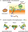
FIGURE 1
FIGURE 1
Structure and function of Tom20 and its hypothetical molecular mechanism to enable the promiscuous recognition. (a) The Tom20 protein resides in the mitochondrial outer membrane and recognizes the targeting signal embedded in the presequences, which are the extra N‐terminal segments of mitochondrial preproteins. The presequences are removed by limited proteolysis after transportation into the matrix. (b) Schematic diagram of the three poses of the presequence helix (depicted as a cylinder) in a dynamic exchange. The three poses were obtained as snapshots in previous crystallographic studies., Three poses were assumed, but more poses could be involved in the dynamic equilibrium. Tom20 is equipped with two hydrophobic pockets: the ϕ15 site and the ϕ4 site
FIGURE 2
FIGURE 2
The concept of the crystal contact‐free space (CCFS) method. (a) The fusion with MBP (maltose‐binding protein) with a connector α‐helix creates CCFS in crystals. The rigid connection ensures the creation of sufficient room, independent of the packing mode of the protein molecules in the crystal. The covalent tethering guarantees the full occupancy of the presequence (magenta oval) in CCFS. (b) The truncation of high‐angle diffraction spots is effective as a low‐pass filter to improve the signal‐to‐noise ratio of the Fo–Fc difference electron density map, for visualization of a highly mobile presequence in CCFS
FIGURE 3
FIGURE 3
Stereo views of the electron density of the wild‐type pALDH presequence in CCFS. (a) Electron density contoured at 3σ corresponding to the presequence (orange mesh) and spacer (blue mesh) in the CCFS of the MBP‐Tom20‐SS‐pALDH(wt) crystal. (b) Crystal structure of Tom20‐SS‐pALDH(wt) (PDB entry 2V1T) as a reference. The presequence trapped in a pose in the binding groove of Tom20 is depicted in the cartoon model. In (a) and (b), the cysteine residue of Tom20 used as the tethering point is red‐colored
FIGURE 4
FIGURE 4
Stereo views of the electron densities of the mutated pALDH presequences in CCFS. (a)–(d) Electron densities contoured at 3σ corresponding to the presequences (magenta mesh) and spacer segment (yellow‐green mesh) in the CCFS of the MBP‐Tom20‐SS‐pALDH(mut) crystals. The electron density corresponding to the presequence and spacer in the CCFS of the MBP‐Tom20‐SS‐pALDH(wt) crystal is superimposed as a reference (gray mesh). (e) All electron densities and Tom20 structures were superimposed. The root‐mean‐square deviations (RMSDs) after superimposing the Cα atoms of the Tom20 part (residues 369–400) are as small as 0.13–0.22 Å
FIGURE 5
FIGURE 5
Comparison of the packing modes of the MBP‐Tom20 molecules in the C2 and P21 crystals. (a) MBP‐Tom20‐SS‐pALDH(L15F) crystal as a representative of the C2 crystals. (b) MBP‐Tom20‐SS‐pALDH(L15W) crystal as a representative of the P21 crystals. The orange and purple symbols mark the positions of the crystallographic two‐fold rotation axis in the C2 crystal, and the non‐crystallographic pseudo two‐fold axis in the P21 crystal, respectively. The magenta squares depict the frame of the ASU units
FIGURE 6
FIGURE 6
Interpretation of the positional shifts of the electron densities in the CCFSs of the MBP‐Tom20‐SS‐pALDH(L15F) and MBP‐Tom20‐SS‐pALDH(L15W) crystals. (a) Molecular basis of the electron density changes observed in CCFS, induced by a mutation in the targeting signal. The sizes of the ovals represent the volumes of the side chains. The flat blue and concave red circles represent the hydrophobic binding sites on Tom20. The large side chain at the ϕ1 position in the Tom20‐binding consensus pushes the presequence peptide up in poses 2 and 3. (b) The overlapping volume (transparent magenta) of the electron densities of the moving α‐helical presequence in CCFS moves upward and tilts up at the C‐terminal side. The horizontal dotted lines indicate the positional relations of the three poses. (c) Changes in the size of the mutated side chains (the blue arrows) and those in the positions of the mass centers of the electron densities in the CCFS (the yellow arrows)

References

    1. Neupert W. A perspective on transport of proteins into mitochondria: A myriad of open questions. J Mol Biol. 2015;427:1135–1158. 10.1016/j.jmb.2015.02.001. - DOI - PubMed
    1. Wiedemann N, Pfanner N. Mitochondrial machineries for protein import and assembly. Annu Rev Biochem. 2017;86:685–714. - PubMed
    1. Schatz G, Dobberstein B. Common principles of protein translocation across membranes. Science. 1996;271:1519–1526. - PubMed
    1. von Heijne G. Mitochondrial targeting sequences may form amphiphilic helices. EMBO J. 1986;5:1335–1342. - PMC - PubMed
    1. Araiso Y, Imai K, Endo T. Role of the TOM complex in protein import into mitochondria: Structural views. Annu Rev Biochem. 2022;91:1–25. - PubMed

Publication types

MeSH terms

Substances