. 2022 Dec 8;7(23):e158378.
doi: 10.1172/jci.insight.158378.

A heterozygous LAMA5 variant may contribute to slowly progressive, vinculin-enhanced familial FSGS and pulmonary defects

Affiliations

A heterozygous LAMA5 variant may contribute to slowly progressive, vinculin-enhanced familial FSGS and pulmonary defects

Jun-Ya Kaimori et al. JCI Insight. .

Abstract

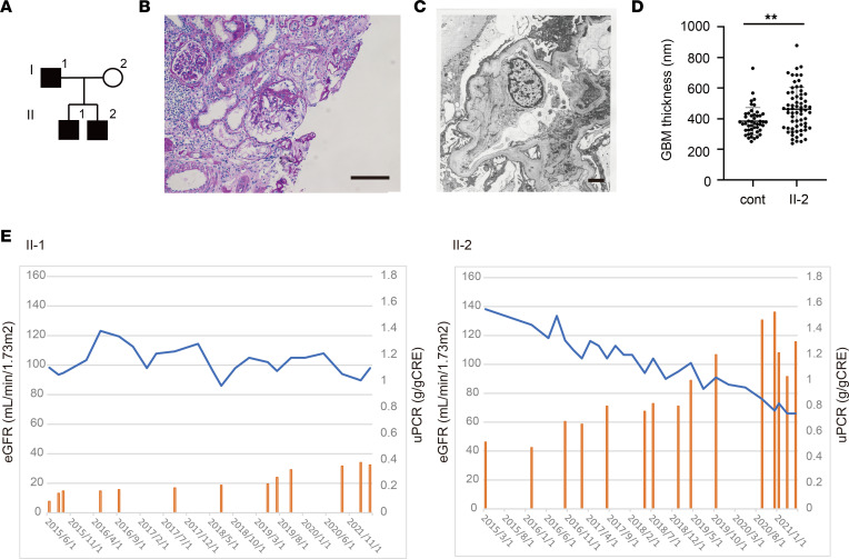
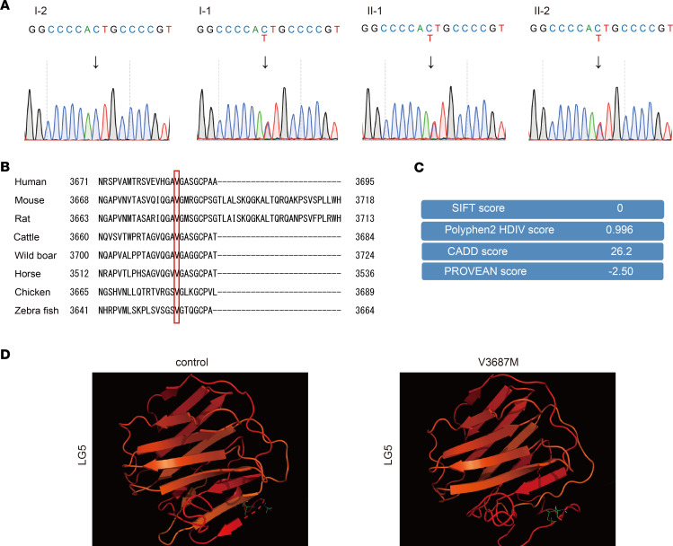
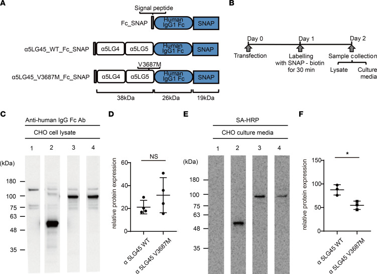
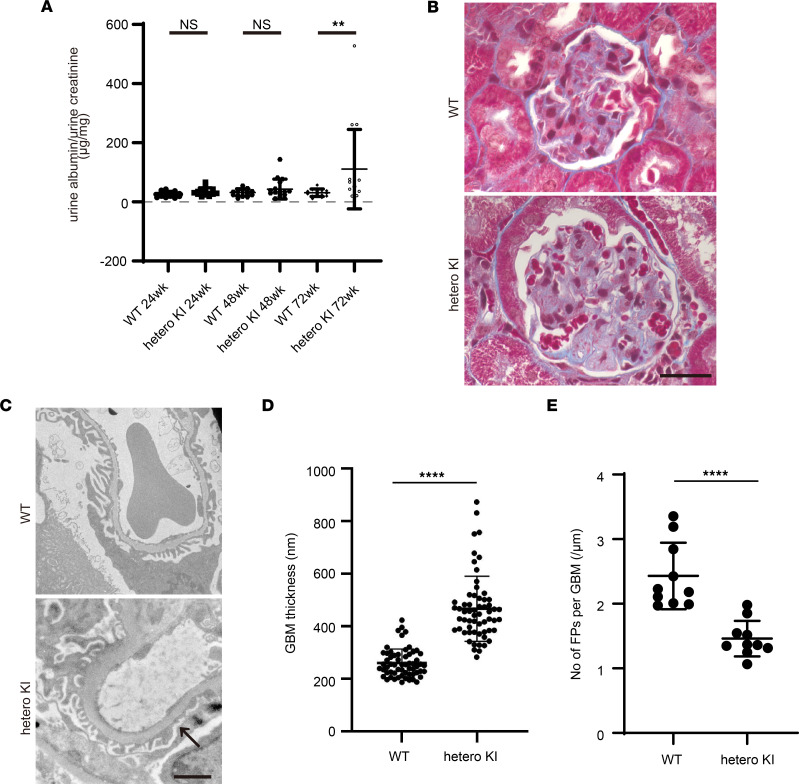
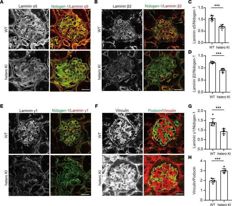
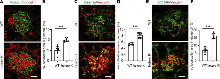
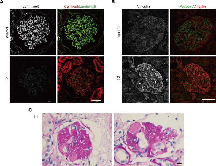
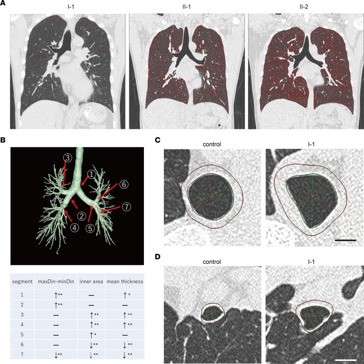
The LAMA5 gene encodes laminin α5, an indispensable component of glomerular basement membrane and other types of basement membrane. A homozygous pathological variant in LAMA5 is known to cause a systemic developmental syndrome including glomerulopathy. However, the roles of heterozygous LAMA5 gene variants in human renal and systemic diseases have remained unclear. We performed whole-exome sequencing analyses of a family with slowly progressive nephropathy associated with hereditary focal segmental glomerulosclerosis, and we identified what we believe to be a novel probable pathogenic variant of LAMA5, NP_005551.3:p.Val3687Met. In vitro analyses revealed cell type-dependent changes in secretion of variant laminin α5 laminin globular 4-5 (LG4-5) domain. Heterozygous and homozygous knockin mice with a corresponding variant of human LAMA5, p.Val3687Met, developed focal segmental glomerulosclerosis-like pathology with reduced laminin α5 and increased glomerular vinculin levels, which suggested that impaired cell adhesion may underlie this glomerulopathy. We also identified pulmonary defects such as bronchial deformity and alveolar dilation. Reexaminations of the family revealed phenotypes compatible with reduced laminin α5 and increased vinculin levels in affected tissues. Thus, the heterozygous p.Val3687Met variant may cause a new syndromic nephropathy with focal segmental glomerulosclerosis through possibly defective secretion of laminin α5. Enhanced vinculin may be a useful disease marker.

Keywords: Extracellular matrix; Genetic diseases; Genetics; Nephrology.


Conflict of interest statement

Conflict of interest: The authors have declared that no conflict of interest exists.

Figures

Figure 1
Figure 1. Clinical analysis of a family with hereditary FSGS.
(A) Pedigree of family with hereditary FSGS. (B) Periodic acid–Schiff staining images of renal biopsy sample from patient I-1. Scale bar: 100 μm. (C) Electron microscopy image of GBM in patient II-2. Scale bar: 2 μm. (D) GBM thickness in patient II-2. **P < 0.01; Mann-Whitney U test. Cont, data from a similarly aged, sex-matched patient with minimal-change NS. (E) Clinical courses of patients II-1 and II-2 in the past 5 years. Blue lines represent estimated glomerular filtration rate (eGFR). Yellow bars show urine protein/creatinine ratio (uPCR).
Figure 2
Figure 2. Identification of a potentially novel LAMA5 heterozygous variant for familial FSGS.
(A) Sanger sequence chromatograms of the patients’ family members. Arrows in chromatograms indicate mutated sites. (B) Amino acid sequence alignment of laminin α5 proteins from multiple species. (C) Scores of variant pathogenicity by SIFT, Polyphen2, CADD, and PROVEAN. (D) Predicted 3D structures of control and variant V3687M laminin α5 LG5 modules. The amino acid molecular models indicated the positions Val3687 (control) and Val3687 (V3687M), respectively.
Figure 3
Figure 3. The fate of this variant laminin α5 protein in vitro.
(A) Structures of transfected expression vectors. (B) Study design for this in vitro experiment. (C) IB analysis of variant laminin α5LG4–5 fragment/Fc-SNAP fusion proteins in lysates of CHO-K1 cells. Cell lysates of negative control (lane 1), Fc-SNAP control (lane 2), WT (lane 3), and V3687M (lane 4) were used for IB analysis. Recombinant proteins were detected using anti-human IgG Fc Ab and quantified by densitometry, as described in the Methods. (D) Density of Fc-SNAP band was defined as 100%. Each column represents the mean of triplicate assays. Bars show SDs. Both variant and WT proteins were expressed. Student’s t test. (E) Pulse-chase analysis of variant laminin α5LG4–5 fragment/Fc-SNAP fusion proteins secreted from CHO-K1 cells. Transfected cells were labeled with SNAP-Biotin for 30 minutes, then cultured in serum-free medium for 1 day. Biotinylated proteins in the conditioned media were visualized and quantified. (F) Density of biotinylated Fc-SNAP band was defined as 100%. *P < 0.05; Student’s t test. In CHO-K1 cells, the secretion of both variant proteins was significantly lower than the secretion of WT protein.
Figure 4
Figure 4. Renal phenotypes of heterozygous V3684M KI mice at 72 weeks of age.
(A) Urine albumin excretion (urine albumin/creatinine ratio) of WT and heterozygous V3684M KI mice at 24, 48, and 72 weeks of age. **P < 0.01; Mann-Whitney U test. (B) Masson’s trichrome staining images of kidney tissues from WT and V3684M KI mice. Scale bar: 20 μm. (C) Electron micrographs of GBM from WT and heterozygous KI mice. Scale bar: 2 μm. The arrow indicates partial effacement of podocyte foot processes. (D) GBM thickness of WT and heterozygous V3684M KI mice at 72 weeks of age. ****P < 0.0001; Mann-Whitney U test. (E) Number of foot processes of podocyte per micrometer GBM length from WT and heterozygous V3684M KI mice at 72 weeks of age. ****P < 0.0001; Mann-Whitney U test.
Figure 5
Figure 5. Protein expression of laminin α5, β2, γ1, and vinculin in the glomeruli in heterozygous V3684M KI mice at 72 weeks of age.
Immunofluorescence staining images of laminin α5 (A), β2 (B), γ1 (E), and nidogen-1 in kidney glomeruli from WT and heterozygous KI mice. Scale bar: 20 μm. Quantification of laminin α5 (C), β2 (D), and γ1 (G) relative to nidogen-1 intensity in the glomerular area. Mean fluorescence intensity of each stain was calculated by Zen software. ***P < 0.001; Mann-Whitney U test. (F) Immunofluorescence staining images of podocin and vinculin in kidney glomeruli from WT and heterozygous KI mice. Scale bar: 20 μm. The tissues were fixed in acetone before the incubation with primary Abs. (H) Quantification of vinculin relative to podocin intensity in the glomerular area. ***P < 0.001; Mann-Whitney U test.
Figure 6
Figure 6. Increased vinculin localization in various glomerular components of heterozygous V3684M KI mice at 72 weeks of age.
(A) Immunofluorescence staining images of podocin and vinculin in kidney glomeruli from WT and heterozygous KI mice. Scale bar: 20 μm. No fixation was performed before the incubation with primary Abs. (B) Quantification of vinculin and podocin colocalization area per glomerular area. ***P < 0.001; Mann-Whitney U test. (C) Immunofluorescence staining images of desmin and vinculin in kidney glomeruli from WT and heterozygous KI mice. Scale bar: 20 μm. (D) Quantification of vinculin and desmin colocalization area per glomerular area. ***P < 0.001; Mann-Whitney U test. (E) Immunofluorescence staining images of CD146 and vinculin in kidney glomeruli from WT and heterozygous KI mice. Scale bar: 20 μm. (F) Quantification of CD146 and desmin colocalization area per glomerular area. ***P < 0.001; Mann-Whitney U test.
Figure 7
Figure 7. Extrarenal phenotypes of heterozygous V3684M KI mice at 72 weeks of age.
(A) Lower magnified images of Masson’s trichrome staining of lung tissues from WT and heterozygous V3684M KI mice. Bronchial tube deformities and enlarged alveolar area were observed in the lung tissue from heterozygous KI mice at 72 weeks of age. Scale bar: 1,000 μm. (B) Quantification of alveolar area in lung tissues from WT and heterozygous KI mice. Alveolar area was quantified using ImageJ software (NIH). ***P < 0.001; Mann-Whitney U test. (C) Immunofluorescence staining images of laminin α5 in BMs in lung bronchial tubes from WT and heterozygous KI mice at 72 weeks of age. Laminin α5 staining was greatly reduced in BM from heterozygous KI mice. Scale bar: 50 μm. (D) Quantification of mean laminin α5 fluorescence intensity along BM from WT and heterozygous KI mice at 72 weeks of age. Mean laminin α5 fluorescence intensity along BM was calculated using Zen software. **P < 0.01; Mann-Whitney U test. (E) Immunofluorescence staining images of vinculin in BM of lung bronchial tubes from WT and heterozygous KI mice at 72 weeks of age. Vinculin staining was greatly enhanced in BM from heterozygous KI mice. Scale bar: 50 μm. (F) Quantification of mean vinculin fluorescence intensity along BM from WT and heterozygous KI mice at 72 weeks of age. ***P < 0.001; Mann-Whitney U test.
Figure 8
Figure 8. Renal phenotypes of patients.
(A) Immunofluorescence staining images of laminin α5 and α5 chain of type IV collagen in glomeruli from a healthy control volunteer and from patients II-1 and II-2. Laminin α5 staining in GBM was lower in patients than in the healthy control. Scale bar: 50 μm. (B) Immunofluorescence staining images of a mechanosensory protein, vinculin, and a podocyte marker, podocin, in glomeruli from a healthy control volunteer and from patient II-2. Vinculin staining was greatly enhanced in podocin-positive cells and endothelial cells in patient II-2. Scale bar: 50 μm. (C) Periodic acid–Schiff staining image of kidney tissue from patient I-1. Glomerular endothelial cell detachments were observed. Scale bar: 50 μm. Arrows indicate the area of endothelial cell detachments.
Figure 9
Figure 9. Extrarenal phenotypes of patients.
(A) Emphysematous changes in patients. Low-attenuation areas (LAAs) were calculated under the threshold of –950 Hounsfield Units and are indicated in red in each chest CT image. Patients II-1 and II-2 had slight and moderate emphysematous changes in their lungs, respectively. Patient I-1 had no emphysematous change. (B) Detailed 3D analyses of bronchial tubes using chest CT. 3D bronchial tree of patient I-1 with indicated measured segments and analysis results. maxDin-minDin: difference between maximum inner diameter and minimum inner diameter at each segment. *P < 0.05; **P < 0.01; Mann-Whitney U test. –, not significant. , increase; ↓, decrease. (C) Cross-sectional images of main bronchi (segment 1) of patient I-1 and an age- and sex-matched healthy control volunteer showing a deformed and thickened bronchus in patient I-1. Scale bar: 1 cm. (D) Cross-sectional images of bronchial tubes (segment 3) of the healthy control volunteer and patient I-1 showing lost bronchial tubes, as well as thickened bronchial tubes, in patient I-1. Scale bar: 1 cm.

Similar articles

Cited by

References

    1. Miner JH. The glomerular basement membrane. Exp Cell Res. 2012;318(9):973–978. doi: 10.1016/j.yexcr.2012.02.031. - DOI - PMC - PubMed
    1. Funk SD, et al. Alport syndrome and Pierson syndrome: diseases of the glomerular basement membrane. Matrix Biol. 2018;71–72:250–261. doi: 10.1016/j.matbio.2018.04.008. - DOI - PMC - PubMed
    1. Hallmann R, et al. Expression and function of laminins in the embryonic and mature vasculature. Physiol Rev. 2005;85(3):979–1000. doi: 10.1152/physrev.00014.2004. - DOI - PubMed
    1. Oakes PW, Gardel ML. Stressing the limits of focal adhesion mechanosensitivity. Curr Opin Cell Biol. 2014;30:68–73. doi: 10.1016/j.ceb.2014.06.003. - DOI - PMC - PubMed
    1. Spenle C, et al. Laminin α5 guides tissue patterning and organogenesis. Cell Adh Migr. 2013;7(1):90–100. doi: 10.4161/cam.22236. - DOI - PMC - PubMed

Publication types