

Generation of vessel co-option lung metastases mouse models for single-cell isolation of metastases-derived cells and endothelial cells
- PMID: 36173713
- PMCID: PMC9526223
- DOI: 10.1016/j.xpro.2022.101691
Generation of vessel co-option lung metastases mouse models for single-cell isolation of metastases-derived cells and endothelial cells
Abstract
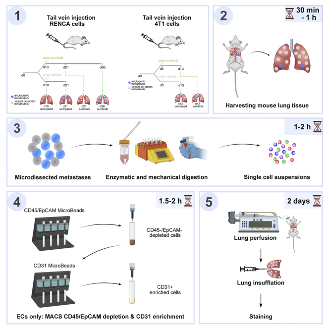
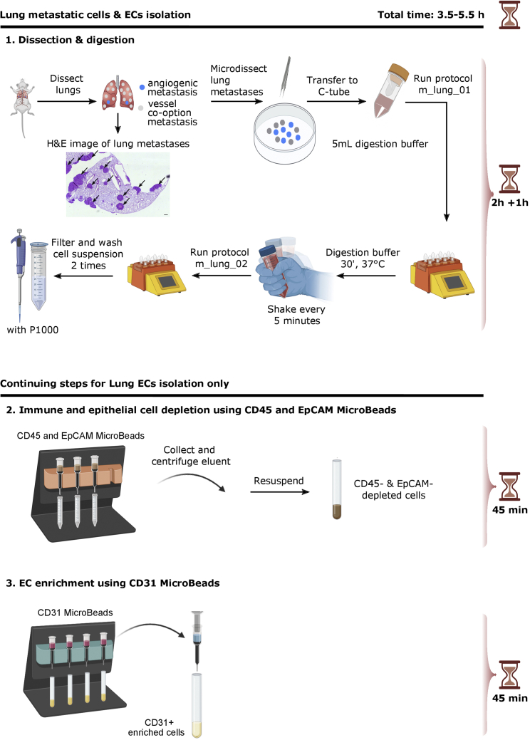
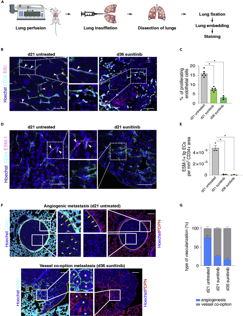
Tumor vessel co-option, a process in which cancer cells "hijack" pre-existing blood vessels to grow and invade healthy tissue, is poorly understood but is a proposed resistance mechanism against anti-angiogenic therapy (AAT). Here, we describe protocols for establishing murine renal (RENCA) and breast (4T1) cancer lung vessel co-option metastases models. Moreover, we outline a reproducible protocol for single-cell isolation from murine lung metastases using magnetic-activated cell sorting as well as immunohistochemical stainings to distinguish vessel co-option from angiogenesis. For complete details on the use and execution of this protocol, please refer to Teuwen et al. (2021).
Keywords: Antibody; Cancer; Cell isolation; Model organisms; Single cell.
Copyright © 2022 The Author(s). Published by Elsevier Inc. All rights reserved.
Conflict of interest statement
Declaration of interests The authors declare no competing interests.
Figures

Similar articles
-
Vessel co-option is common in human lung metastases and mediates resistance to anti-angiogenic therapy in preclinical lung metastasis models.J Pathol. 2017 Feb;241(3):362-374. doi: 10.1002/path.4845. Epub 2016 Dec 29. J Pathol. 2017. PMID: 27859259 Free PMC article.
-
Angiogenesis in NSCLC: is vessel co-option the trunk that sustains the branches?Oncotarget. 2017 Jun 13;8(24):39795-39804. doi: 10.18632/oncotarget.7794. Oncotarget. 2017. PMID: 26950275 Free PMC article. Review.
-
The Inhibition of Vessel Co-Option as an Emerging Strategy for Cancer Therapy.Int J Mol Sci. 2024 Jan 11;25(2):921. doi: 10.3390/ijms25020921. Int J Mol Sci. 2024. PMID: 38255995 Free PMC article. Review.
-
Tumor microenvironment conditions that favor vessel co-option in colorectal cancer liver metastases: A theoretical model.Semin Cancer Biol. 2021 Jun;71:52-64. doi: 10.1016/j.semcancer.2020.09.001. Epub 2020 Sep 10. Semin Cancer Biol. 2021. PMID: 32920126 Review.
-
Tumor vessel co-option probed by single-cell analysis.Cell Rep. 2021 Jun 15;35(11):109253. doi: 10.1016/j.celrep.2021.109253. Cell Rep. 2021. PMID: 34133923
Cited by
-
Antiangiogenic-immune-checkpoint inhibitor combinations: lessons from phase III clinical trials.Nat Rev Clin Oncol. 2024 Jun;21(6):468-482. doi: 10.1038/s41571-024-00886-y. Epub 2024 Apr 10. Nat Rev Clin Oncol. 2024. PMID: 38600370 Review.
References
-
- Bridgeman V.L., Vermeulen P.B., Foo S., Bilecz A., Daley F., Kostaras E., Nathan M.R., Wan E., Frentzas S., Schweiger T., et al. Vessel co-option is common in human lung metastases and mediates resistance to anti-angiogenic therapy in preclinical lung metastasis models. J. Pathol. 2017;241:362–374. doi: 10.1002/path.4845. - DOI - PMC - PubMed
-
- Conchinha N.V., Sokol L., Teuwen L.A., Veys K., Dumas S.J., Meta E., García-Caballero M., Geldhof V., Chen R., Treps L., et al. Protocols for endothelial cell isolation from mouse tissues: brain, choroid, lung, and muscle. STAR Protoc. 2021;2:100508. doi: 10.1016/j.xpro.2021.100508. - DOI - PMC - PubMed
Publication types
MeSH terms
LinkOut - more resources
Full Text Sources
Medical
Research Materials
Miscellaneous
