Review
. 2023 Jan 1;103(1):919-956.
doi: 10.1152/physrev.00060.2021. Epub 2022 Sep 29.

The choroid plexus: a missing link in our understanding of brain development and function

Affiliations
Review

The choroid plexus: a missing link in our understanding of brain development and function

Norman R Saunders et al. Physiol Rev. .

Abstract

Studies of the choroid plexus lag behind those of the more widely known blood-brain barrier, despite a much longer history. This review has two overall aims. The first is to outline long-standing areas of research where there are unanswered questions, such as control of cerebrospinal fluid (CSF) secretion and blood flow. The second aim is to review research over the past 10 years where the focus has shifted to the idea that there are choroid plexuses located in each of the brain's ventricles that make specific contributions to brain development and function through molecules they generate for delivery via the CSF. These factors appear to be particularly important for aspects of normal brain growth. Most research carried out during the twentieth century dealt with the choroid plexus, a brain barrier interface making critical contributions to the composition and stability of the brain's internal environment throughout life. More recent research in the twenty-first century has shown the importance of choroid plexus-generated CSF in neurogenesis, influence of sex and other hormones on choroid plexus function, and choroid plexus involvement in circadian rhythms and sleep. The advancement of technologies to facilitate delivery of brain-specific therapies via the CSF to treat neurological disorders is a rapidly growing area of research. Conversely, understanding the basic mechanisms and implications of how maternal drug exposure during pregnancy impacts the developing brain represents another key area of research.

Keywords: blood-brain barrier; cerebrospinal fluid; choroid plexus; development; drug penetration.


Conflict of interest statement

S.A.L. maintains a financial interest in AstronauTx, Ltd., and is on the Scientific Advisory Board of RM Global.

Figures

None
Graphical abstract
FIGURE 1.
FIGURE 1.
Historical publication data for choroid plexus and vascular endothelial cell barriers in the brain. Although minimal work was published from 1834 to the mid-1900s, an explosion in publications focusing on the blood-brain barrier and brain endothelial barrier properties began around 1945. Data collected from PubMed May 14, 2022 with the following search terms: “choroid plexus,” “cerebral blood vessels,” “blood-brain barrier,” “blood cerebrospinal fluid barrier.”
FIGURE 2.
FIGURE 2.
Gross morphology of cerebral ventricles and choroid plexuses within them in the mammalian brain.
FIGURE 3.
FIGURE 3.
Schematic cross section of 2 choroid plexus villi. The mammalian and nonmammalian vertebrate choroid plexuses consist of numerous fronds projecting into the cerebrospinal fluid (CSF), which are composed of several villous processes. An outer simple cuboidal epithelium lies on a basal lamina surrounding an inner stromal core of connective tissue that is highly vascularized. It is derived from the adjacent ependyma lining the ventricle walls. The plexus differs from the ependyma by the presence of apical tight junctions. The epithelial cells are polarized. The apical membrane facing the CSF has uneven borders of irregular microvilli and numerous groups of cilia. The lateral membranes of the epithelial cells display complex infoldings at their basal ends. Each villus contains a large fenestrated capillary consisting of very thin endothelial cells. The stromal connective tissue is composed of a loose network of collagen fibers, secreted by occasional elongated fibroblasts (yellow). Globular macrophages (green), rich in phagolysosomes, are also present in the stromal core; these are distinct from the star-shaped dendritic cells. A few of the dendritic cells lie between the basal lamina and the choroid epithelium, extending processes between epithelial cells. Kolmer cells (epiplexus cells, dark blue) lie on the ventricular surface of the epithelial cells, closely associated with the microvillous border. Adapted from Ref. , with permission from Journal of Neuropathology & Experimental Neurology.
FIGURE 4.
FIGURE 4.
Changing fluid sources and targets during neurulation and early neurogenesis. Developing brain schema derived from mouse development. Major changes occur throughout neural tube closure (NTC) and early brain development. A: the neuroectoderm of the open neural tube folds to generate neuroepithelium, and the fluid environment changes from amniotic fluid (AF) to cerebrospinal fluid (CSF). Ages are shown for mouse development at embryonic days (E)8.5, E10.5, and E12.5. Scale bars, 1 mm. B: before NTC, the fluid environment of the neuroectodermal progenitors is AF that contains evidence of high levels of translation and glycolysis that occurs in the progenitors during this time. Early CSF before choroid plexus formation contains unique markers of brain parenchyma development, as well as some components that have been shown to signal to neural progenitors. Later in development, CSF components can arise from the neural progenitors or from the choroid plexus. CSF also mixes and flows to move components to different regions of the ventricular system. AF composition includes organelle components (e.g., ribosomal or mitochondrial proteins). CSF contents include free molecules [e.g., 5-hydroxytryptamine (5-HT)], molecules bound to carrier proteins (e.g., RA+ RBP4); organelle components (e.g., ribosomal or mitochondrial proteins), free proteins or peptides (e.g., WNT3A or cytokines), or membrane-bound particles (e.g., exosomes) that act as carriers for additional proteins (e.g., SHH) or nucleic acid (e.g., miRNA loops). These different components have unique signaling modalities including receptor binding to cells or fusion with cells in direct contact with CSF, including neural progenitors or choroid plexus cells (epithelial cells, mesenchymal cells, endothelial cells, immune cells, neurons).
FIGURE 5.
FIGURE 5.
Development of plexus epithelial cells and transcription factors involved in development of the lateral ventricular choroid plexus. The choroid plexus epithelial cells (CPECs) develop from modified neuroepithelium and are added to the structure from the dorsal (upper) surface. Experiments using Monodelphis domestica (South American gray short-tailed opossum) after a single injection of 5-bromo-2-deoxyuridine (BrdU) have been used to identify plexus growth in vivo. Positive nuclei initially seen in CPECs at the root of the plexus (top), with additionally added new cells pushing the plexus structure out from the neuroependymal wall, causing the complex invaginated structure to form. In this way, new epithelial cells are added along the stalk of the plexus, away from the root, displaced by newly dividing cells. No cells are described as being added from the ventral surface. Specific transcription factors involved in the promotion (green) or inhibition (red) of plexus epithelial development and growth are also shown. CSF, cerebrospinal fluid. Adapted from Ref. , with permission from Cerebrospinal Fluid Research, and Ref. , with permission from Frontiers in Neuroscience.
FIGURE 6.
FIGURE 6.
Localization of proteins for ion transporters, channels, and associated enzymes and identification of their corresponding genes in adult and embryonic (E15) rat choroid plexus. Data for localization of the proteins are from Refs. –. Cerebrospinal fluid (CSF) secretion results from coordinated transport of ions and water from basolateral membrane to cytoplasm, then sequentially across apical membrane into ventricles [for review see Davson and Segal (118)]. On the plasma-facing membrane there is parallel Cl/HCO3 exchange (AE2, Slc4a2) and Na+-HCO3 cotransport (NBC1, Slc4a4), with net effect of bringing Cl into cells in exchange for HCO3 (119). Na+-dependent Cl/HCO3 exchange (NCBE, Slc4a10) at the basolateral membrane modulates pH and perhaps CSF formation (114,115, 120). Apical Na+ influx by NHE5 (Slc9a5) and ATB1 (Atb1b1, Na+-K+-ATPase, asterisk) maintains a low cell Na+ concentration ([Na+]) that sets up a basolateral gradient to drive Na+ uptake (121). Na+ is extruded into CSF mainly via the Na+-K+-ATPase pump (ATB1, Atb1b1) and, under some conditions, the Na+-K+-Cl cotransporter NKCC1 (Slc12a2; see Ref. for review). Overall cell volume is maintained by the K+-Cl cotransporters NCCT (Slc12a3) and KCC1 (Slc12a4). Aquaporin (AQP1/3/4) channels on CSF-facing membrane (123) mediate water flux into ventricles (124). AQP1 is also localized to the basolateral membrane (125). Polarized distribution of carbonic anhydrase (CAR) and Na+-K+-ATPase, and aquaporins, enable net ion and water translocation to CSF (see Refs. , for review). The gene Slc4a7 (NBCN1) was not detected by RNA sequencing (RNA-seq), although it has been reported in both rat and mouse choroid plexus (114). The genes for Clir (chloride inwardly rectifying) channels has not been previously identified but are probably Clica and Clicb. The gene for VRAC (volume-regulated anion channel) is not known (127). The carbonic anhydrases CAR2 and CAR8 have an intracellular distribution; CAR8 has been shown to lack the characteristic enzyme activity of these proteins (128). It is not known whether it is functional in the embryo. The CLIC chloride channels are also intracellular (129), but their location is unknown. Insets show the fold differences (FDs) for genes expressed at a higher level in the embryonic (red) or the adult (blue) choroid plexus. Adapted from Ref. , with permission from PLoS One.
FIGURE 7.
FIGURE 7.
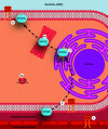
Proposed transepithelial pathway for albumin through choroid plexus epithelial cells. Glycophorins A/C (GYPA/C) in the endothelial cells deliver albumin to the basement membrane (1) that then can be taken up into plexus epithelial cells by GYPA/C or Secreted protein acidic and rich in cysteine (SPARC) (2). From here, albumin moves along a SPARC-specific pathway through tubulocisternal endoplasmic reticulum (3) and Golgi (4a) or via a VAMP-mediated pathway in vacuoles, lysosomes, or multivesicular bodies (4b). On the apical surface of the plexus epithelium, GYPA/C may be involved in efflux of protein from the cell into the cerebrospinal fluid (CSF) (5), as validated by extensive GYPA immunoreactivity in embryonic plexus. In the adult, the lack of immunoreactivity in the endoplasmic reticulum and Golgi along with increased expression of gene products for VAMP molecules suggest that the majority of transport occurs via VAMP-mediated vesicular/lysosomal transport (4b). Adapted from Ref. , with permission from PLoS One.
FIGURE 8.
FIGURE 8.
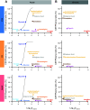
Mean cerebrospinal fluid (CSF)-to-plasma ratios for 3H-labeled drugs compared to lipid solubility (log10Doctanol partition coefficient) following acute and chronic treatment. A: permeability studies in rats at embryonic day (E)19, postnatal day (P)4, and adult. Intraperitoneal drug injection to fetuses (except cimetidine via mother) samples taken at 30 min (acute treatment). Drugs were compared with 3 molecules of increasing lipid solubility (sucrose, l-glucose, glycerol) that enter the CSF by diffusion. The similarity of results for sucrose and l-glucose at the 3 ages shows that the marked increase in CSF turnover that occurs during brain development (13) does not account for the decline in CSF-to-plasma ratios with age for all the drugs tested. Ratios for all drugs were <100%, indicating restricted entry into CSF that was different for each drug and not related to lipid solubility. Age-related changes likely to be due to development of efflux mechanisms (ABC transporters or related metabolic enzymes). Log10Doctanol computed from Tetko et al. (361). B: chronic measurements of CSF penetration of same drugs in A. Thirty-minute intraperitoneal drug exposure with 3H-labeled drugs after 4- to 5-day treatment with clinically relevant doses (chronic treatment). All ratios were <100%, indicating some degree of restriction. Compared with acute doses (A) at E19 there was an increase in ratios for acetaminophen/paracetamol, cimetidine, and digoxin, suggesting that the capacity of the efflux system was exceeded. At P4, the drugs had similar ratios in the 2 treatment groups. In adults the ratio for acetaminophen/paracetamol was reduced, suggesting upregulation of an efflux transporter in response to longer-term treatment. There was a significant increase in expression of Abcb1b (P-glycoprotein) in adult choroid plexus, which could account for this (362). Note change of the x-axis to log10Doctanol positive values only in right-hand column (chronic). Data from Ref. , with permission from Scientific Reports; additional data from Ref. , with permission from F1000Research, and Ref. , with permission from ACS Pharmacology & Translational Science.
FIGURE 9.
FIGURE 9.
Localization of ABC efflux transporters in choroid plexus epithelial cells of the fetus and adult. Data for localization of the proteins and developmental gene expression changes are from Refs. , , –. See also FIGURE 10 for an overview of efflux mechanisms at the blood-cerebrospinal fluid (CSF) barrier. Most transporters require movement of the molecule through the entire phospholipid bilayer, either under control of diffusion gradients or by energy (ATP)-mediated transfer. An exception to this is PGP (Abcc1), which collects molecules for efflux directly from the bilayer itself before entry into the cell cytoplasm.
FIGURE 10.
FIGURE 10.
Efflux mechanisms in the apical membrane of a choroid plexus cell. Multiple mechanisms exist for removal of molecules out of the cerebrospinal fluid (CSF). Example transporters given [e.g., PGP (Abcb1), MRPs (ABCC family transporters, see FIGURE 9), BCRP (Abcg2)]. “Others ”covers organic anion and cation acid transporters in FIGURE 9. Based on efflux transporter functions in brain barrier cells and adapted from Ref. , with permission from Frontiers in Pharmacology. ADP, adenosine diphosphate; ATP, adenosine triphosphate.
FIGURE 11.
FIGURE 11.
Age comparison of drug entry into cerebrospinal fluid (CSF). CSF-to-plasma concentration ratios (%) of valproate (A) or lamotrigine (B) at embryonic day (E)19 and postnatal day (P)4 and in nonpregnant female adult rats collected 30 min after a single intraperitoneal injection of 100 mg/kg valproate or 20 mg/kg lamotrigine. Points are results from a single animal. Means ± SD; n = 3–7 individual animals. **P < 0.01, 1-way ANOVA with multiple comparisons. ns, Not significant. Data replotted from Ref. , with permission from F1000Research.

Similar articles

Cited by

References

    1. Weed LH. The cerebrospinal fluid. Physiol Rev 2: 171–203, 1922. doi:10.1152/physrev.1922.2.2.171. - DOI
    1. Saunders NR, Dreifuss JJ, Dziegielewska KM, Johansson PA, Habgood MD, Møllgård K, Bauer HC. The rights and wrongs of blood-brain barrier permeability studies: a walk through 100 years of history. Front Neurosci 8: 404, 2014. doi:10.3389/fnins.2014.00404. - DOI - PMC - PubMed
    1. Stern L, Gautier R. Recherches sur le liquide cephalo-rachidien. Arch Int Physiol 17: 138–192, 1921. doi:10.3109/13813452109146211. - DOI
    1. Stern L, Gautier R. Le passage dans le liquide cephalo-rachidien de substances introduites dans la circulation et leur action sur le systeme nerveux central chez les differentes especes animales. RCR Soc Phys Hist Nat Genève 35: 91–94, 1918.
    1. Liddelow SA. Fluids and barriers of the CNS: a historical viewpoint. Fluids Barriers CNS 8: 2, 2011. doi:10.1186/2045-8118-8-2. - DOI - PMC - PubMed

Publication types

LinkOut - more resources