. 2022 Sep 29;18(9):e1010417.
doi: 10.1371/journal.pgen.1010417. eCollection 2022 Sep.

Gap junctions mediate discrete regulatory steps during fly spermatogenesis

Affiliations

Gap junctions mediate discrete regulatory steps during fly spermatogenesis

Yanina-Yasmin Pesch et al. PLoS Genet. .

Abstract

Gametogenesis requires coordinated signaling between germ cells and somatic cells. We previously showed that Gap junction (GJ)-mediated soma-germline communication is essential for fly spermatogenesis. Specifically, the GJ protein Innexin4/Zero population growth (Zpg) is necessary for somatic and germline stem cell maintenance and differentiation. It remains unknown how GJ-mediated signals regulate spermatogenesis or whether the function of these signals is restricted to the earliest stages of spermatogenesis. Here we carried out comprehensive structure/function analysis of Zpg using insights obtained from the protein structure of innexins to design mutations aimed at selectively perturbing different regulatory regions as well as the channel pore of Zpg. We identify the roles of various regulatory sites in Zpg in the assembly and maintenance of GJs at the plasma membrane. Moreover, mutations designed to selectively disrupt, based on size and charge, the passage of cargos through the Zpg channel pore, blocked different stages of spermatogenesis. Mutations were identified that progressed through early germline and soma development, but exhibited defects in entry to meiosis or sperm individualisation, resulting in reduced fertility or sterility. Our work shows that specific signals that pass through GJs regulate the transition between different stages of gametogenesis.


Conflict of interest statement

The authors have declared that no competing interests exist.

Figures
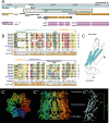
Fig 1
Fig 1. The genomic locus of zpg, its protein structure, and the identification of residues of interest.
(A) Overview of the genetic locus of zpg (inx4) on chromosome 3L, showing genes (blue), transcripts (orange) and coding sequence (magenta) of Zpg and the neighboring genes. The DNA stretch that is included in the rescue construct used in this study is indicated by a grey box. (B) Sequence alignment of Zpg (Inx4) with other Drosophila innexins and C. elegans INX-6, which was used as basis for in silico 3D structure homology modeling. The N-terminal portions of the proteins are depicted (approximately amino acid 1–80, see numbers on the right) and the degree of conservation is indicated in bar graphs. Polar amino acids are shown in grey, hydrophobic in yellow, positively charged in magenta and negatively charged in cyan. The residues D21, D50 and D59 and well as the first (C1) and second cysteine (C2) in Zpg, which were used as targets for mutagenesis, are part of this stretch of the protein and their location is indicated. Note that D21, D50 and the cysteine residues show a high degree of conservation among the innexins, whereas D59 does not. (C-C”’) Predicted structure of Drosophila Zpg reveals octameric arrangement around a central pore. Simplified view in C. Top view in C’. Side view in C”. Each subunit is labeled in a different color. Single Zpg subunit is depicted in C”’ and as a cartoon in C. The first extracellular domain as well as the entire N-terminus are facing inside the channel pore. Potentially functionally relevant residues within the channel opening are labeled in magenta (D21, D50, D59). While D21 and D50 are conserved among innexins, D59 was chosen as target for mutagenesis due to its predicted location at the narrow opening of the channel.
Fig 2
Fig 2. Zpg function can be fully restored by introducing a GFP-tagged genomic rescue construct into the zpg null mutant background.
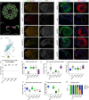
Zpg staining (green in A-C; single channels depicted in grey in A’-C’) is strongly enriched at the soma-germline boundary in the wild type (A, A’), but cannot be detected in the rudimentary testes of zpg null mutants (B, B’). Wild type like distribution of Zpg can be seen in flies having the zpg::GFP GR (Genomic rescue) construct in the zpg null mutant background (C, C’). Hubs are marked by DN-Cadherin in red, nuclei are labeled in blue (A-C). Heterozygous expression of zpg::GFP GR (D, D’) reveals strong colocalization of the transgenic construct (GFP, green) and endogenous Zpg (red). Fas3 labels the hub. D’ shows single channel GFP signal at germ cell membranes in grey. Compared to wt (E, G), zpg null (H, J) mutants show a strong reduction of mitotic Vasa+ germ cells (green), indicating an early arrest in germ cell differentiation. In contrast, the number of germ cells (green) in zpg::GFP GR rescue flies (K, M) is indistinguishable from wt. The number of early somatic cells labeled by markers Zfh-1 (cyst stem cells and immediate daughter cells; magenta) and Tj (grey) is, compared to wt (E, F), greater in testes of zpg null mutants (H, I), but unaffected in zpg::GFP GR (K, L). Number of cells expressing the late somatic cell marker Eya (magenta) is, compared to wt (G), lower in zpg null mutants (J), but unaffected in zpg::GFP GR testes (M). Quantification of (N) the number of germline stem cells (GSCs, defined as single Vasa+ cells contacting the hub), (O) Zfh-1-positive cells, (P) Tj-positive cells, (Q) Eya-positive cells, (R) spermatid bundles, and (S) fertility in wildtype, zpg null mutant, and zpg::GFP GR rescue flies. Scale bars represent 30–100 μm, as indicated above them. p-values are for difference from wildtype and indicated by asterisks with *p<0.05, **p<0.01, ***p<0.001.
Fig 3
Fig 3. The C-terminus of Zpg is crucial for protein function, while C-terminal phosphorylation sites are dispensable.
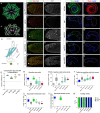
Zpg staining (green in A-D; single channels in grey in A’-D’) is absent in zpg mutants rescued with Zpg containing a C-terminal deletion (zpg deltaCT::GFP) (B, B’), as the antibody binding site is deleted. zpg deltaCT::GFP mutant testes are severely reduced in size and the hub (DN-Cadherin, red) is enlarged. In zpg mutants expressing a genomic rescue construct containing mutations in phosphorylation sites (C, C’: zpg Y352F, D, D’: zpg Y352F/S356A), Zpg staining normally localizes to the germline-soma boundaries (as indicated by arrows). Wt control is shown in A, A’. Nuclei are highlighted in blue. (E-F) localization of the GFP tag in testes when zpg deltaCT::GFP is expressed in the null mutant background (E-E’) and in flies with one copy of endogenous zpg (F-F’). In testes of both genotypes, the GFP signal accumulates intracellularly. Compared to wt (G, I), significantly less Vasa+ early germ cells (green) can be detected in testes of zpg deltaCT::GFP flies (J, L), whereas no difference to wt was seen in zpg Y352F (M, O) and zpg Y352F/S356A mutants (P, R). The number of Zfh-1+ cells (wt shown in F) and Tj+ cells (wt shown in H) was higher in zpg deltaCT::GFP testes (J, K), but not in the phospho mutants (M-Q). Less cells expressing the late somatic marker Eya were detected in testes of zpg deltaCT::GFP flies (L) than in wt (I), but no change was found in the phosphorylation mutants (O, R). This indicates defective germ cell and somatic cell differentiation in zpg deltaCT::GFP flies, but not in the two phosphorylation mutants. Quantification of (S) germline stem cells (GSCs), (T) Zfh-1-positive cells, (U) Tj-positive cells, (V) Eya-positive cells, (W) spermatid bundles, and (X) fertility, shows loss of function upon deletion of the C-terminus, but no defects in phosphorylation mutants. Scale bars represent 30–50 μm, as indicated above them. p-values are for difference from wildtype and indicated by asterisks with *p<0.05, **p<0.01, ***p<0.001.
Fig 4
Fig 4. Zpg does not function as a hemichannel.
(A-D) zpg mutants rescued with genomic rescue constructs in which one or more cysteine residues were mutated, hindering the formation of gap junctions, have rudimentary testes and no Zpg is detected by antibody staining (green in A-D; single channels depicted in grey in A’-D’; wt in A-A’; zpg C6S, B-B’:, zpg C145S, C-C’; zpg C236S, D-D’). Hubs are marked with DNCad in red, nuclei are highlighted in blue. (E-E”’) In testes of zpg mutants expressing GFP-tagged versions of the cysteine mutation constructs (zpg C6S::GFP in E, E’, zpg C26S::GFP in E”, E”’), a strong intracellular accumulation of the GFP signal can be detected, while Zpg antibody staining is very weak. Cysteine mutations in zpg cause a strong defect in early stages of germ cell differentiation as detected by Vasa staining (F, H: wt; I, K: zpg C6S, L, N: zpg C145S; O, Q: zpg C236S). Compared to wt (F, G), the expression of the early somatic markers Zfh-1 (magenta; I, L, O) and Tj (grey; J, M, P) was increased in all three cysteine mutants, whereas the number of cells expressing the late marker Eya (K, N, Q) was decreased. Quantification of (R) germline stem cells (GSCs), (S) Zfh-1-positive cells, (T) Tj-positive cells, (U) Eya-positive cells, (V) spermatid bundles, and (W) fertility shows null mutant-like phenotypes in cysteine mutants, leading to complete sterility. Scale bars represent 30–50 μm, as indicated above them. p-values are for difference from wildtype and indicated by asterisks with *p<0.05, **p<0.01, ***p<0.001.
Fig 5
Fig 5. Mutations in the Zpg D50 channel pore residue result in germ cell differentiation defects.
(A) Homology model of Zpg showing the position of aspartate 50 (D50) within the channel pore. (A’) D50 is predicted to form a hydrogen bond with glutamine 49 (Q49) of the adjacent Zpg subunit. (A”’) Simplified model highlighting the location of D50 (marked in pink) in the first extracellular loop. (B-F) Colocalization of wildtype endogenous Zpg and GFP-tagged, mutated Zpg. Flies heterozygous for a null allele of zpg but also containing one copy of the wildtype genomic zpg rescue construct (zpg GFP::GR; B-B’), no rescue construct (C-C’), one copy of the zpg D50A mutant rescue construct (D-D’), one copy of the zpg D50R mutant rescue construct (E-E’) and one copy of the zpg D50K mutant rescue construct (F-F’), the GFP-tagged D50 mutants show strong colocalization with the endogenous Zpg at the membrane. This high degree of colocalization is also revealed by the quantification of the Pearson coefficient between the GFP and Zpg antibody staining (G). (H-Q) Staining for the mitotic germ cell marker Vasa and the late-stage germ cell marker Boule. In the wildtype, Vasa staining is mostly concentrated in the apical part of the testis (H), whereas Boule marks meiotic cysts and long, parallel bundles of spermatids (I). In zpg null mutant testes little Vasa (J) and no Boule (K) signal is detected. In both zpg D50A (L) and zpg D50R (N) testes, the Vasa signal is strong and broadly localized. However, in zpg D50A testes, Vasa-positive cysts are abnormally found throughout the entire testis (L), and defective cysts can be observed in both mutants (circled in L, arrowhead in N). In addition, Boule staining in testes of zpg D50A (M) and zpg D50R (O) mutants reveals disorganized spermatid bundles. While zpg D50K mutant testes have a larger number of germ cells and larger mitotic cysts compared to zpg null mutants (P), they fail to reach meiosis (Q). Quantification of (R) germline stem cells (GSCs), (S) Zfh-1-positive cells, (T) Tj-positive cells, (U) Eya-positive cells, (V) spermatid bundles, and (W) fertility data. The data indicates late germ cell differentiation defects in zpg D50A and zpg D50R mutants and a stronger phenotype closer to the null mutant in zpg D50K mutants. Hubs are either encircled or indicated by asterisks. Scale bars represent 50 μm, as indicated above them. n>30 single crosses per genotype for fertility tests. p-values are for difference from wildtype and indicated by asterisks with *p<0.05, **p<0.01, ***p<0.001.
Fig 6
Fig 6. Mutating the residue D59 of Zpg in the channel pore results in late-stage defects in germ cell differentiation.
(A) Homology model of Zpg showing the position of Aspartate 59 (D59) at the narrowest part of the channel pore, constricting its diameter. (A’) D59 is predicted to form a hydrogen bond with lysine (K58) of the neighboring Zpg subunit. (A”) Simplified model highlighting the location of D59 in the first extracellular loop of Zpg, facing inside the pore. (B-F) Colocalization of wildtype endogenous Zpg and GFP-tagged, mutated Zpg. Flies heterozygous for a null allele of zpg but also containing one copy of the wildtype genomic zpg rescue construct (zpg GFP::GR; B-B’), no rescue construct (C-C’), one copy of the zpg D59A mutant rescue construct (D-D’), one copy of the zpg D59N mutant rescue construct(E-E’), and one copy of the zpg D59H mutant rescue construct (F-F’). The GFP-tagged D59 mutants show strong colocalization with the endogenous Zpg at the membrane. This high degree of colocalization is also revealed by the quantification of the Pearson coefficient between the GFP and Zpg antibody staining (G). (H-Q) Staining for the mitotic germ cell marker Vasa and the late-stage germ cell marker Boule. In the wildtype, Vasa staining is mostly concentrated in the apical part of the testis (H), whereas Boule marks meiotic cysts and long, parallel bundles of spermatids (I). In zpg null mutant testes little Vasa (J) and no Boule (K) signal is detected. Early germ cell differentiation defects are detected in zpg D59A (L, M), but not in zpg D59N (N, O) or zpg D59H (P,Q) mutants. Impaired entry to meiosis is seen in testes of zpg D59A mutants, since the Vasa signal (L) takes up the entire testis and the Boule signal mostly labels late-stage GC cysts, with very few spermatids (M). Weaker phenotypes are seen in testes of zpg D59N and zpg D59H mutants, with wildtype Vasa staining (N, P, respectively). However, abnormal cysts are occasionally seen (for example see P, arrowhead). Although Boule staining is rescued in zpg D59N and zpg D59H mutants they show some disorganization of sperm bundles (O, Q). Quantification of (R) germline stem cells (GSCs), (S) Zfh-1-positive cells, (T) Tj-positive cells, (U) Eya-positive cells, (V) spermatid bundles, and (W) fertility reveals late germ cell differentiation defects in all mutants, with the strongest phenotype seen in zpg D59A. Hubs are either encircled or indicated by asterisks. Scale bars represent 50 μm, as indicated above them. n>30 single crosses per genotype for fertility tests. p-values are for difference from wildtype and indicated by asterisks with *p<0.05, **p<0.01, ***p<0.001.
Fig 7
Fig 7. Mutations in the channel-gating N-terminus of Zpg lead to loss of function.
(A) Homology model of Zpg showing the position of Aspartate 21 (D21) residue within the Zpg channel reveals a localization within the channel pore. (A’) The side chains of D21 are not in proximity to any other amino acids. This makes a direct interaction with the cargo likely. (A”) The introduced N-terminal mutations are highlighted in pink in a schematic view of the Zpg protein structure. In the deletion mutant zpg delta2-5, the highly flexible N-terminal domain was shortened, while D21 sits at the hinge between the N-terminal chain and the first transmembrane domain. Due to the limited number of germ cells in the N-terminal mutants, which makes it hard to assess subcellular localization, the localization of Zpg (GFP-tagged; green in B-F, single channels in B’-F’) was analyzed in testes of flies harboring one copy of the respective mutation and once copy of endogenous Zpg (red in B-F). In the control (zpg::GFP GR, B-B’), the mutated and endogenous Zpg colocalize at the plasma membrane. In non-rescued flies heterozygous for zpg (C-C’), Zpg localizes to the membrane and no GFP-tagged construct is expressed. Testes of zpg D21A (D-D’) and zpg D21N (E’E’) mutants display weak membrane localization of the mutated proteins (see arrows), while the majority of the signal is cytoplasmic. In contrast, strong colocalization is found in zpg delta2-5 mutants (F-F’). (G) Measurement of the Pearson coefficient for colocalization of Zpg and GFP in testes of heterozygotes with one copy of endogenous and one copy of mutated Zpg. Strong colocalization in zpg delta2-5 mutants, similar to the zpg::GFP GR control, is observed, but only weak colocalization in zpg D21A and zpg D21N mutants. (H-Q) Staining for the mitotic germ cell marker Vasa and the late-stage germ cell marker Boule. In the wildtype, Vasa staining is mostly concentrated in the apical part of the testis (H), whereas Boule marks meiotic cysts and long, parallel bundles of spermatids (I). In zpg null mutant testes little Vasa (J) and no Boule (K) signal is detected. Testes of zpg D21A (L-M), zpg D21N (N-O), and zpg delta2-5 (P-Q) mutants exhibit a zpg null mutant-like phenotype. Quantification of (R) germline stem cells (GSCs), (S) Zfh-1-positive cells, (T) Tj-positive cells, (U) Eya-positive cells, (V) spermatid bundles, and (W) fertility reveals null mutant-like phenotypes in all three N-terminal mutants, the exception being partial rescue in the number of Zfh-1 positive cells in zpg D21A mutants. Hubs are either encircled or indicated by asterisks. Scale bars represent 50 μm, as indicated above them. n>30 single crosses per genotype for fertility tests. Unless otherwise indicated p-values are for difference from wildtype and indicated by asterisks with *p<0.05, **p<0.01, ***p<0.001.
Fig 8
Fig 8. Defects in the spermatid stage are detected in a subset of zpg channel pore mutants.
(A-F) Phase contrast imaging of “onion stage” spermatids in live testes in wt and in the subset of zpg mutants that show intermediate germline differentiation defects. In the wildtype (A), spermatids appear as round cells with large nuclei with a small nebenkern (black dot inside nuclei). Major defects such as multinucleation and abnormal nucleus-to-nebenkern ratios are seen in spermatids of zpg mutants rescued with the D50A (B), D50R (C), D59A (D), and D59H (E) mutant zpg genomic rescue constructs, as indicated by red arrows. Spermatids of zpg D59N mutants (F) do not show nuclear defects. (H-L) Spermatid individualization complexes (ICs) in testes of freshly eclosed flies were stained with rhodamine phalloidin (labels actin cones, magenta) and TO-PRO-3 (labels nuclei, green). In the wildtype (H), the actin cones are tightly associated with the elongated nuclei of 64 developing spermatids. In testes of zpg D50A (H), zpg D50R (I), zpg D59A (J), zpg D59H (K) and zpg D59N (L) mutants, the association of actin can be disrupted and the overall IC structure appears disorganized. Scale bars indicate 10 μm. (M) Quantification of the angle of association between the actin cones and the nuclei in ICs. The wildtype has a 180° angle between the actin and the nuclei due to the linear organization of the IC. In all analyzed mutants, but in particular in zpg D50A and zpg D59A, the angle is smaller, indicating disorganization. n = 45 for all mutants except zpg D59A (n = 25 due to lower abundance of spermatid bundles). (N) Simplified model summarizing of the function of Zpg during spermatogenesis. Zpg is required for all major developmental transitions in fly spermatogenesis. Schematic depicts the process of spermatogenesis starting at stem cell the stem cell niche at the apical tip of the testis and ending at differentiated mature sperm (GSCs, dark green; CySCs and cyst cells, magenta; hub; pink). The gap junction (cyan) consisting of Zpg and Inx2 is found at the soma-germline interface and allows bi-directional passage of cargo (yellow arrows) between soma and germline. Soma-germline communication is required for the first, mitotic steps of germ cell division, as zpg null mutants fail to enter the transit-amplifying stages. Zpg-mediated soma-germline communication is also required in later stages of germ cell differentiation, since germ cells in hypomorphic zpg mutants generated in this study failed to enter and properly execute meiosis and/or spermatid individualization. The different stages of spermatogenesis are indicated by a colour code (Green: early stages, yellow: mid stages, red: late stages).

References

    1. Boyle M, Stephen D. n.d. ‘Specification, Migration and Assembly of the Somatic Cells of the Drosophila Gonad’, 11. doi: 10.1242/dev.121.6.1815 - DOI - PubMed
    1. Kiger A. ‘Stem Cell Self-Renewal Specified by JAK-STAT Activation in Response to a Support Cell Cue’. Science. 2001; 294 (5551): 2542–45. doi: 10.1126/science.1066707 - DOI - PubMed
    1. Boussouar F, Benahmed M. ‘Lactate and Energy Metabolism in Male Germ Cells’. Trends in Endocrinology & Metabolism. 2004; 15 (7): 345–50. doi: 10.1016/j.tem.2004.07.003 - DOI - PubMed
    1. Hudry B, Goeij E, Mineo A, Gaspar P, Hadjieconomou D, Studd C, et al.. ‘Sex Differences in Intestinal Carbohydrate Metabolism Promote Food Intake and Sperm Maturation’. Cell. 2019; 178 (4): 901–918.e16. doi: 10.1016/j.cell.2019.07.029 - DOI - PMC - PubMed
    1. Gilboa L, Lehmann R. ‘Soma–Germline Interactions Coordinate Homeostasis and Growth in the Drosophila Gonad’. Nature. 2006; 443 (7107): 97–100. doi: 10.1038/nature05068 - DOI - PubMed

Publication types

Grants and funding