. 2023 Jan;68(1):90-102.
doi: 10.1165/rcmb.2022-0131OC.

Chronic Obstructive Pulmonary Disease and Cigarette Smoke Lead to Dysregulated Mucosal-associated Invariant T-Cell Activation

Affiliations

Chronic Obstructive Pulmonary Disease and Cigarette Smoke Lead to Dysregulated Mucosal-associated Invariant T-Cell Activation

Megan E Huber et al. Am J Respir Cell Mol Biol. 2023 Jan.

Abstract

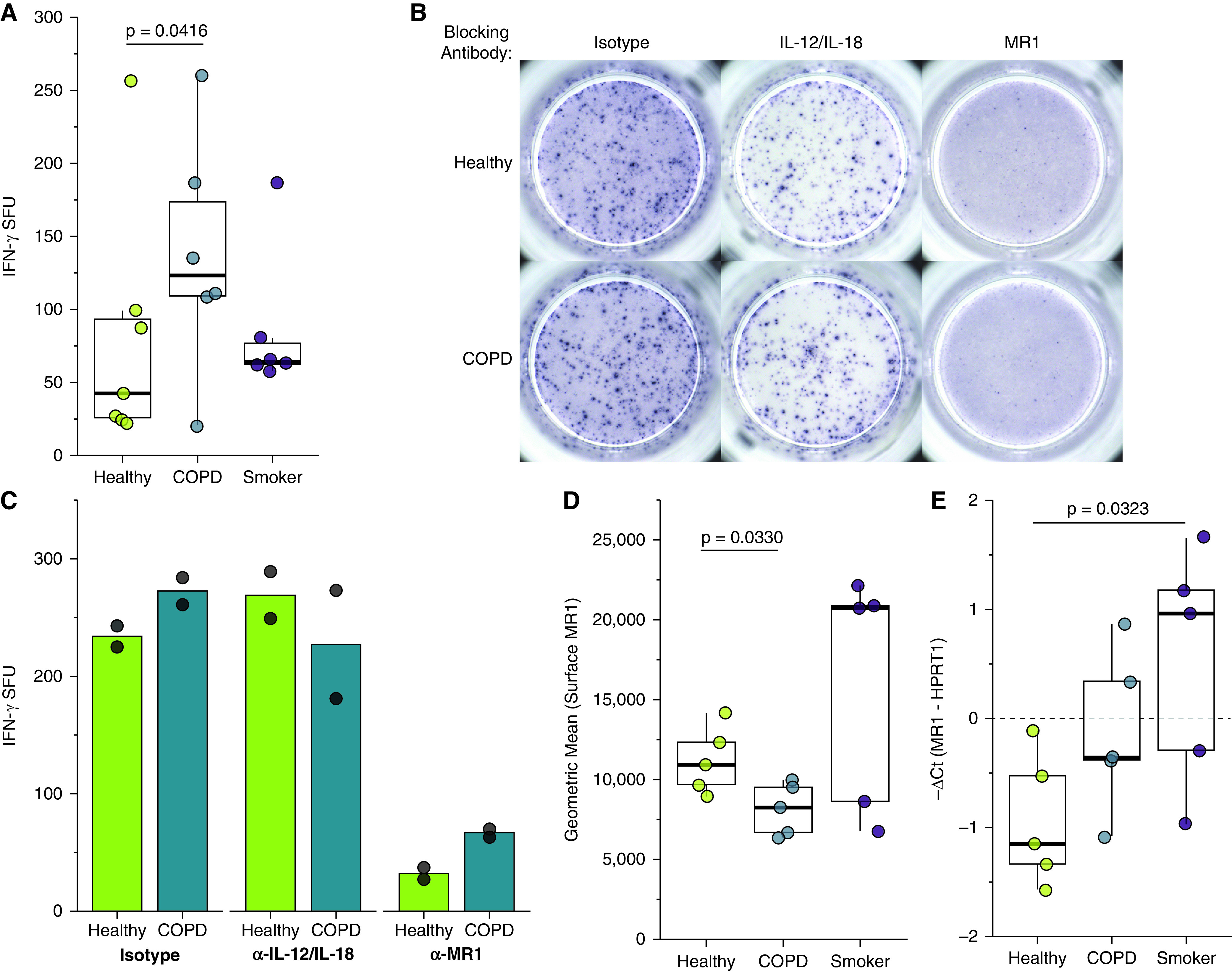
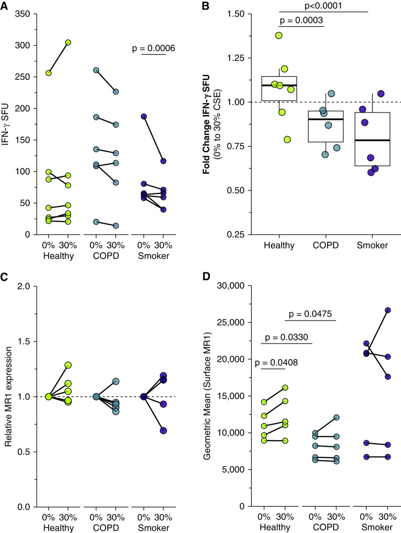
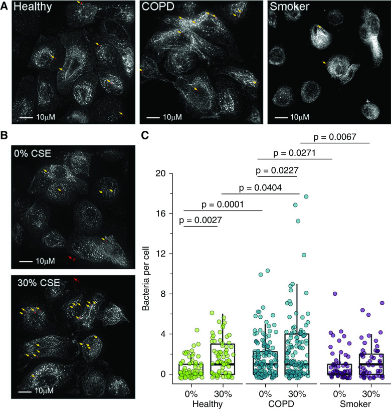
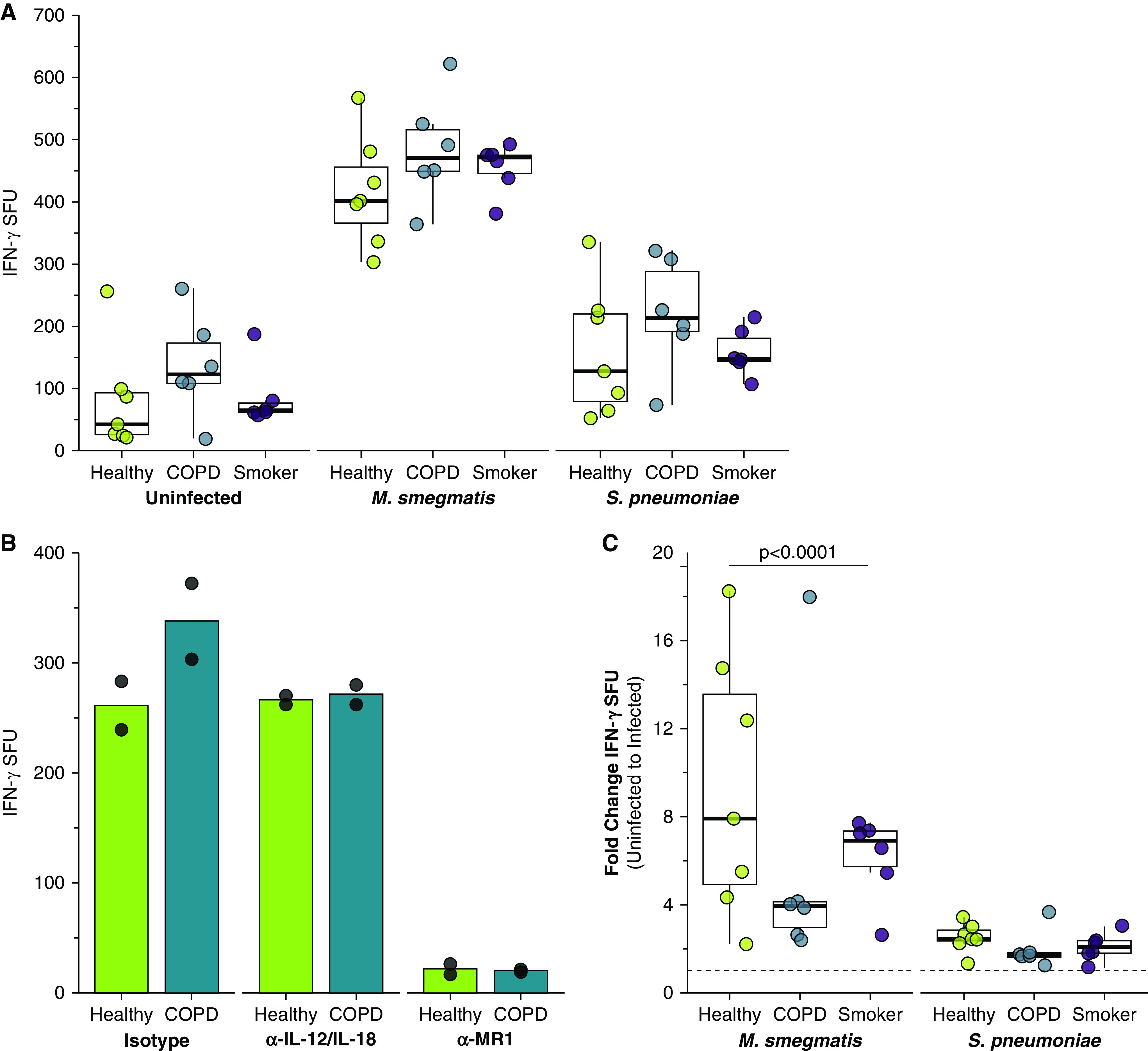
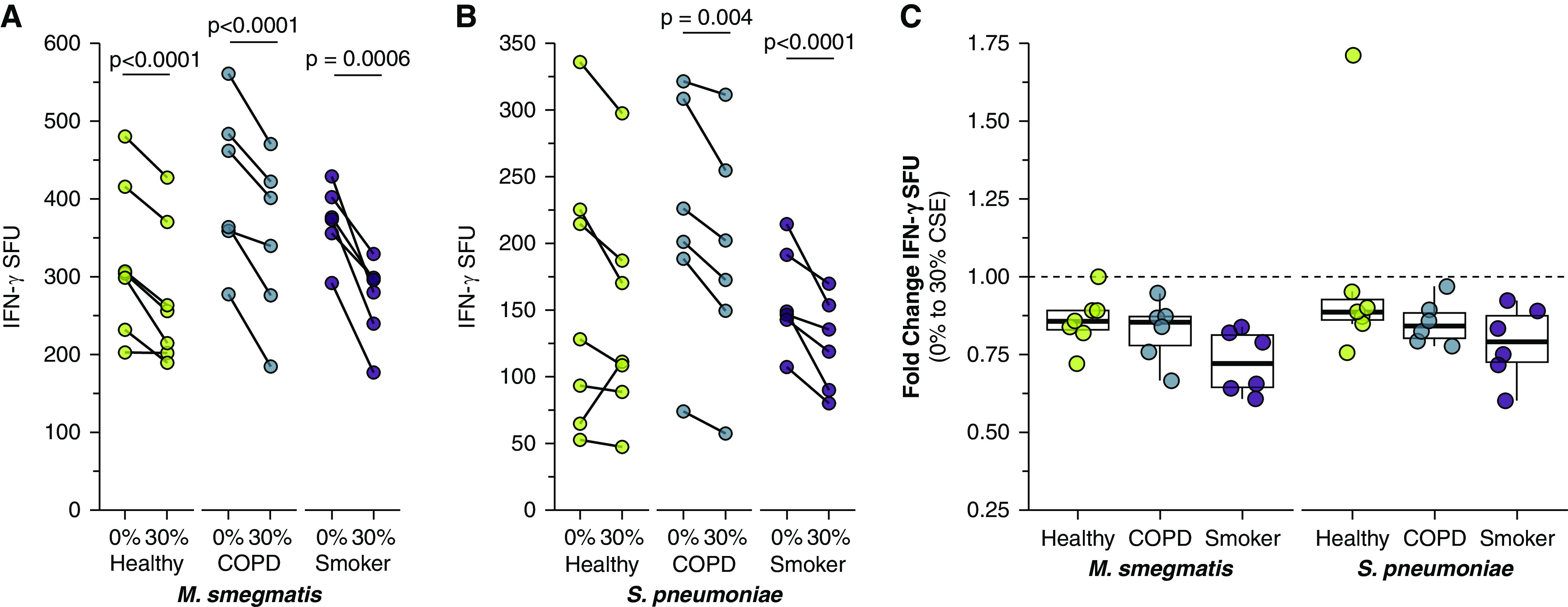
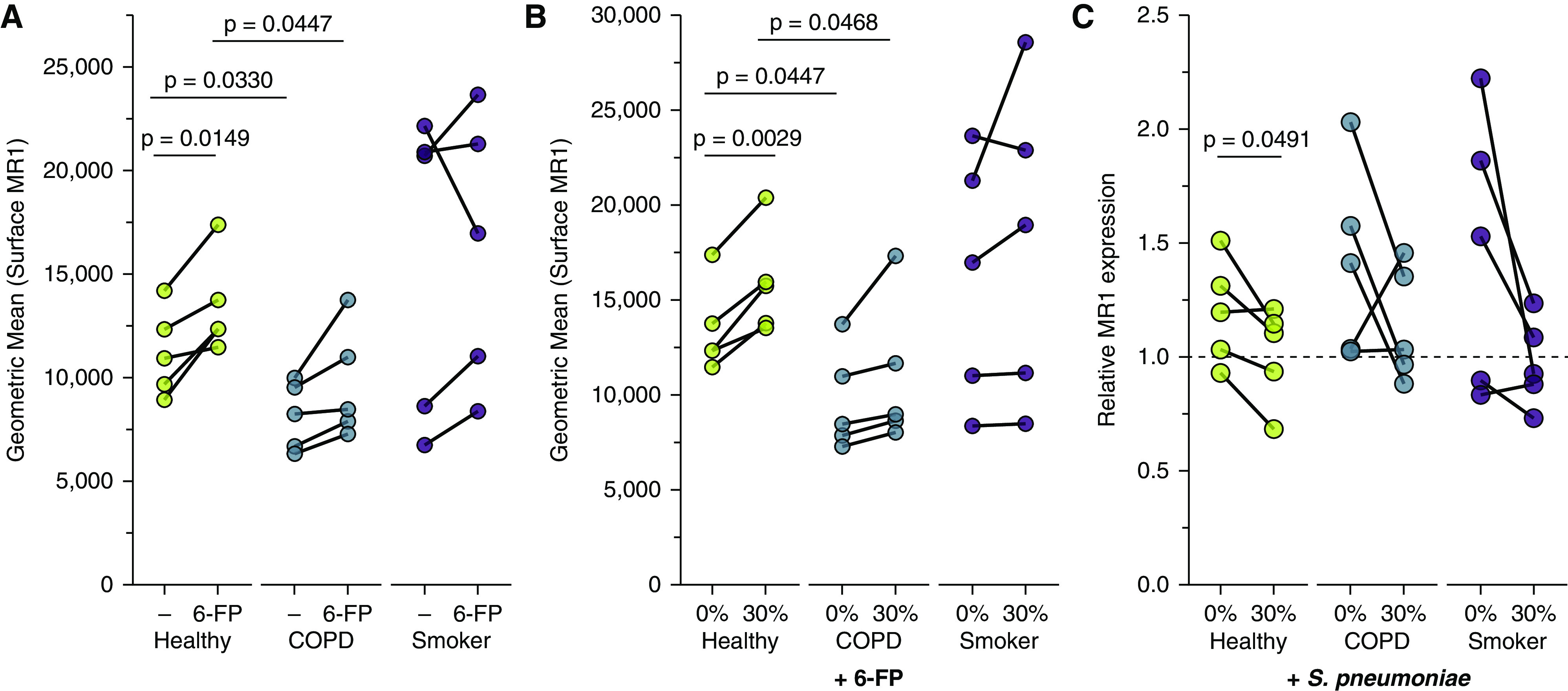
Chronic obstructive pulmonary disease (COPD) is associated with airway inflammation, increased infiltration by CD8+ T lymphocytes, and infection-driven exacerbations. Although cigarette smoke is the leading risk factor for COPD, the mechanisms driving the development of COPD in only a subset of smokers are incompletely understood. Lung-resident mucosal-associated invariant T (MAIT) cells play a role in microbial infections and inflammatory diseases. The role of MAIT cells in COPD pathology is unknown. Here, we examined MAIT cell activation in response to cigarette smoke-exposed primary human bronchial epithelial cells (BECs) from healthy, COPD, or smoker donors. We observed significantly higher baseline MAIT cell responses to COPD BECs than healthy BECs. However, infected COPD BECs stimulated a smaller fold increase in MAIT cell response despite increased microbial infection. For all donor groups, cigarette smoke-exposed BECs elicited reduced MAIT cell responses; conversely, cigarette smoke exposure increased ligand-mediated MR1 surface translocation in healthy and COPD BECs. Our data demonstrate that MAIT cell activation is dysregulated in the context of cigarette smoke and COPD. MAIT cells could contribute to cigarette smoke- and COPD-associated inflammation through inappropriate activation and reduced early recognition of bacterial infection, contributing to microbial persistence and COPD exacerbations.

Keywords: MAIT cells; MR1; Streptococcus pneumoniae; chronic obstructive pulmonary disease; cigarette smoke.


Figures

Figure 1.
Figure 1.
Primary bronchial epithelial cells (BECs) elicit microbe-independent, MR1-dependent responses by mucosal‐associated invariant T (MAIT) cells. (A) Primary BECs from healthy (n = 7), chronic obstructive pulmonary disease (COPD) (n = 6), or smoker (n = 6) donors were incubated with the D426 G11 MAIT cell clone in an enzyme-linked immunospot (ELISPOT) assay with IFN-γ production as the readout. Data points are the mean IFN-γ spot-forming units (SFUs) of two technical replicates per donor. Statistical analysis was performed as described in the data supplement and is summarized in Table 2. (B and C) BECs from the healthy and COPD donors that induced the greatest IFN-γ SFUs in A were incubated with blocking antibodies to IL-12/IL-18 or MR1 5 hours before the addition of the MAIT cells in an IFN-γ ELISPOT assay. Results are presented as (B) ELISPOT well images from one representative experiment and (C) the mean of two experimental replicates. Control IgG1 and IgG2a isotype antibodies are pooled from one representative experiment each. (D) Primary BECs from healthy, COPD, or smoker donors (n = 5) were stained for surface expression of MR1 by flow cytometry. (E) RNA was isolated from healthy, COPD, or smoker donor BECs (n = 5), and real-time quantitative PCR (RT-PCR) was performed to detect amplification of MR1 and the internal control, HPRT1. Data points are the mean of three technical replicates per donor. Two-tailed unpaired t tests were performed to determine statistical significance for D and E. MR1 = MHC class I related molecule.
Figure 2.
Figure 2.
Decreased MAIT cell responses to primary BECs after treatment with cigarette smoke extract (CSE). (A and B) Primary BECs from healthy (n = 7), COPD (n = 6), or smoker (n = 6) donors were infected with media containing 0% or 30% CSE for 3 hours before the addition of D426 G11 MAIT cells in an IFN-γ ELISPOT assay. Statistical analysis was performed as described in the data supplement and is summarized in Tables 2 and 3. (A) Data points are the mean IFN-γ SFUs of two experimental replicates paired by an individual donor. (B) Fold change IFN-γ SFUs between 0% and 30% CSE-treated primary BECs from healthy, COPD, or smoker donors, calculated pairwise by donor. (C and D) Primary BECs from healthy, COPD, or smoker donors (n = 5) were incubated with 0% or 30% CSE for 3 hours. (C) RNA was isolated from BECs, and real-time quantitative PCR (RT-PCR) was performed to detect amplification of MR1 and the internal control, HPRT1. MR1 expression was calculated by 2ΔΔCt method, relative to 0% CSE pairwise control and HPRT1 expression. (D) Cells were washed, incubated overnight, then stained for surface expression of MR1 by flow cytometry. Data points are mean fluorescence intensities paired by individual donor. Statistical significance was determined by two-tailed paired t tests for same-donor 0% and 30% CSE treatment in C and D and unpaired t tests for donor group comparison in D.
Figure 3.
Figure 3.
Increased infection of primary BECs from COPD donors or after CSE treatment. (A and B) BECs from representative healthy, COPD, or smoker donors were incubated with media containing 0% or 30% CSE for 3 hours where indicated and infected with fluorescently labeled Streptococcus pneumoniae for 3 hours. Fixed cells were stained with α-HLA-A,B,C antibody to label the cell surface and NucBlue nuclear stain. Approximately 20 fields per donor were selected without bias on the basis of nuclear stain, and whole cells within these fields were then analyzed by Imaris to enumerate the number of bacteria associated with individual cells. Shown are representative images of S. pneumoniae-infected primary BECs for each condition. White = surface stain and red = fluorescent S. pneumoniae pseudocolor. Arrows indicate adherent bacteria (yellow) enumerated for analysis or extracellular bacteria (red) excluded from the analysis. Scale bar, 10μM. (C) Quantification of S. pneumoniae per cell. Data points indicate individual cells, analyzed by one-way ANOVA.
Figure 4.
Figure 4.
Increased microbe-dependent, MR1-dependent MAIT cell responses to infected primary BECs from COPD donors. (A) Primary BECs from healthy (n = 7), COPD (n = 6), or smoker (n = 6) donors were infected with media control, Mycobacterium smegmatis (0.1 μl/well), or S. pneumoniae (multiplicity of infection [MOI] = 20) for 1 hour before the addition of D426 G11 MAIT cells in an IFN-γ ELISPOT assay. Data points are the mean IFN-γ SFUs of two technical replicates per donor. Statistical analysis was performed as described in the data supplement and is summarized in Tables 2 and 3. (B) BECs from the healthy and COPD donors that induced the greatest IFN-γ SFUs in Figure 1A were incubated with blocking antibodies to IL-12/IL-18 or MR1 for 4 hours and infected with S. pneumoniae (MOI = 20) for 1 hour before the addition of the MAIT cells in an IFN-γ ELISPOT assay. Results are presented as the mean of two experimental replicates. (C) Fold change IFN-γ SFUs between uninfected and microbial-infected BECs from healthy, COPD, or smoker donors, calculated pairwise by donor from raw data in A. Statistical analysis was performed as described in the data supplement and is summarized in Table 2.
Figure 5.
Figure 5.
Reduced MAIT cell responses to infected BECs after treatment with CSE. (A and B) Primary BECs from healthy (n = 7), COPD (n = 6), or smoker (n = 6) donors were incubated with media containing 0% or 30% CSE for 3 hours. BECs were infected with (A) M. smegmatis (0.05 μl/well) or (B) S. pneumoniae (20 MOI) for 1 hour before the addition of D426 G11 MAIT cells in an IFN-γ ELISPOT assay. (C) Fold change IFN-γ SFUs between 0% and 30% CSE-treated primary BECs infected with M. smegmatis or S. pneumoniae, calculated pairwise by donor from data in A and B. Statistical analysis was performed as described in the data supplement and is summarized in Tables 2 and 3.
Figure 6.
Figure 6.
Increased MR1 expression in primary BECs exposed to cigarette smoke. (A and B) Primary BECs from healthy, COPD, or smoker donors (n = 5) were incubated with media containing 0% or 30% CSE for 3 hours where indicated, then incubated overnight with the MAIT cell ligand 6-FP (6-formylpterin) before harvest and staining for surface expression of MR1 by flow cytometry. Data points are mean fluorescence intensities paired by individual donor. (C) Primary BECs from healthy, COPD, or smoker donors were incubated with media containing 0% or 30% CSE for 3 hours, washed, then infected with S. pneumoniae for 3 hours. RNA was isolated from BECs, and real-time quantitative PCR (RT-PCR) was performed to detect amplification of MR1 and the internal control, HPRT1. MR1 expression was calculated by 2ΔΔCt method, relative to no-treatment pairwise control and HPRT1 expression. Statistical significance was determined by two-tailed paired t tests for same-donor treatment analyses or unpaired t tests for donor group comparison.

Comment in

References

    1. World Health Organization. 2019.
    1. Sopori M. Effects of cigarette smoke on the immune system. Nat Rev Immunol . 2002;2:372–377. - PubMed
    1. Stämpfli MR, Anderson GP. How cigarette smoke skews immune responses to promote infection, lung disease and cancer. Nat Rev Immunol . 2009;9:377–384. - PubMed
    1. Nuorti JP, Butler JC, Farley MM, Harrison LH, McGeer A, Kolczak MS, et al. Active Bacterial Core Surveillance Team Cigarette smoking and invasive pneumococcal disease. N Engl J Med . 2000;342:681–689. - PubMed
    1. Aghapour M, Raee P, Moghaddam SJ, Hiemstra PS, Heijink IH. Airway epithelial barrier dysfunction in chronic obstructive pulmonary disease: role of cigarette smoke exposure. Am J Respir Cell Mol Biol . 2018;58:157–169. - PubMed

Publication types

MeSH terms