. 2023 Feb;68(2):176-185.
doi: 10.1165/rcmb.2021-0123OC.

A Novel MIP-1-Expressing Macrophage Subtype in BAL Fluid from Healthy Volunteers

Affiliations

A Novel MIP-1-Expressing Macrophage Subtype in BAL Fluid from Healthy Volunteers

Paul A Reyfman et al. Am J Respir Cell Mol Biol. 2023 Feb.

Abstract

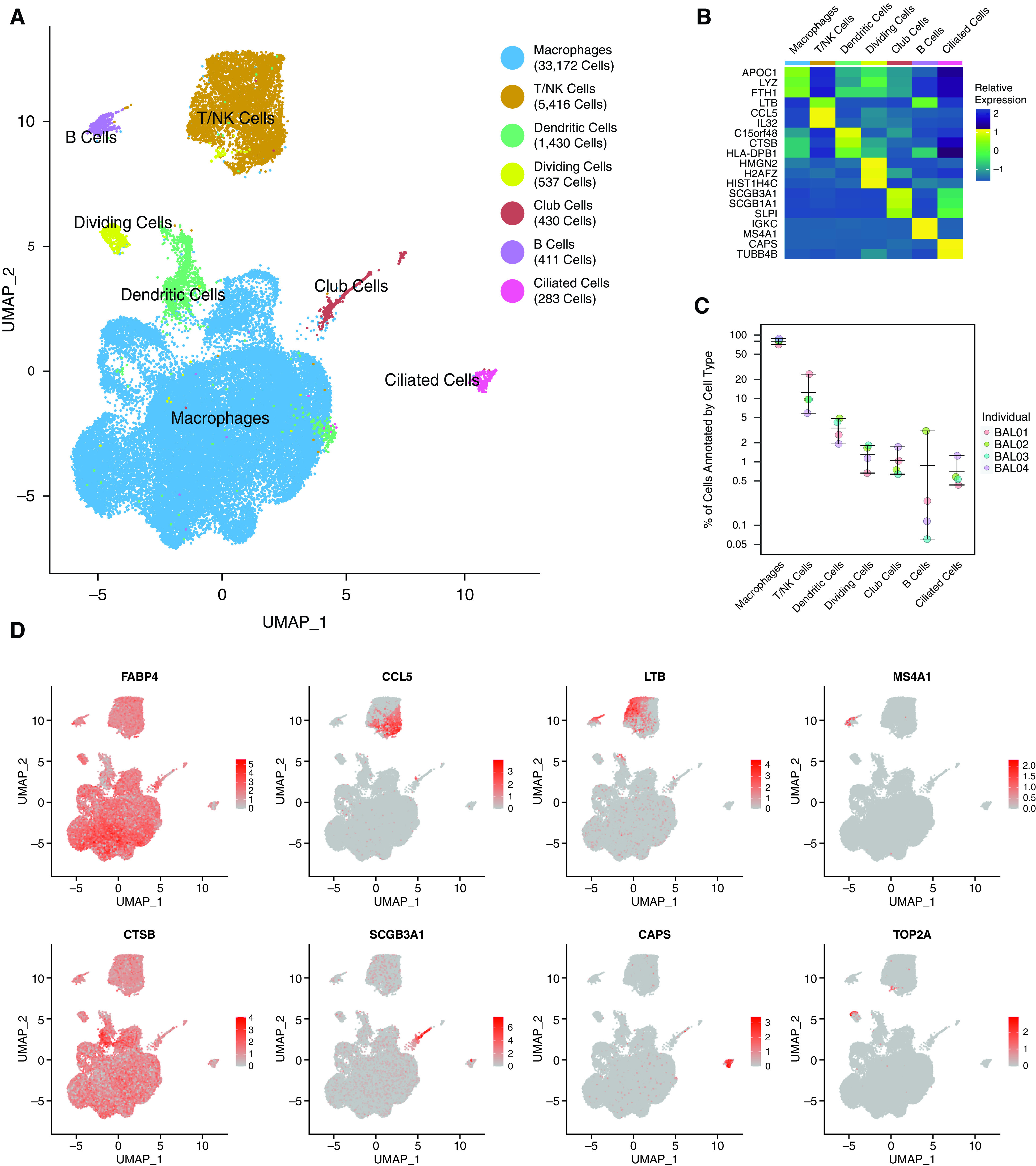
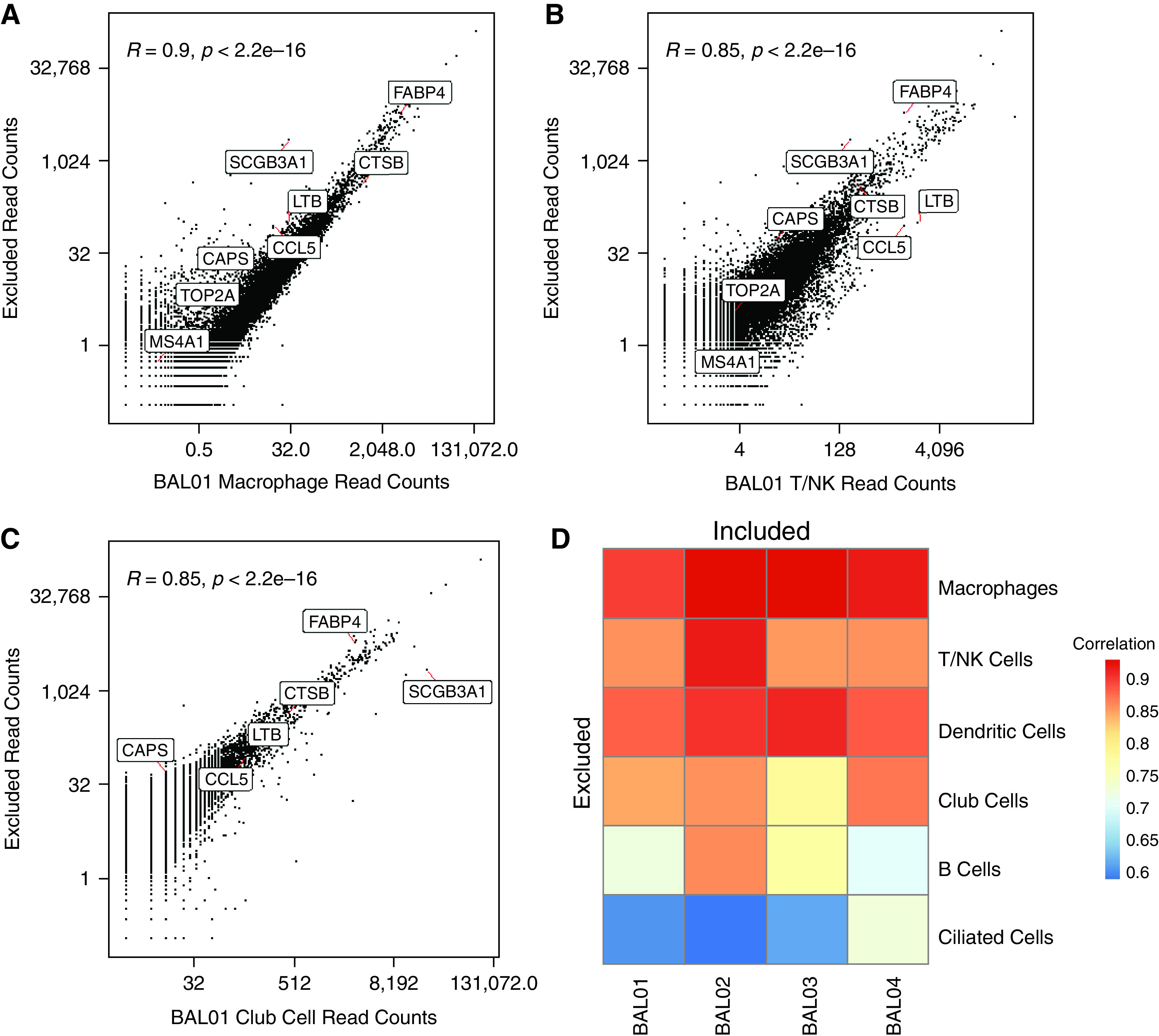
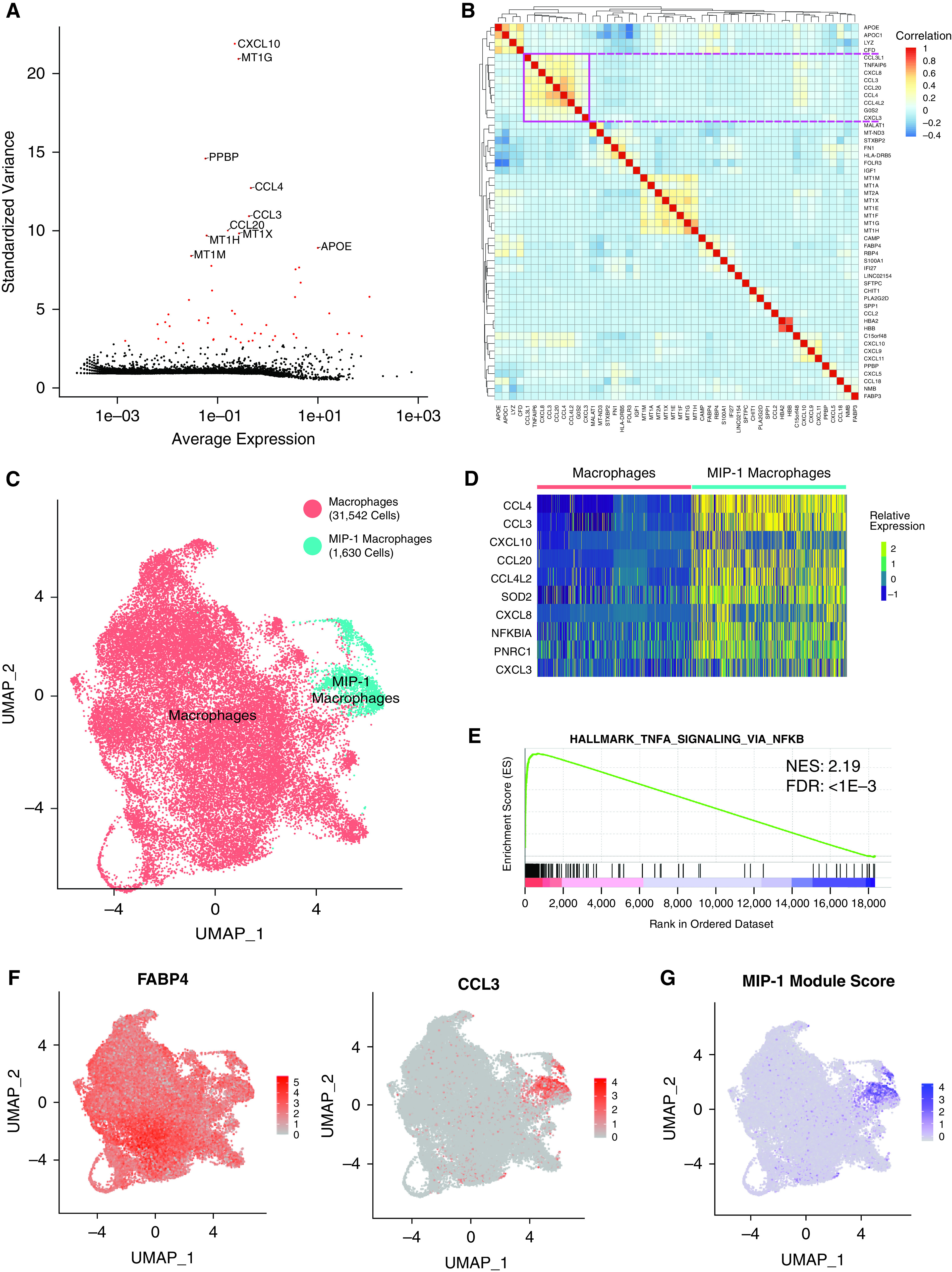
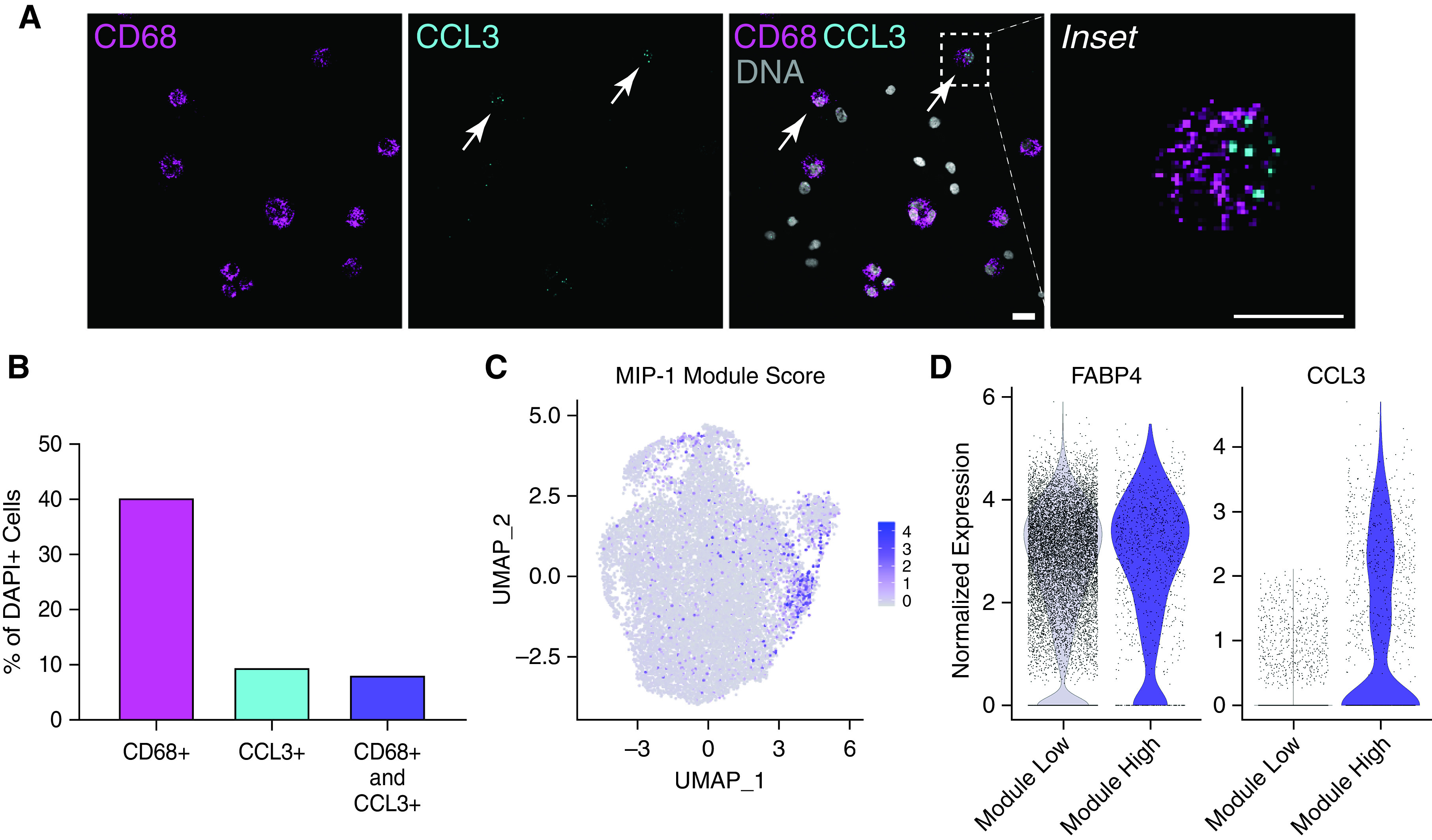
Tissue availability remains an important limitation of single-cell genomic technologies for investigating cellular heterogeneity in human health and disease. BAL represents a minimally invasive approach to assessing an individual's lung cellular environment for diagnosis and research. However, the lack of high-quality, healthy lung reference data is a major obstacle to using single-cell approaches to study a plethora of lung diseases. Here, we performed single-cell RNA sequencing on over 40,000 cells isolated from the BAL of four healthy volunteers. Of the six cell types or lineages we identified, macrophages were consistently the most numerous across individuals. Our analysis confirmed the expression of marker genes defining cell types despite background signals because of the ambient RNA found in many single-cell studies. We assessed the variability of gene expression across macrophages and defined a distinct subpopulation of cells expressing a set of genes associated with Macrophage Inflammatory Protein 1 (MIP-1). RNA in situ hybridization and reanalysis of published lung single-cell data validated the presence of this macrophage subpopulation. Thus, our study characterizes lung macrophage heterogeneity in healthy individuals and provides a valuable resource for future studies to understand the lung environment in health and disease.

Keywords: BAL; genomics; heterogeneity; lung immunology; macrophage.


Figures

Figure 1.
Figure 1.
Single-cell RNA sequencing identifies diverse cell populations in BAL fluid from healthy control volunteers. (A) UMAP representation of the transcriptional profile of BAL cells manually annotated as one of six cell types or dividing cells. The total number of cells in each cell type is indicated in the legend. (B) Relative expression of the top two to three marker genes (by fold change) across cell types. (C) Percentage (in log scale) of cells in each cell type by individual sample. Whiskers represent the range of values. (D) Visualization of normalized expression for canonical cell type genes across cells. All figures represent data from BAL01-04. NK = natural killer; UMAP = uniform manifold approximation and projection.
Figure 2.
Figure 2.
Comparison of empty droplets with cell droplets to assess ambient RNA. Scatterplot of the read counts for each gene in empty droplets that are excluded from the analysis versus cells that are annotated as (A) macrophages, (B) T/NK cells, and (C) club cells in BAL01. The Pearson correlations, R, and associated P values are indicated. A few example genes representing different cell types are annotated. (D) Pearson correlation between the read counts for genes from excluded droplets versus included and annotated as one of six cell types in individual BAL samples (BAL01–04).
Figure 3.
Figure 3.
Analysis of macrophage heterogeneity reveals a MIP-1 (macrophage inflammatory protein 1) subpopulation. (A) Scatterplot showing the average normalized expression of each gene by its standardized variance across macrophage cells. The top 10 most variable genes are annotated, and the top 50 most variable genes are shown in red. (B) Pairwise Pearson correlation between the expression profiles of the top 50 most variable genes across macrophages. The pink square denotes a large clade of coregulated genes. (C) UMAP representation of the transcriptional profile of macrophage cells with a subset annotated as belonging to the MIP-1 cluster. The total number of cells in each cluster is indicated in the legend. (D) Relative expression of top 10 differentially expressed marker genes (by fold change) for the MIP-1 macrophage cluster. These largely overlap but do not exactly match those in B. (E) Representative-enriched pathway resulting from GSEA analysis of marker gene list ranked by fold-change in MIP-1 compared with other macrophages. The NES and adjusted P value for the FDR are shown. (F) Visualization of normalized expression across cells for macrophage marker, FABP4, and MIP-1 gene, CCL3. (G) Visualization of MIP-1 module score across macrophage cells. All figures represent data from BAL01–04. FDR = false discovery rate; NES = Normalized Enrichment Score.
Figure 4.
Figure 4.
Validation of MIP-1 subpopulation in BAL fluid and another lung dataset. (A) Representative image of RNA in situ hybridization (RNA-ISH) on a BAL fluid sample from an additional individual (BAL05). Staining for macrophage marker (CD68), MIP-1 marker (CCL3), DNA (DAPI), and all channels merged (overlay). Scale bar, 40 μm. Arrows indicate CD68+ CCL3+ cells. (B) Quantification of RNA-ISH experiment above by percentage of cells from the total number of DAPI+ cells. Data represent 292 cells across four images. (C) Visualization of MIP-1 module score across cells annotated as macrophages in published data from donor transplant lung tissue (18). (D) Violin plot showing the distribution of normalized expression for FABP4 and CCL3 across macrophage cells annotated in C as either low (<0.3) or high (>0.3) module score. (C and D) Represent data from eight donor lungs.

References

    1. Franks TJ, Colby TV, Travis WD, Tuder RM, Reynolds HY, Brody AR, et al. Resident cellular components of the human lung: current knowledge and goals for research on cell phenotyping and function. Proc Am Thorac Soc . 2008;5:763–766. - PubMed
    1. Ardain A, Marakalala MJ, Leslie A. Tissue-resident innate immunity in the lung. Immunology . 2020;159:245–256. - PMC - PubMed
    1. Agostini C, Chilosi M, Zambello R, Trentin L, Semenzato G. Pulmonary immune cells in health and disease: lymphocytes. Eur Respir J . 1993;6:1378–1401. - PubMed
    1. Hussell T, Bell TJ. Alveolar macrophages: plasticity in a tissue-specific context. Nat Rev Immunol . 2014;14:81–93. - PubMed
    1. Guilliams M, De Kleer I, Henri S, Post S, Vanhoutte L, De Prijck S, et al. Alveolar macrophages develop from fetal monocytes that differentiate into long-lived cells in the first week of life via GM-CSF. J Exp Med . 2013;210:1977–1992. - PMC - PubMed

Publication types