Review
. 2022 Nov 2;110(21):3627-3644.
doi: 10.1016/j.neuron.2022.09.003. Epub 2022 Sep 28.

Bioelectronic medicine: Preclinical insights and clinical advances

Affiliations
Review

Bioelectronic medicine: Preclinical insights and clinical advances

Valentin A Pavlov et al. Neuron. .

Abstract

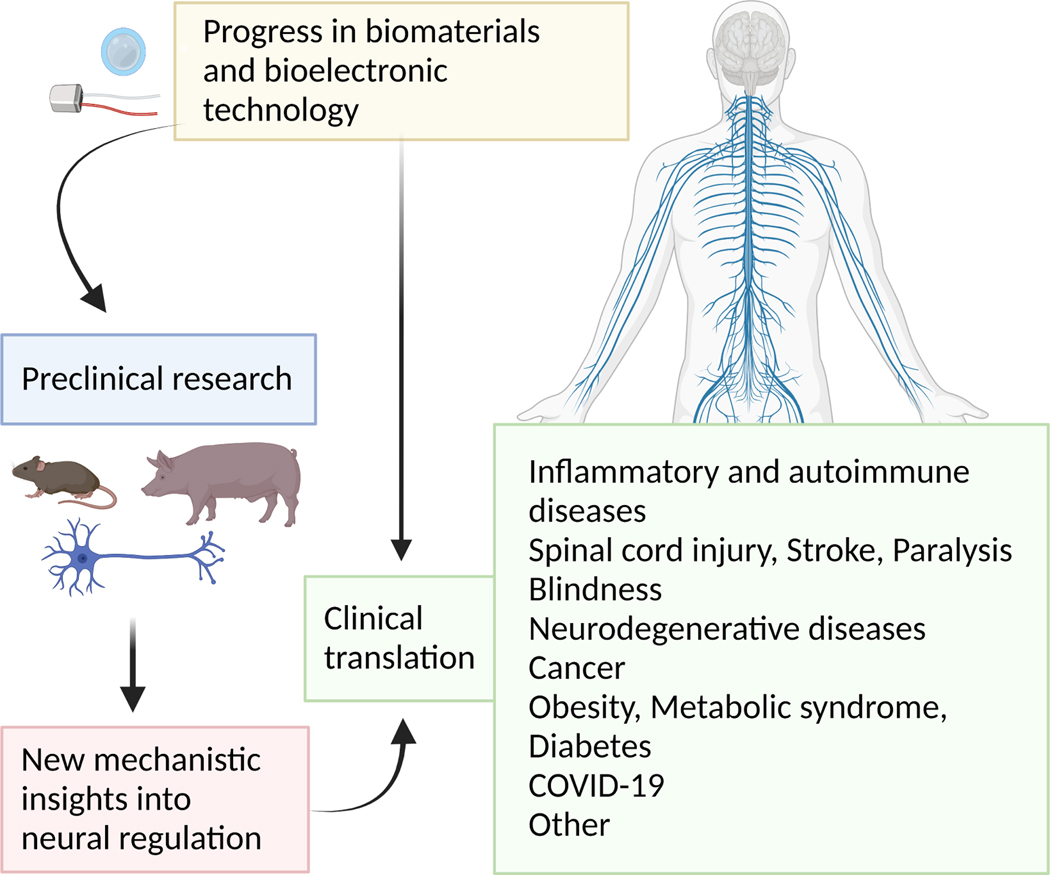
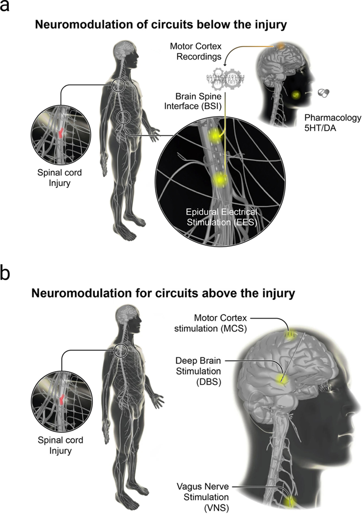
The nervous system maintains homeostasis and health. Homeostatic disruptions underlying the pathobiology of many diseases can be controlled by bioelectronic devices targeting CNS and peripheral neural circuits. New insights into the regulatory functions of the nervous system and technological developments in bioelectronics drive progress in the emerging field of bioelectronic medicine. Here, we provide an overview of key aspects of preclinical research, translation, and clinical advances in bioelectronic medicine.


Conflict of interest statement

Declaration of interests K.J.T. and V.A.P. have co-authored patents broadly related to the content of this review. They have assigned their rights to the Feinstein Institutes for Medical Research. K.J.T. also declares that he is a consultant to Setpoint Medical.

Figures

Figure 1.
Figure 1.. Bioelectronic medicine constituents.
In bioelectric medicine there is a symbiotic relationship between preclinical and clinical research that is driven and facilitated by progress in biomaterials and developing and implementing new interfaces and devices for neuromodulation and monitoring homeostatic alterations. Improved mechanistic understanding of nervous system regulation and identifying lead therapeutic modalities in preclinical studies are utilized in clinical studies evaluating new bioelectronic approaches in disease treatment.
Figure 2.
Figure 2.. The inflammatory reflex.
Pathogen invasion and tissue injury cause innate immune responses and inflammation with the release of cytokines through interaction of pathogen associated molecular patterns and danger associated molecular patterns with TLRs, NLRs, and other pattern-recognition receptors (not shown) on macrophages and other immune cells. These interactions trigger intracellular cascade mechanisms, including NF-κB nuclear translocation and inflammasome activation with the release of TNF, IL-1β, IL-6 and other cytokines. These cytokines activate signals along afferent (sensory) vagus nerve neurons (in green) residing in the nodose ganglion and terminating in the brainstem NTS. Pathogens can also directly trigger sensory neuronal activation (Chiu et al., 2013). The signals are further communicated to DMN with the generation of efferent (motor) vagus nerve (in blue) signaling with the release of ACh in innervated organs, including the liver, pancreas, gastrointestinal tract, heart, and lungs. Another source of efferent vagus neurons is NA. Efferent vagus neurons interact with splenic neurons in the celiac-superior mesenteric ganglion complex. Activation of the vagus nerve – splenic nerve axis results in the release of NE that binds β2AR on a subset of T cells, containing ChAT, the ACh synthesizing enzyme (T-ChAT cells). ACh released from post-ganglionic vagus neurons or from T-ChAT cells interacts with α7nAChR on macrophages and other immune cells. Activation of this receptor results in inhibition of NF-κB nuclear translocation leading to suppression of TNF and other pro-inflammatory cytokine synthesis. Other intracellular mechanisms downstream of α7nAChR include activation of JAK2/STAT3 (de Jonge et al., 2005), inflammasome inhibition (Lu et al., 2014a), and adenylyl cyclase 6 signaling (Tarnawski et al., 2018) (not shown). The inflammatory reflex can be activated by VNS or using α7nAChR agonists. Efferent vagus nerve signaling in the inflammatory reflex can also be activated through brain mAChR -mediated mechanisms by galantamine or mAChR ligands. ACh, acetylcholine; α7nAChR, alpha7 nicotinic acetylcholine receptor; βAR, beta adrenergic receptor; ChAT, choline acetyltransferase; DMN, dorsal motor nucleus of the vagus; IL-1β, interleukin 1 betta, IL-6, interleukin 6; JAK2/STAT3, Janus kinase 2/signal transducer and activator of transcription 3; mAChR, muscarinic acetylcholine receptor; NE, norepinephrine; NA, nucleus ambiguus; NF-κB, nuclear factor kappa-B; NTS, nucleus tractus solitarius; NLRs, nucleotide-binding oligomerization domain-like receptors; TLRs, toll like receptors; TNF, tumor necrosis factor; VNS, vagus nerve stimulation. See text for details. Figure created using Biorender based on a relevant author’s figure previously published in Pharmacology &Therapeutics (Pavlov, 2021).
Figure 3.
Figure 3.. Advancing bioelectronic neuromodulation technology in mice and humans
(a) Core functionality of a generalizable fully implantable system. The wired interface is replaced by an implanted system capable of stimulation/sensing and energy storage and wireless communication. (b) Miniaturized vagus nerve stimulation system. The MicroRegulator implanted on the left cervical vagus nerve is designed to be fully self-contained, including a pulse generator, an application-specific integrated circuit, integrated electrodes, a rechargeable battery, and a near-field antenna used both for telemetry and battery recharging. The positioning and orientation device (POD), consisted of a flexible silicone, encases the MicroRegulator, ensuring close contact with the nerve. The POD additionally served to electrically insulate the device from surrounding tissue. The charging band (wireless charger) contains a radio frequency coil and is used to recharge the battery inside the MicroRegulator and to also communicate via Bluetooth with an iPad-based control application enabling the health-care provider to prescribe doses on the implanted Micro-Regulator. Figure created with Biorender (a) using a previously published figure in Bioelectronic Medicine (Datta-Chaudhuri, 2021) and (b) adapting a figure previously published in Lancet Rheumatology (Genovese et al., 2020).
Figure 4.
Figure 4.. Schematic illustrations and images of a fully implantable, soft optoelectronic system for wireless, closed-loop optogenetic modulation of bladder function.
(a) The platform consists of an optoelectronic stimulation and sensing (OESS) module and a low-modulus, stretchable strain gauge (SG) with integrated μ-ILEDs that wraps around the bladder to monitor changes in its volume and to provide optogenetic stimulation to the neurons that innervate the bladder. The WCP module records the response of the strain gauge, controls operation of the μ-ILEDs and provides power management. Wireless data communication to and from the WCP module relies on Bluetooth protocols and a tablet computer. Power is delivered wirelessly by resonant magnetic coupling through an antenna transmitter. (b) Photograph of optoelectronic stimulation and sensing module including the strain gauge, μ-ILEDs and wireless base station for data communication. (c) Schematic illustrations showing the placement of the strain gauge around the bladder, with an implanted, wired connection to the WCP module subcutaneously implanted anterior to the bladder. Images created by and used with permission of Janet SinnHanlon, The DesignGroup@VetMed, University of Illinois at UrbanaChampaign. (d) Rat implanted with the complete system (a green μ-ILED indicator on the WCP module verifies function). (e) Computed tomography image of a device implanted for 1 month. Previously published in Nature (Mickle et al., 2019) and used with permission.
Figure 5.
Figure 5.. Neuromodulation approaches in spinal cord injury.
(a) Neuromodulation strategies to engage circuits below the lesion after spinal cord injury (SCI). The reactivation or modulation of spinal circuits for locomotion has been demonstrated with the use of epidural electrical stimulation (EES) combined with the oral or intrathecal administration of serotonergic and dopaminergic agonists. EES can also be used to optimize autonomic function post-SCI (i.e., blood pressure management). Brain-spine interfaces (BSIs) also provide an alternative strategy for locomotion through bypassing the injury. (b) Neuromodulation strategies to engage circuits above the lesion after SCI. Neuromodulation therapies have been delivered to the mesencephalic locomotor region using deep brain stimulation (DBS) to facilitate locomotion. Motor cortex stimulation (MCS) has been applied for extensive periods of time daily to promote the growth and sprouting of cortico-spinal tract fibers. In preclinical SCI studies VNS has been applied to augment motor recovery learning and plasticity during motor rehabilitation (Ganzer et al., 2018). Previously published in Bioelectronic Medicine (Cho et al., 2019).
Figure 6.
Figure 6.. Advanced bionic limb technologies.
The most advanced technologies for the mechanical and neural interfacing of bionic limbs with the body are targeted muscle reinnervation (1), osseointegration (2), implanted sensors (3), and advanced neural-decoding algorithms (4) that can be combined with modern multi-articulating prosthetic limbs (5) Credit: Aron Cserveny. Previously published in Nature Biomedical Engineering (Farina et al., 2021) and used with permission.

Similar articles

Cited by

References

    1. 2020. Building a bioelectronic medicine movement 2019: insights from leaders in industry, academia, and research. Bioelectron Med, 6, 1. - PMC - PubMed
    1. ADAIR D, TRUONG D, ESMAEILPOUR Z, GEBODH N, BORGES H, HO L, DOUGLAS BREMNER J, BADRAN BW, NAPADOW V, CLARK VP & BIKSON M. 2020. Electrical stimulation of cranial nerves in cognition and disease. Brain Stimul, 13, 717–750. - PMC - PubMed
    1. ADDORISIO ME, IMPERATO GH, DE VOS AF, FORTI S, GOLDSTEIN RS, PAVLOV VA, VAN DER POLL T, YANG H, DIAMOND B, TRACEY KJ & CHAVAN SS 2019. Investigational treatment of rheumatoid arthritis with a vibrotactile device applied to the external ear. Bioelectronic Medicine, 5, 4. - PMC - PubMed
    1. AL-ALY Z, XIE Y. & BOWE B. 2021. High-dimensional characterization of post-acute sequelae of COVID-19. Nature, 594, 259–264. - PubMed
    1. AMINIAN A, BENA J, PANTALONE KM & BURGUERA B. Association of obesity with postacute sequelae of COVID-19. Diabetes, Obesity and Metabolism, n/a. - PMC - PubMed

LinkOut - more resources