. 2022 Jan-Dec;14(1):2126272.
doi: 10.1080/19490976.2022.2126272.

Periodontitis-related salivary microbiota aggravates Alzheimer's disease via gut-brain axis crosstalk

Affiliations

Periodontitis-related salivary microbiota aggravates Alzheimer's disease via gut-brain axis crosstalk

Jiangyue Lu et al. Gut Microbes. 2022 Jan-Dec.

Abstract

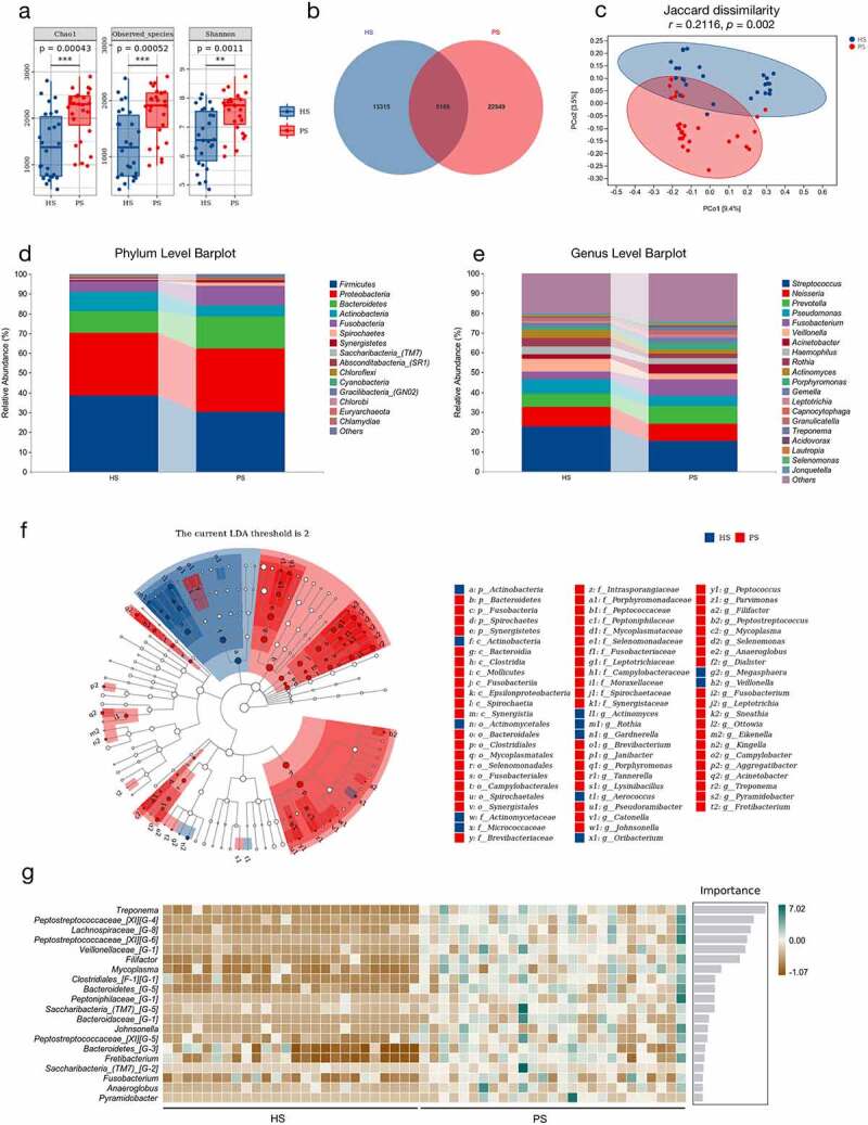
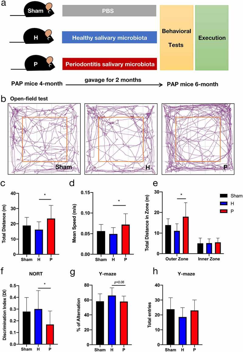
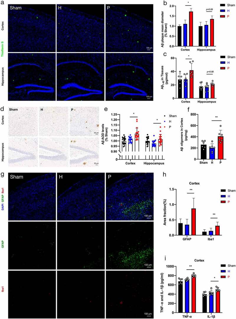
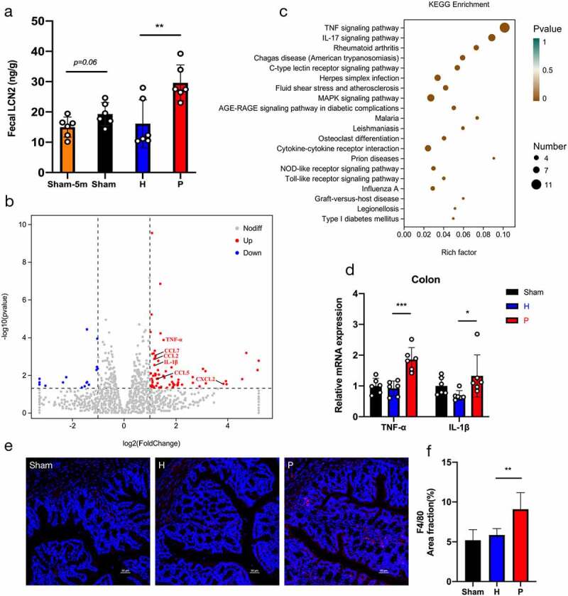
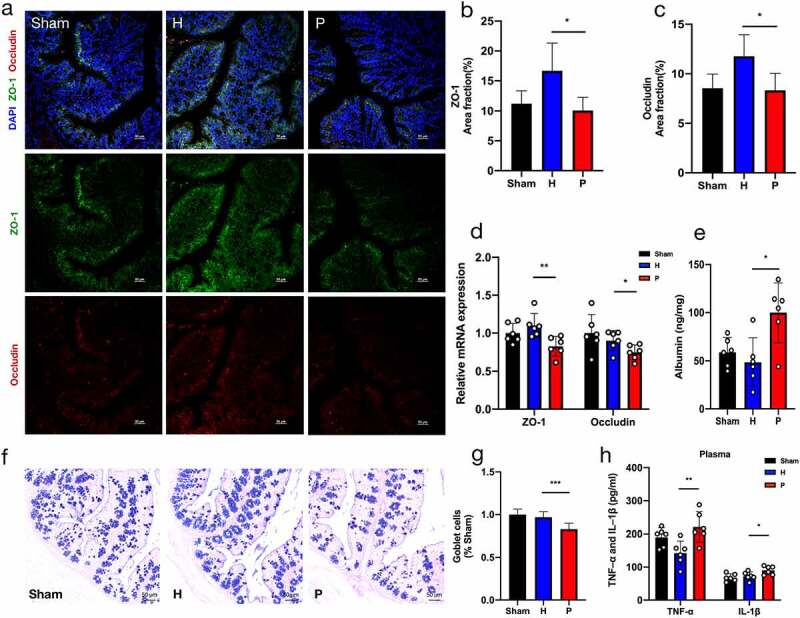
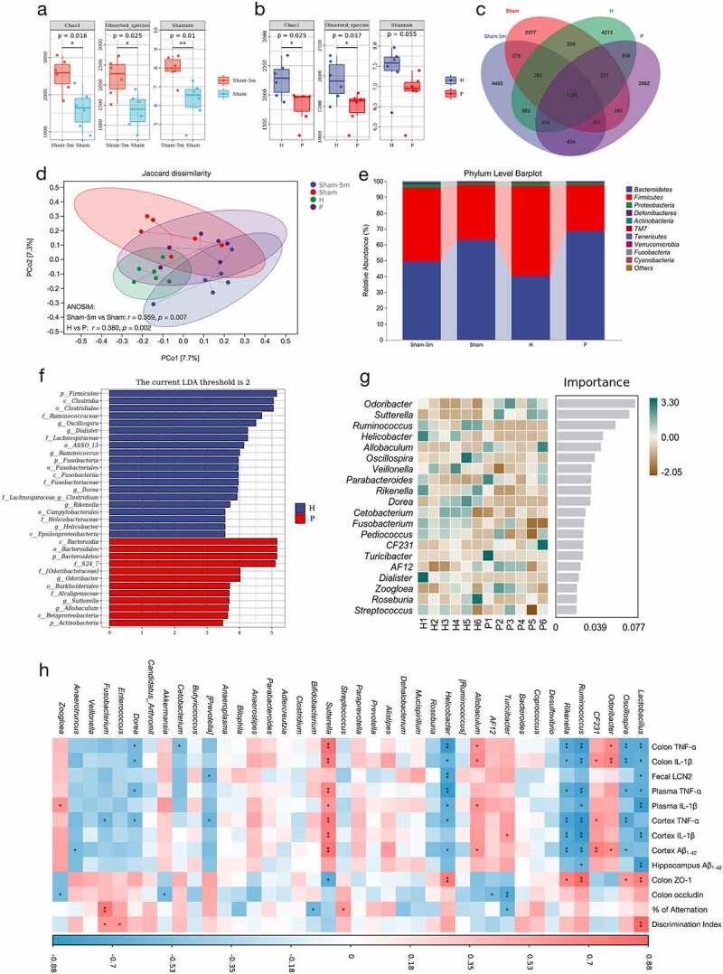
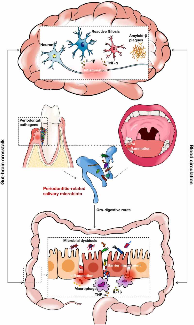
The oral cavity is the initial chamber of digestive tract; the saliva swallowed daily contains an estimated 1.5 × 10¹² oral bacteria. Increasing evidence indicates that periodontal pathogens and subsequent inflammatory responses to them contribute to the pathogenesis of Alzheimer's disease (AD). The intestine and central nervous system jointly engage in crosstalk; microbiota-mediated immunity significantly impacts AD via the gut-brain axis. However, the exact mechanism linking periodontitis to AD remains unclear. In this study, we explored the influence of periodontitis-related salivary microbiota on AD based on the gut-brain crosstalk in APPswe/PS1ΔE9 (PAP) transgenic mice. Saliva samples were collected from patients with periodontitis and healthy individuals. The salivary microbiota was gavaged into PAP mice for two months. Continuous gavage of periodontitis-related salivary microbiota in PAP mice impaired cognitive function and increased β-amyloid accumulation and neuroinflammation. Moreover, these AD-related pathologies were consistent with gut microbial dysbiosis, intestinal pro-inflammatory responses, intestinal barrier impairment, and subsequent exacerbation of systemic inflammation, suggesting that the periodontitis-related salivary microbiota may aggravate AD pathogenesis through crosstalk of the gut-brain axis. In this study, we demonstrated that periodontitis might participate in the pathogenesis of AD by swallowing salivary microbiota, verifying the role of periodontitis in AD progression and providing a novel perspective on the etiology and intervention strategies of AD.

Keywords: Alzheimer’s disease; Periodontitis; gut microbiota; gut-brain axis; inflammation; saliva.


Conflict of interest statement

No potential conflict of interest was reported by the author(s).

Figures

Figure 1.
Figure 1.
The differences of salivary microbiota in patients with periodontitis and healthy controls according to the 16S rRNA data. (a) Alpha-diversity indices of bacterial species in periodontitis saliva (PS) and healthy saliva (HS), including Chao1, Observed species, and Shannon index. Each box plot represented the median, interquartile range, minimum, and maximum values (n = 26–27). (b) Venn diagram indicating amplicon sequence variants (ASVs) between HS and PS. (c) Principal coordinate analysis (PCoA) of salivary microbiota on the basis of Jaccard dissimilarity. ANOSIM, r = 0.2116, p = .002. (d-e) Relative abundance of microbiota at the phylum (d) and genus (e) levels between two groups. (f) LDA Effect Size (LEfSe) Cladograms showing differences in the bacterial taxa between HS and PS (LDA > 2). (g) Random forest analysis indicating the importance score for genus between HS and PS. *p < .05, **p < .01, ***p < .001.
Figure 2.
Figure 2.
Anxiety degree and cognitive impairment in groups of PAP mice after gavaging for two months. (a) Design of the animal experiments. (b-e) The open-field test included representative tracking images of movement in 5 min (b), total distance traveled (c), mean speed (d), and total distance traveled in outer and inner zones (e). (f) Discrimination index (DI) in the new object recognition test (NORT). (g-h) Percentage of spontaneous alternation (g) and the number of total entries (h) in Y-maze task. Data were presented as the mean ± SD (n = 10), *p < .05, **p < .01. H, healthy; P, periodontitis.
Figure 3.
Figure 3.
β-amyloid (Aβ) accumulation and neuroinflammation in groups of PAP mice. (a) Representative images displayed Aβ plaque stained by Thioflavin S. Scale bar = 100 µm. (b) Quantification of Aβ plaque diameter in the cortex and hippocampus (n = 3). (c) Aβ1-42 in cortex and hippocampus by ELISA detection (n = 6). (d-e) Representative images of Aβ immunohistochemistry (d) and quantification of Aβ immunopositivity in the cortex and hippocampus (e) (n = 3 areas in the region of interest from 6 mice per group). Scale bar = 50 µm. (f) Levels of Aβ oligomers in cortex detected by ELISA (n = 6) (g) Representative images of astrogliosis (GFAP) and microgliosis (Iba1). Scale bar = 100 µm. (h) Area fractions of GFAP and Iba1 in the cortex of mice (n = 3). (i) The expressions of TNF-α and IL-1β in cortex using ELISA detection (n = 6). These data were presented as the mean ± SD, *p < .05, **p < .01. H, healthy; P, periodontitis; AOD, average optical density.
Figure 4.
Figure 4.
Periodontitis-related salivary microbiota intensifies intestinal inflammation in PAP mice. (a) Concentrations of fecal lipocalin-2 (LCN2) detected by ELISA in groups of Sham-5 m (at 5 months of age), Sham, H and P (n = 6). (b) Visualization of differentially expressed genes (DEGs) in the colon tissue of mice in P group compared with H group (n = 3). (c) Bubble diagram of the KEGG enrichment analysis based on DEGs between P and H groups. The horizontal axis represented the gene ratio and the size of dots represented the number of genes in the KEGG term. (d) Relative mRNA expressions of TNF-α and IL-1β in the colon tissue (n = 6). (e-f) Representative images of macrophages (F4/80) (e) and quantification of area fractions in the colon tissue (f) (n = 3). Scale bar = 50 µm. Data were presented as the mean ± SD, *p < .05, **p < .01, ***p < .001. H, healthy; P, periodontitis.
Figure 5.
Figure 5.
Periodontitis-related salivary microbiota impairs intestinal barrier in PAP mice. (a-c) Representative images of ZO-1 and occludin immunostaining (a) and quantification of area fractions in the colon of PAP mice (b-c) (n = 3). Scale bar = 50 µm. (d) Relative mRNA expressions of ZO-1 and occludin (n = 6). (e) Concentrations of fecal albumin detected by ELISA (n = 6). (f-g) Alcian blue and periodic acid-Schiff (AB-PAS) staining showing the distribution of goblet cells in colon (f) and the goblet cells per crypts that normalized to the Sham group (g) (n = 3). Scale bar = 50 µm. (h) The ELISA test indicating levels of TNF-α and IL-1β in plasma of mice (n = 6). Data were presented as the mean ± SD in the different experimental groups, *p < .05, **p < .01, ***p < .001. H, healthy; P, periodontitis.
Figure 6.
Figure 6.
Periodontitis-related salivary microbiota alters the composition of gut microbiota in PAP mice. (a-b) Fecal microbial diversity estimated by Chao1, Observed species and Shannon index of PAP mice between the ages of 5 and 6 months in Sham group (a) and between H and P groups (b) (n = 6). (c) A Venn diagram showing the overlaps among groups. (d) PCoA of fecal microbiota on the basis of Jaccard dissimilarity and ANOSIM. Sham-5 m vs Sham, r = 0.359, p = .007; H vs P, r = 0.380, p = .002. (e) Relative abundance of microbiota at the phylum level among groups. (f-g) LEfSe analysis (g) and random forest analysis (g) indicating the fecal taxonomic differences between H and P groups. (h) Correlation analysis of top 40 microbes with AD-related parameters based on the Spearman correlation coefficient test. A color gradient from blue (negative correlation) to red (positive correlation) indicating the correlation effect. *p < .05, **p < .01. H, healthy; P, periodontitis.
Figure 7.
Figure 7.
The schematic diagram for this study. Periodontitis led to significant changes in the composition of salivary microbiota. The oro-digestive translocation of periodontitis-related salivary microbiota induced gut microbial dysbiosis, intestinal proinflammatory responses and intestinal barrier impairment. These gut-associated pathogenesis may interact mutually with the brain lesions of AD through the blood circulation system.

Similar articles

Cited by

References

    1. Page RC, Offenbacher S, Schroeder HE, Seymour GJ, Kornman KS.. Advances in the pathogenesis of periodontitis: summary of developments, clinical implications and future directions. Periodontol. 1997;14:216–19. 10.1111/j.1600-0757.1997.tb00199.x. - DOI - PubMed
    1. Seneviratne CJ, Zhang CF, Samaranayake LP. Dental plaque biofilm in oral health and disease. Chin J Dent Res. 2011;14:87–94. - PubMed
    1. Borgnakke WS, Ylöstalo PV, Taylor GW, Genco RJ. Effect of periodontal disease on diabetes: systematic review of epidemiologic observational evidence. J Clin Periodontol. 2013;40(Suppl 14):S135–152. 10.1111/jcpe.12080. - DOI - PubMed
    1. Sanz M, Marco Del Castillo A, Jepsen S, Gonzalez-Juanatey JR, D’Aiuto F, Bouchard P, Chapple I, Dietrich T, Gotsman I, Graziani F, et al. Periodontitis and cardiovascular diseases: consensus report. J Clin Periodontol. 2020;47(3):268–288. 10.1111/jcpe.13189. - DOI - PMC - PubMed
    1. Keller A, Rohde JF, Raymond K, Heitmann BL. Association between periodontal disease and overweight and obesity: a systematic review. J Periodontol. 2015;86(6):766–776. 10.1902/jop.2015.140589. - DOI - PubMed

Publication types