. 2023 Mar;19(3):275-283.
doi: 10.1038/s41589-022-01149-6. Epub 2022 Sep 29.

Targeted protein S-nitrosylation of ACE2 inhibits SARS-CoV-2 infection

Affiliations

Targeted protein S-nitrosylation of ACE2 inhibits SARS-CoV-2 infection

Chang-Ki Oh et al. Nat Chem Biol. 2023 Mar.

Abstract

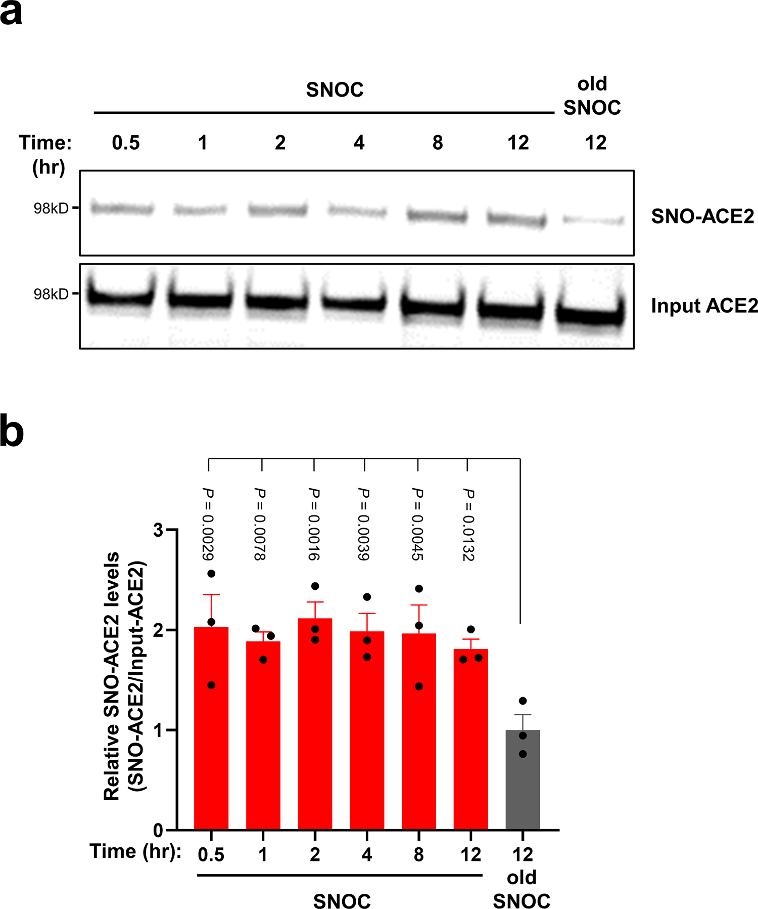
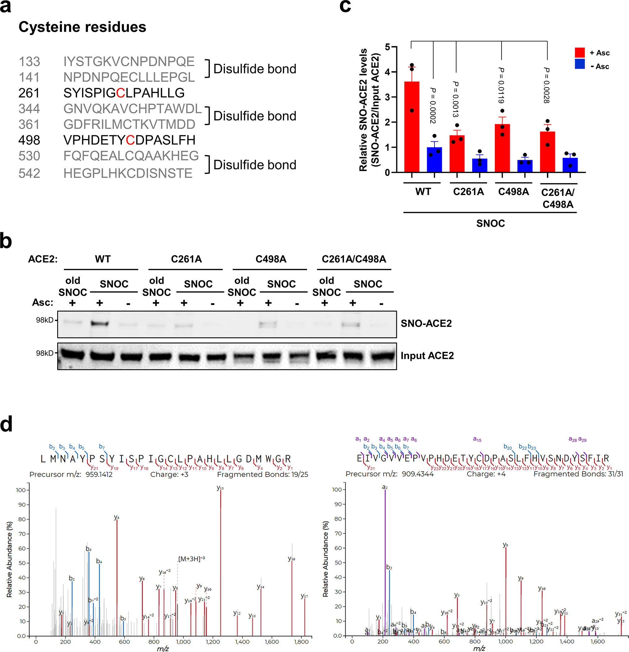
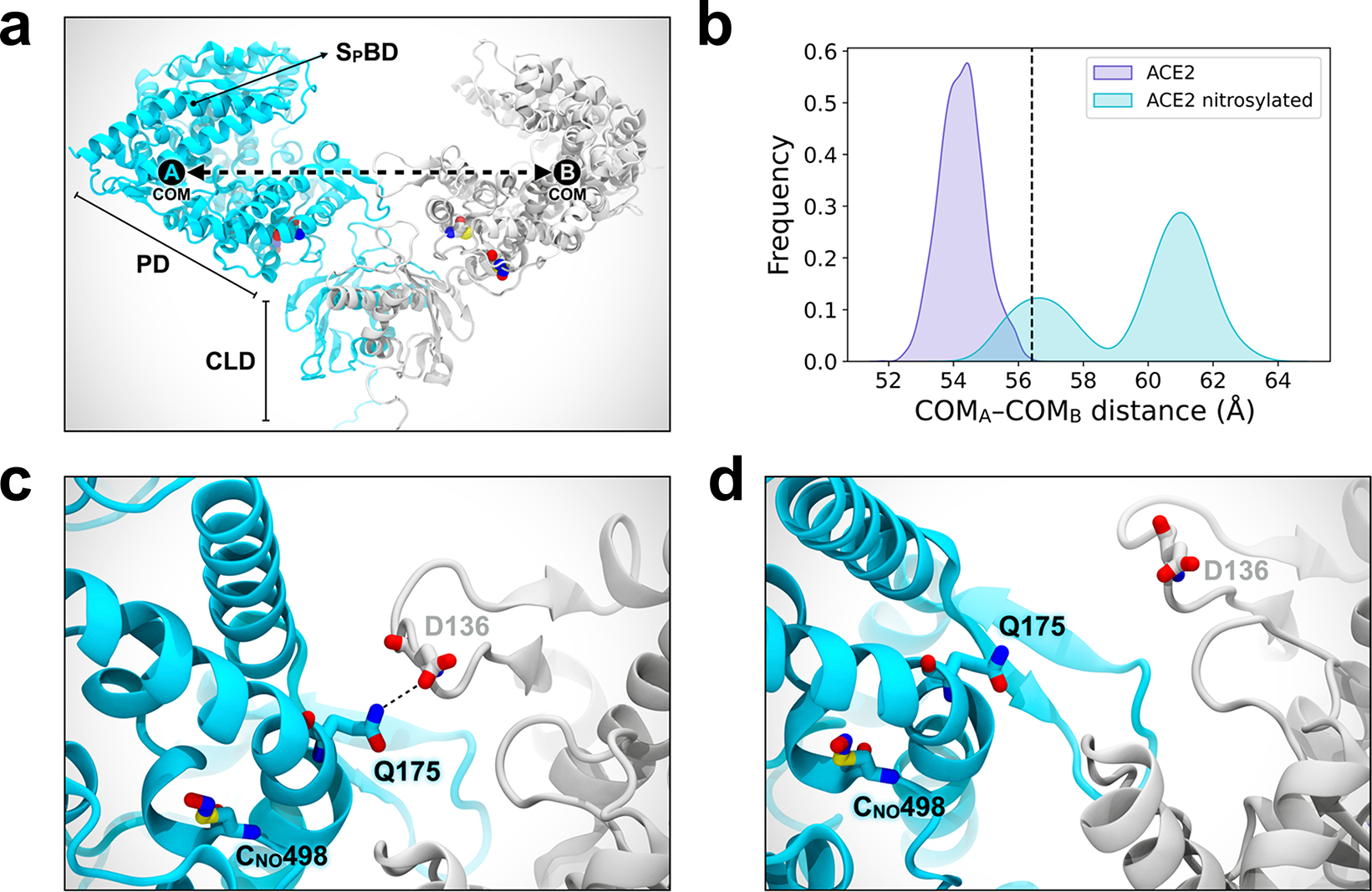
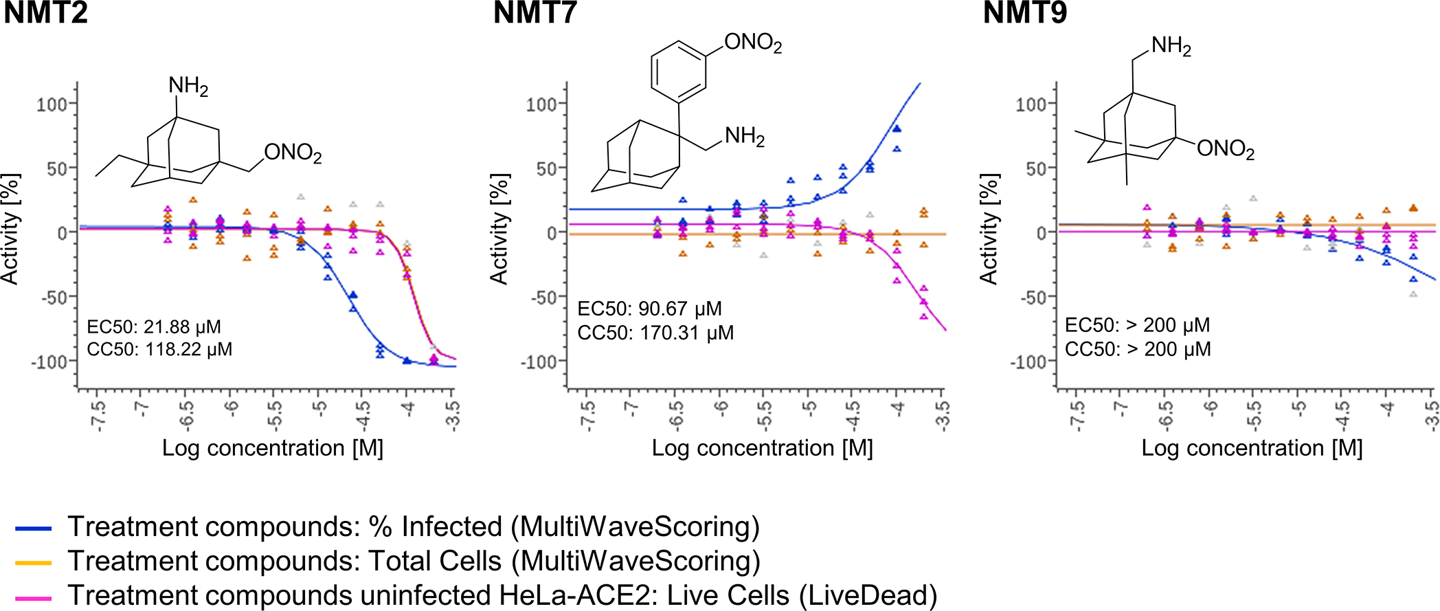
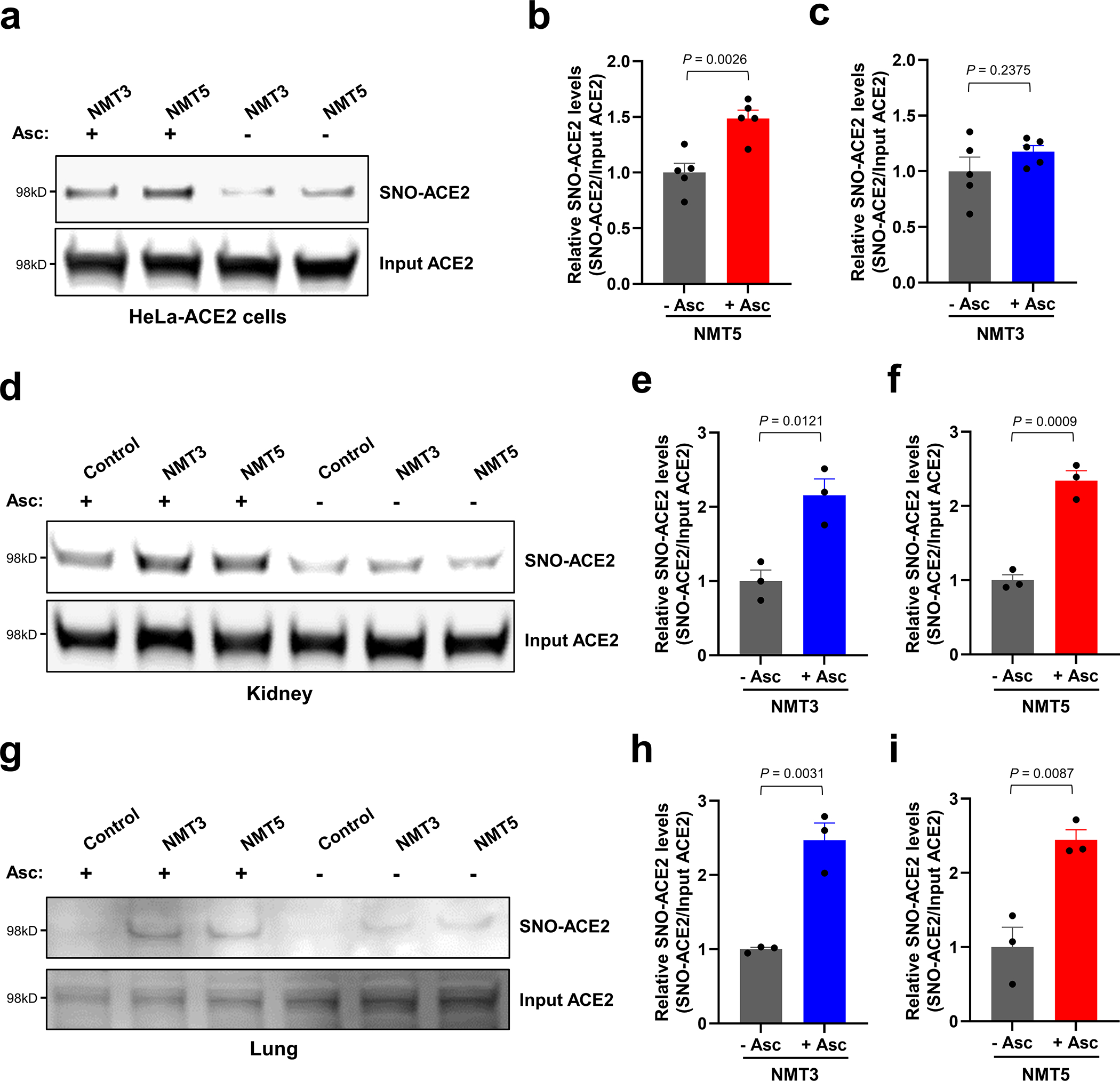
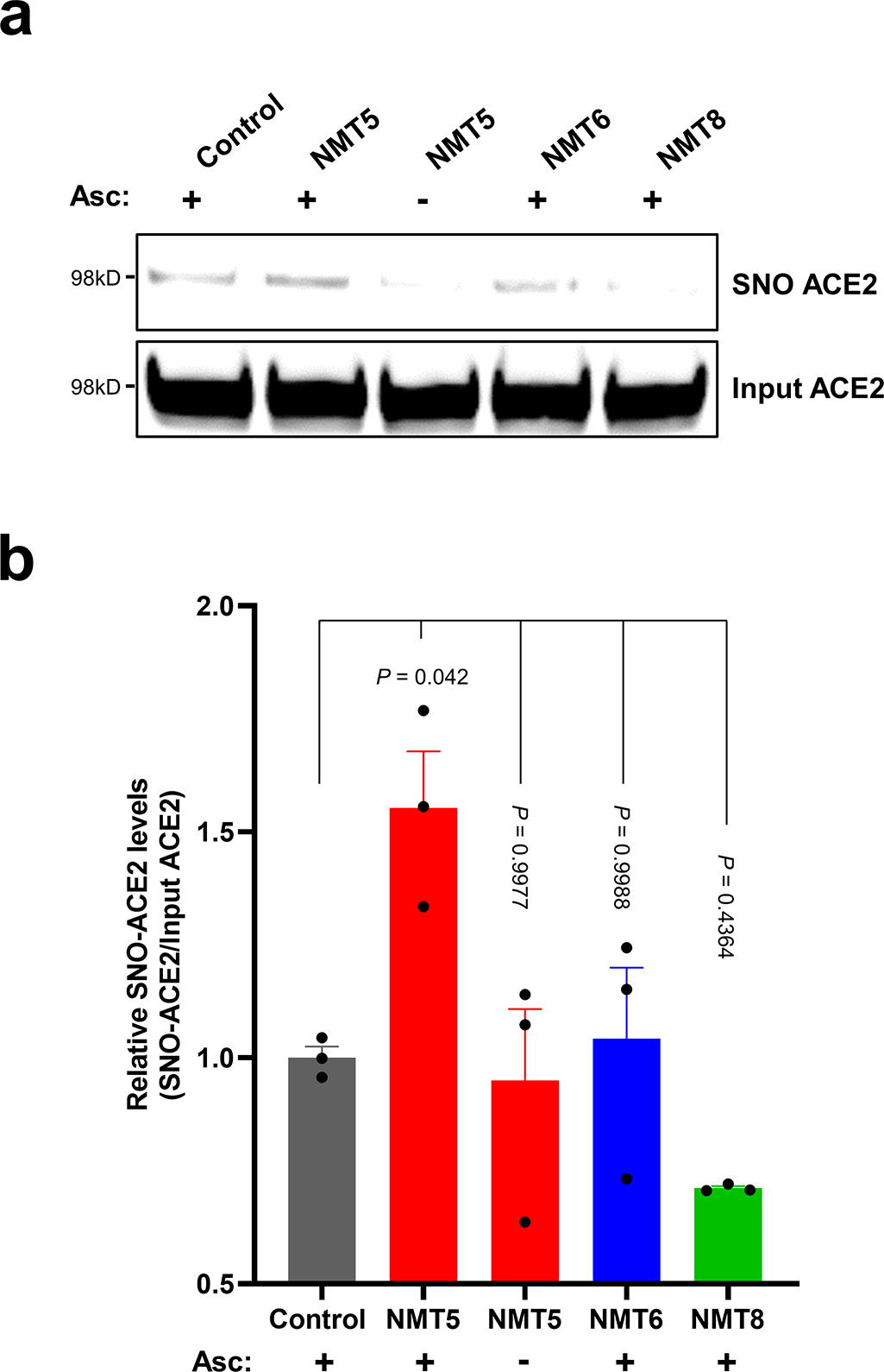
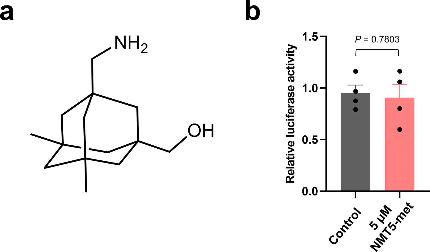
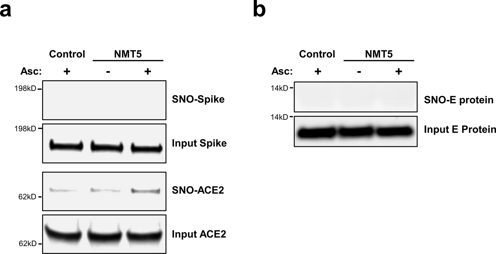
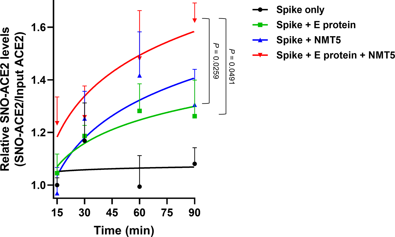
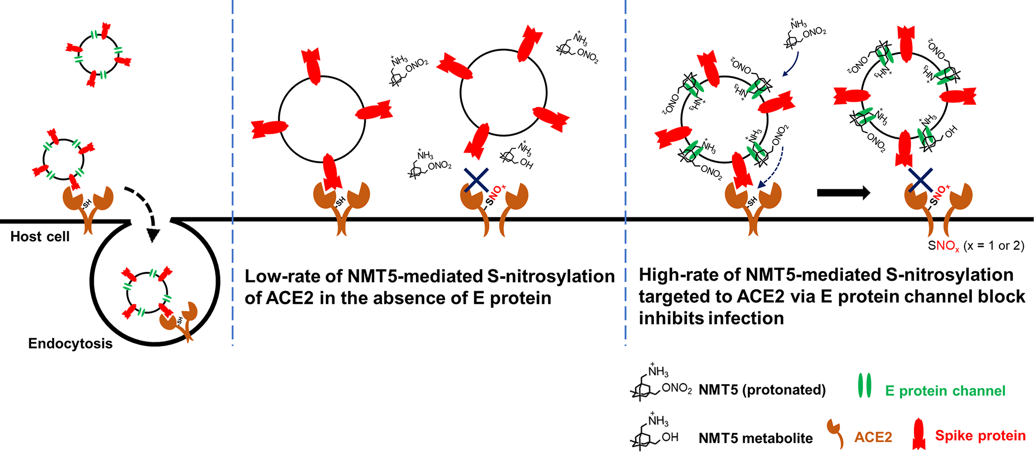
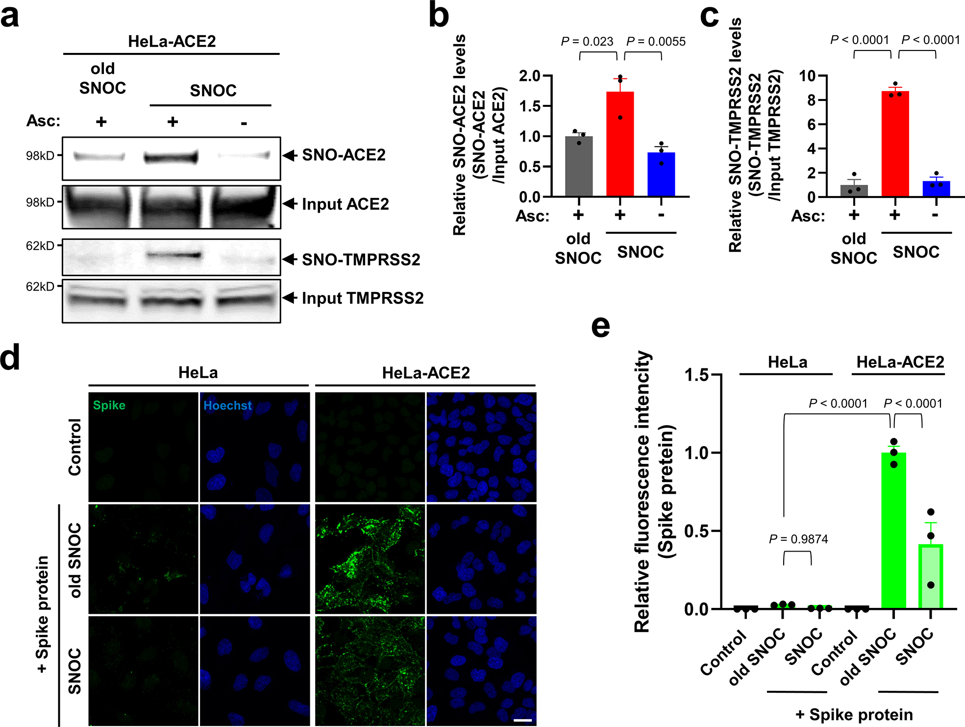
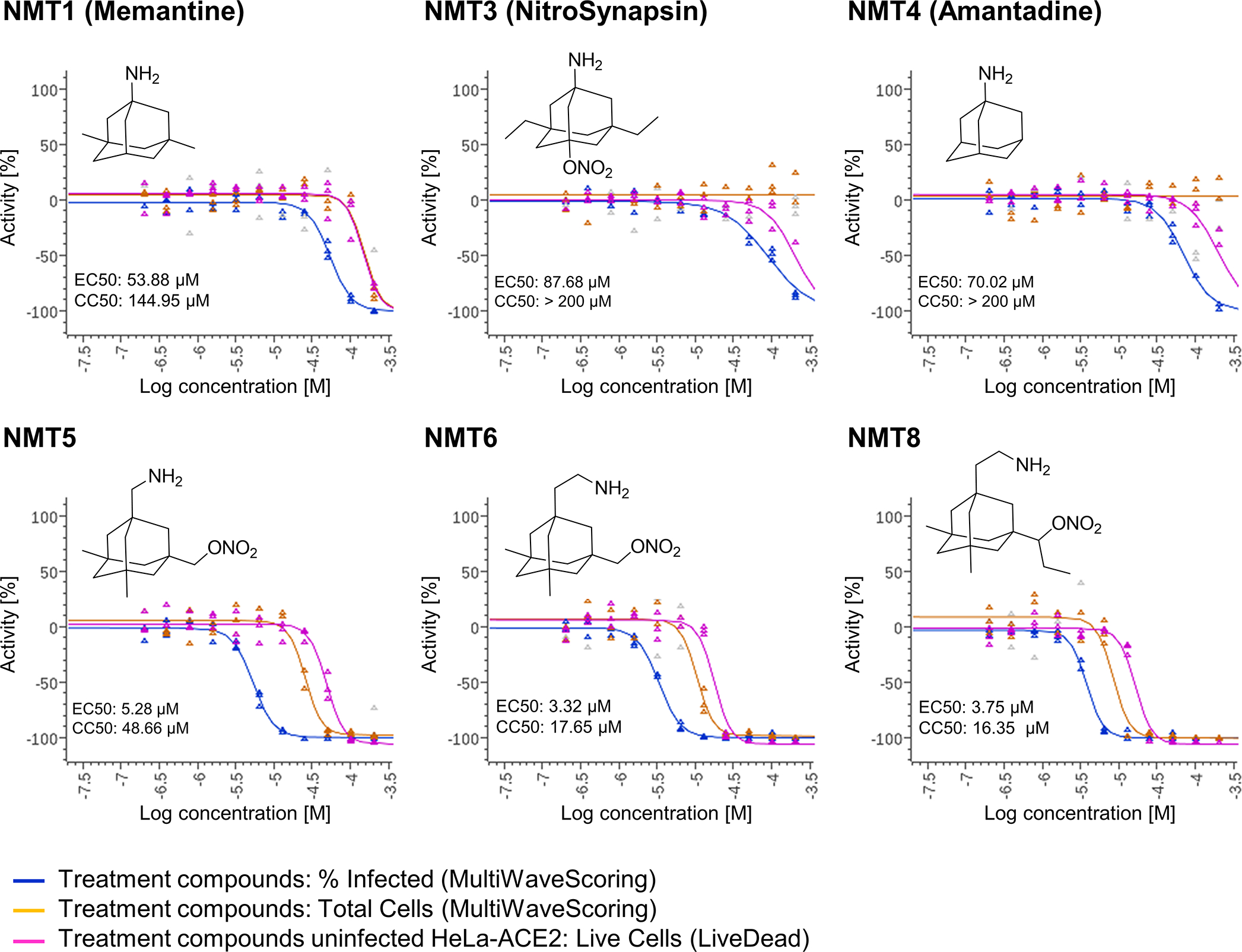
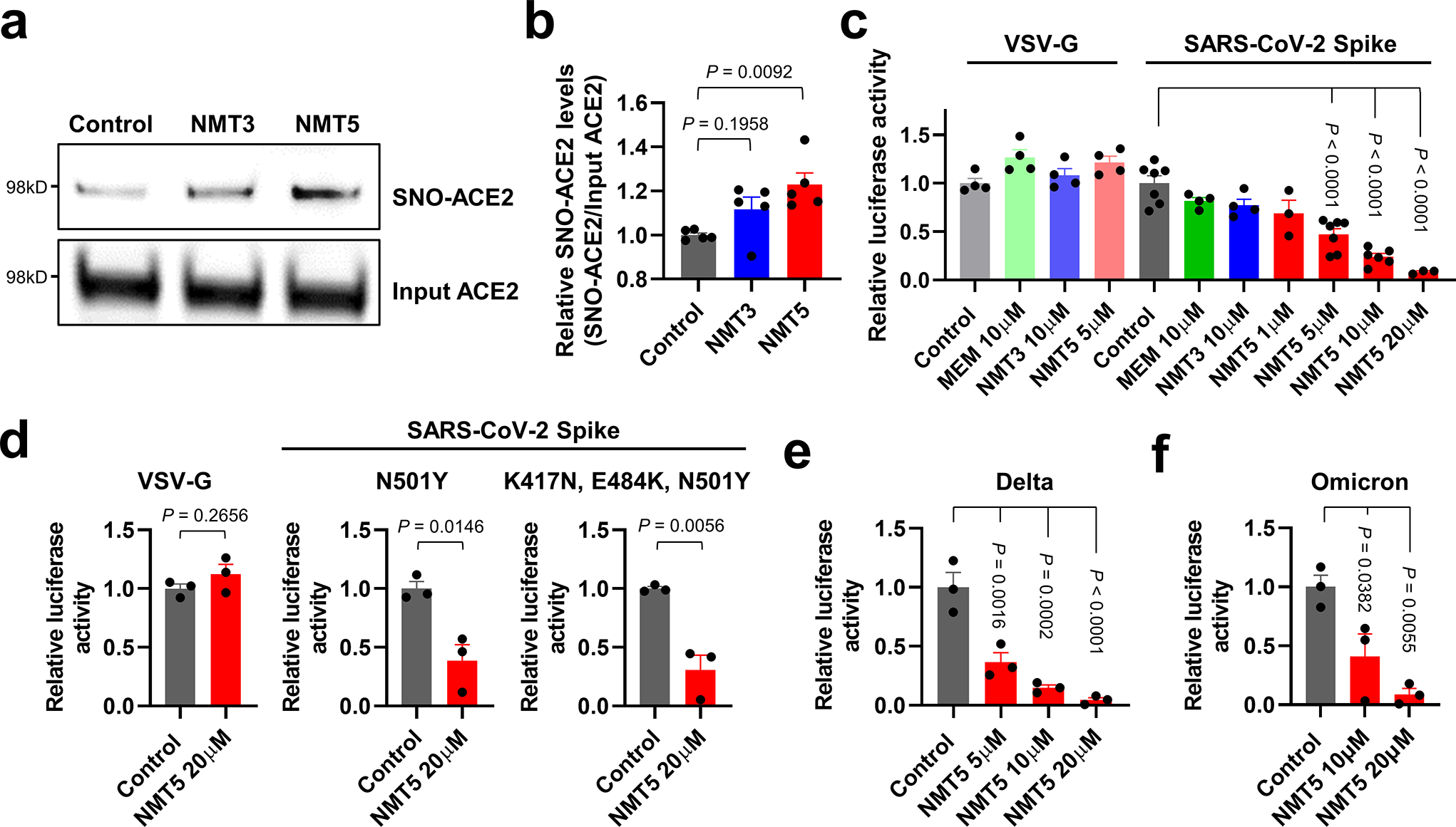
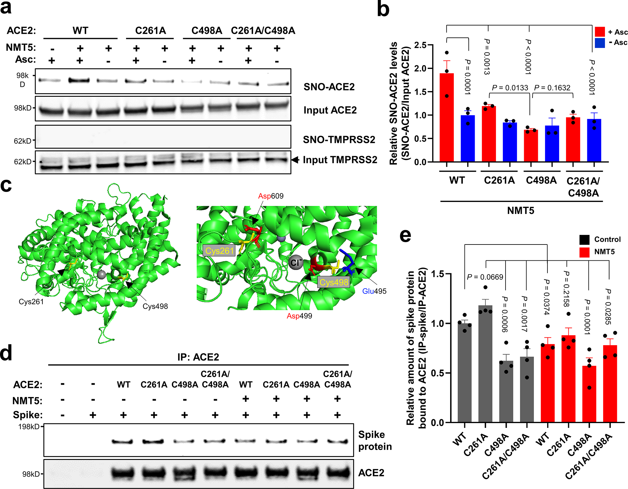
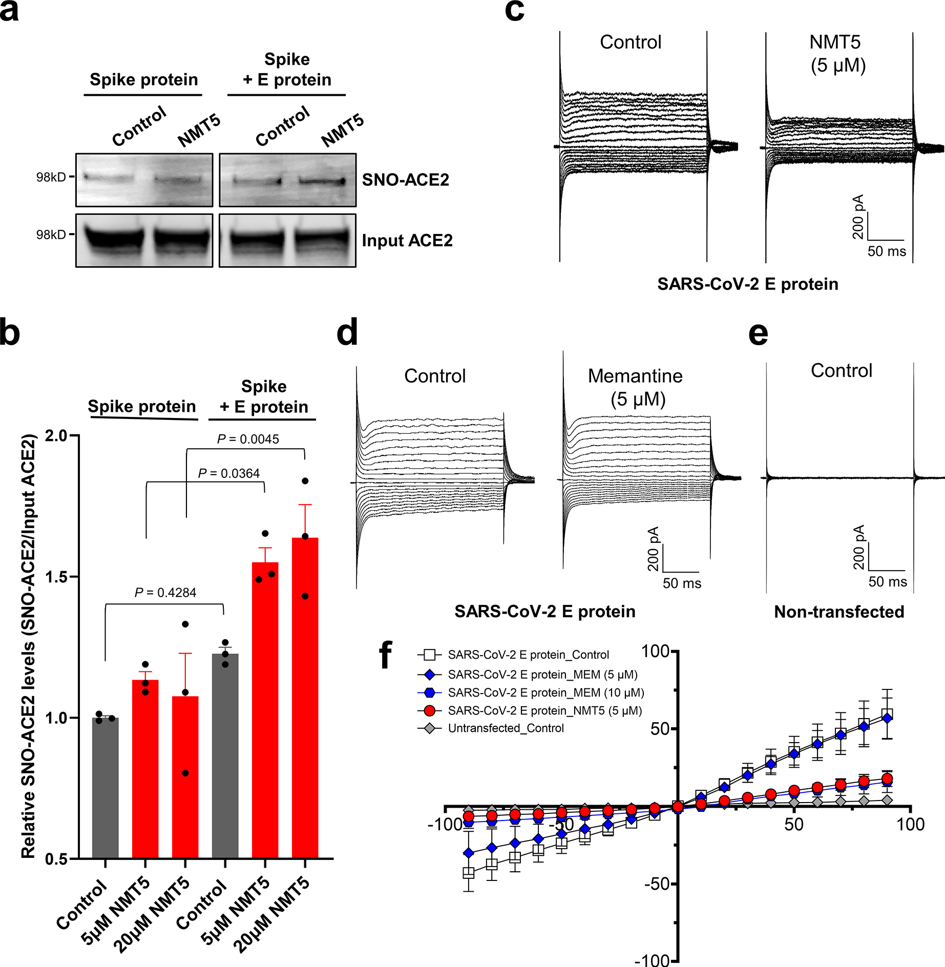
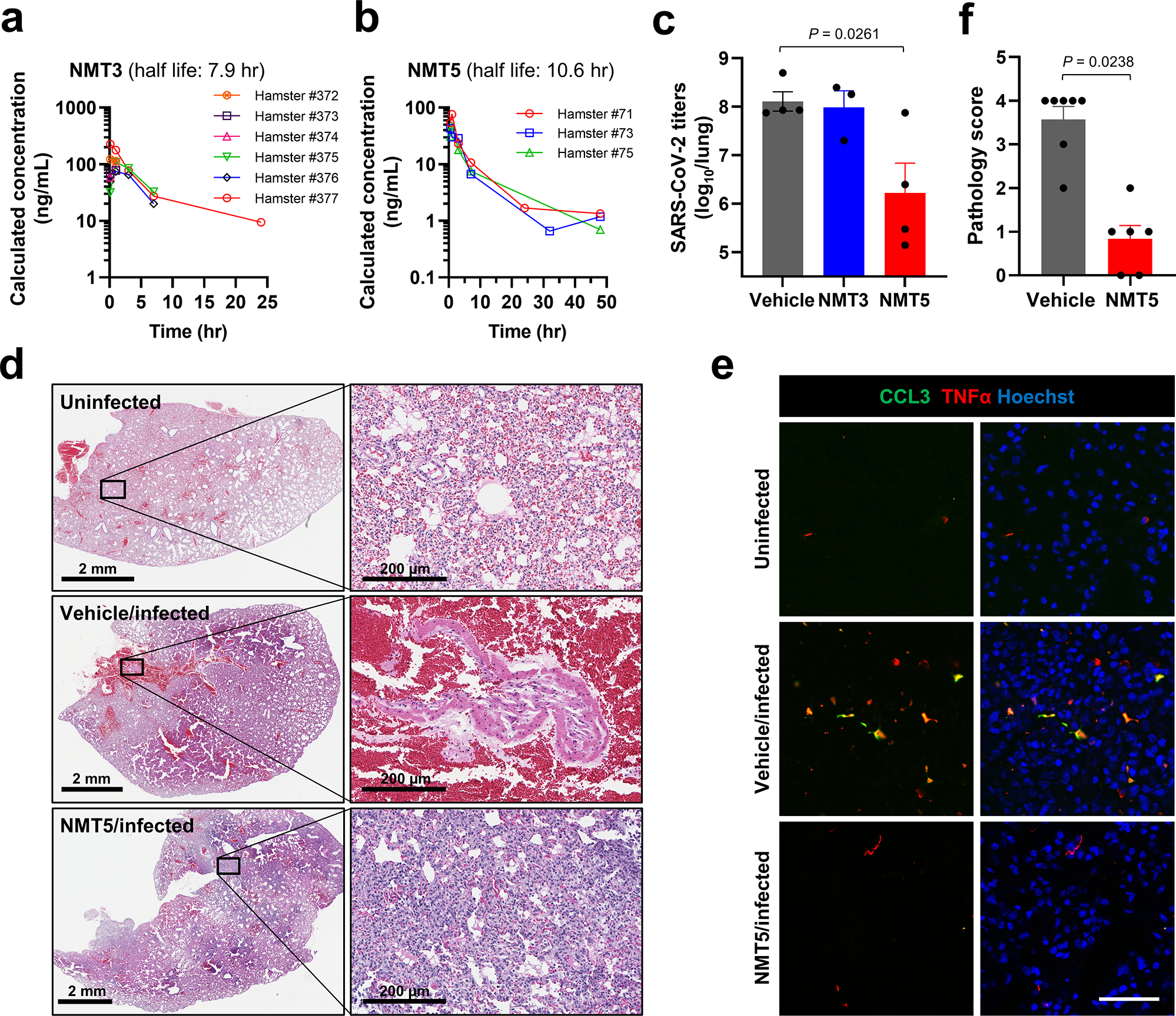
Prevention of infection and propagation of severe acute respiratory syndrome coronavirus 2 (SARS-CoV-2) is a high priority in the Coronavirus Disease 2019 (COVID-19) pandemic. Here we describe S-nitrosylation of multiple proteins involved in SARS-CoV-2 infection, including angiotensin-converting enzyme 2 (ACE2), the receptor for viral entry. This reaction prevents binding of ACE2 to the SARS-CoV-2 spike protein, thereby inhibiting viral entry, infectivity and cytotoxicity. Aminoadamantane compounds also inhibit coronavirus ion channels formed by envelope (E) protein. Accordingly, we developed dual-mechanism aminoadamantane nitrate compounds that inhibit viral entry and, thus, the spread of infection by S-nitrosylating ACE2 via targeted delivery of the drug after E protein channel blockade. These non-toxic compounds are active in vitro and in vivo in the Syrian hamster COVID-19 model and, thus, provide a novel avenue to pursue therapy.


Conflict of interest statement

Competing interests

The authors declare that S.A.L. is an inventor on patents for the use of memantine and various aminoadamantane nitrate compounds for neurodegenerative and neurodevelopmental disorders. He is also an inventor on composition of matter patents and use patents for aminoadamantane nitrate compounds in treating COVID-19 and other viral diseases. Per Harvard University guidelines, S.A.L. participates in a royalty-sharing agreement with his former institution Boston Children’s Hospital/Harvard Medical School, which licensed the drug memantine (Namenda®) to Forest Laboratories, Inc./Actavis/Allergan/AbbVie for use in dementia. The aminoadamantane nitrate compounds have been licensed to EuMentis Therapeutics, Inc. (Newton, Massachusetts). C.B. is a chemist employed at EuMentis Therapeutics. The other authors declare no financial conflicts of interest relevant to this publication.

Figures

Extended Data Fig. 1 |
Extended Data Fig. 1 |. S-Nitrosylation of ACE2 persists for at least 12 hours.
a, HeLa-ACE2 cells were exposed to 100 μM SNOC; 30 min later the cells were incubated in serum free medium for the time periods indicated. Cell lysates were then subjected to biotin-switch assay to assess protein S-nitrosylation, which was detected by immunoblotting with anti-ACE2 antibody. b, Ratio of SNO-ACE2/input ACE2 protein. Data are mean + s.e.m. by one-way ANOVA with Fisher’s LSD multiple comparisons. n = 3 biological replicates.
Extended Data Fig. 2 |
Extended Data Fig. 2 |. Identification of cysteine residues in ACE2 that are S-nitrosylated.
a, List of human ACE2 peptides (± 7 amino acid residues flanking a cysteine residue); gray: peptides involved in disulfide bond formation; black: peptides containing a free cysteine thiol (red) that could potentially be S-nitrosylated. b, HEK293T cells were transiently transfected with plasmids containing human WT ACE2 or cysteine mutant ACE2 (C261A, C498A, or C261/498A). One day after transfection, cells were exposed to 100 μM SNOC. After 20 minutes, cells were subjected to biotin-switch assay. Absence of ascorbate (Asc-) served as a negative control. c, Ratio of SNO-ACE2/input ACE2. Data are mean + s.e.m. by two-way ANOVA with Tukey’s multiple comparisons. n = 3 biological replicates. d, HEK293T cells expressing ACE2 were exposed to SNOC and subjected to biotin switch. The peptides were eluted by reduction for subsequent detection by LC-MS/MS. Representative MS/MS spectra of detected peptides from human ACE2 containing Cys261 (left) or Cys498 (right) are shown.
Extended Data Fig. 3 |
Extended Data Fig. 3 |. Molecular dynamics simulation of S-nitrosylation of ACE2.
a, Molecular representation of the S-nitrosylated-ACE2/RBD model upon transient detachment at the level of the peptidase domain dimeric interface. SNO-Cys261 and SNO-Cys498 are shown with Van der Waals spheres. The black dots indicate qualitative placement of centers of mass (COM) for each ACE2 protomer, and the dashed arrow represents the distance between COMs. Spike’s RBDs and N-glycans, which were included in the simulation, are hidden for image clarity. SpBD, Spike binding domain; CLD, collectrin-like domain; PD, peptidase domain. b, Distribution of the distance between COMs from molecular dynamics simulations of WT ACE2/RBD (purple) vs. nitrosylated-ACE2/RBD (cyan). Dashed black line at approximately 56.5 Å indicates the reference distance between COMs calculated from the cryo-EM structure (PDB: 6M17). S-Nitrosylated-ACE2/RBD shows an overall larger distance between COMs with a bimodal distribution. c, Close-up image illustrating Q175A to D136B interaction present in starting conformations of the S-nitrosylated-ACE2 system. d, Close-up image illustrating the disruption of the interaction between Q175A and D136B occurring along the dynamics of the S-nitrosylated-ACE2 system.
Extended Data Fig. 4 |
Extended Data Fig. 4 |. Dose-response of drugs screened against SARS-CoV-2 infection.
Dose-response curves showing the EC50 of each compound against SARS-CoV-2 (% infected cells, blue), total cell counts (orange) in the infection experiment and the CC50 for uninfected host cell toxicity (magenta), as assessed in HeLa-ACE2 cells. See also Supplementary Data Set 1 for full dataset. Continued from Fig. 2.
Extended Data Fig. 5 |
Extended Data Fig. 5 |. NMT5 S-nitrosylates ACE2 in vitro and in vivo.
a, Detection of SNO-ACE2 in vitro. HeLa-ACE2 cells were exposed to 10 μM NMT3 or 5 μM NMT5. After 1 h, cells were subjected to the biotin-switch assay in the presence or absence of ascorbate. SNO-ACE2 and input ACE2 were detected by immunoblotting with anti-ACE2 antibody. b, c, Ratio of SNO-ACE2/input ACE2. Data are mean + s.e.m. by two-tailed Student’s t test. n = 5 biological replicates. di, Detection of SNO-ACE2 in vivo. Syrian hamsters received 10 mg/kg of NMT3 or of NMT5 by oral gavage and were sacrificed 48 h later. Kidney and lung tissues were subjected to biotin-switch assay in the presence or absence of ascorbate. Note that in some samples, low levels of SNO-ACE2 were observed in control tissue, suggesting endogenous S-nitrosylation of ACE2 may occur at low levels. Graphs show ratio of SNO-ACE2/input ACE2. Data are mean + s.e.m. by two-tailed Student’s t test. n = 3 Syrian hamsters for each condition.
Extended Data Fig. 6 |
Extended Data Fig. 6 |. Protein S-nitrosylation of ACE2 by NMT5.
a, HeLa-ACE2 cells were treated with 10 μM NMT5, NMT6, or NMT8. After 1 h, cell lysates were subjected to biotin-switch assay for protein S-nitrosylation, detected by immunoblotting with anti-ACE2 antibody. The ascorbate minus (Asc-) sample served as a negative control. b, Ratio of SNO-ACE2/input ACE2 protein. Data are mean + s.e.m. by one-way ANOVA with Tukey’s multiple comparisons. n = 3 biological replicates.
Extended Data Fig. 7 |
Extended Data Fig. 7 |. Critical role of nitro group of NMT5 suppressing SARS-CoV-2 infection on pseudovirus entry assay.
a, Chemical structure of NMT5 metabolite (NMT5-met, lacking the nitro group). b, HeLa-ACE2 cells were incubated in the presence and absence of 5 μM NMT5-met with SARS-CoV-2 Spike (D614) pseudovirus particles. After 48 h, viral transduction efficiency was monitored by luciferase activity. Inhibitory activity was lost in the absence of the nitro group (compare with Fig. 3c, e). Data are mean + s.e.m. by two-tailed Student’s t test. n = 4 biological replicates.
Extended Data Fig. 8 |
Extended Data Fig. 8 |. Lack of S-nitrosylation of Spike protein and E protein.
a, Purified recombinant SARS-CoV-2 Spike (S1 + S2) protein and ACE2 protein were exposed to 100 μM SNOC; 30 min later, samples were subjected to biotin-switch assay in the presence or absence of ascorbate (Asc) to assess protein S-nitrosylation. n = 3 biological replicates. b, Lack of E protein S-nitrosylation by NMT5. HA-tagged E protein plasmid was transiently transfected into HEK293 cells. One day after transfection, cells were exposed to 10 μM NMT5. After 1 hour, the cells were harvested and subjected to biotin-switch assay in the presence or absence of ascorbate (Asc) to assess protein S-nitrosylation, which was detected by immunoblotting with anti-HA antibody. n = 3 biological replicates.
Extended Data Fig. 9 |
Extended Data Fig. 9 |. Targeted S-nitrosylation of ACE2 by NMT5 in the presence of envelope (E) viroporin protein.
Kinetic analysis of S-nitrosylation of ACE2. E protein plasmid was transiently transfected into HEK293 cells stably-expressing Spike protein cells. Subsequently, the cells were harvested and plated onto ACE2-expressing HeLa cells in the presence or absence of 20 μM NMT5. At the indicated timepoints, cells were subjected to biotin-switch assay. Ordinate shows ratio of SNO-ACE2/total input ACE2 protein above the baseline value, defined as 1.0. Data are mean + s.e.m. by two-way ANOVA with Fisher’s LSD test. n = 5 biological replicates.
Extended Data Fig. 10 |
Extended Data Fig. 10 |. Schematic of NMT5 targeting of SNO-ACE2 via SARS-CoV-2 E protein.
S-Nitrosylation of ACE2 on the host cell inhibits SARS-CoV-2 entry and thus infection. Note that at physiological pH, the bridgehead amine of NMT5, like other aminoadamantanes, is generally protonated to NH3+.
Fig. 1 |
Fig. 1 |. SNOC increases S-nitrosylation of ACE2 and inhibits binding of SARS-CoV-2 Spike (S) protein.
a, Assay for SNO-ACE2 and SNO-TMPRSS2 in HeLa-ACE2 cells. Cells were exposed to 100 μM SNOC or, as a control, ‘old’ SNOC (from which NO had been dissipated). After 20 minutes, cell lysates were subjected to biotin-switch assay to assess S-nitrosylated (SNO-) and input (total) proteins detected by immunoblotting with cognate antibody. The ascorbate minus (Asc-) sample served as a negative control. b, c, Ratio of SNO-ACE2/input ACE2 protein and SNO-TMPRSS2/input TMPRSS2 protein. Data are mean + s.e.m.by one-way ANOVA with Tukey’s multiple comparisons. n = 3 biological replicates. d, HeLa and HeLa-ACE2 cells were pre-exposed to 100 μM SNOC or old SNOC. After 30 minutes, 10 μg/ml of purified recombinant SARS-CoV-2 Spike (S1 + S2) protein was incubated with the cells. After 1 h, cells were fixed with 4% PFA for 15 minutes, and bound Spike protein was detected by anti-Spike protein antibody; nuclei stained with Hoechst. Scale bar, 20 μm. e, Quantification of relative fluorescence intensity. Data are mean + s.e.m. by two-way ANOVA with Tukey’s multiple comparisons. n = 3 biological replicates.
Fig. 2 |
Fig. 2 |. Dose-response of drugs screened against SARS-CoV-2.
Dose-response curves showing the EC50 of each compound against SARS-CoV-2 (% infected cells, blue), total cell counts (orange) in the infection experiment and the CC50 for uninfected host cell toxicity (magenta), as assessed in HeLa-ACE2 cells. See also Supplementary Data Set 1 for full dataset.
Fig. 3 |
Fig. 3 |. NMT5 inhibits SARS-CoV-2 pseudoviral entry.
a, HeLa-ACE2 cells were treated with 10 μM NMT3 or 5 μM NMT5. After 1 h, cell lysates were subjected to biotin-switch assay for protein S-nitrosylation, detected by immunoblotting with anti-ACE2 antibody. b, Ratio of SNO-ACE2/input ACE2 protein. Data are mean + s.e.m. by one-way ANOVA with Tukey’s multiple comparisons. n = 5 biological replicates. c, HeLa-ACE2 cells were incubated with SARS-CoV-2 Spike (D614) or VSV-G (control) pseudovirus particles in the presence and absence of MEM (memantine), NMT3, or NMT5. After 48 h, viral transduction efficiency was monitored by luciferase activity. Data are mean + s.e.m. by one-way ANOVA with Tukey’s multiple comparisons. n = 3 to 7 biological replicates. df, HeLa-ACE2 cells were incubated in the presence and absence of NMT5 with SARS-CoV-2 N501Y Spike, SARS-CoV-2 K417N/E484K/N501Y Spike, or VSV-G (control) pseudovirus particles (d); or with SARS-CoV-2 delta variant (e) or omicron variant pseudovirus particles (f). After 48 h, viral transduction efficiency was monitored by luciferase activity. Data are mean + s.e.m. by two-tailed Student’s t test (d) or one-way ANOVA with Tukey’s for multiple comparisons (e, f). n = 3 biological replicates.
Fig. 4 |
Fig. 4 |. S-Nitrosylation of ACE2 by NMT5 inhibits binding to Spike protein.
a, HEK293T cells were transfected with plasmids encoding human WT ACE2 or non-nitrosylatable mutant ACE2 (C262A, C498A, or C261A/C498A). Cells were treated with 10 μM NMT5, and subjected to biotin-switch assay for detection of S-nitrosylated proteins by immunoblotting with anti-ACE2 and anti-TMPRSS2 antibodies. The absence of ascorbate (Asc-) served as a negative control. b, Ratio of SNO-ACE2/input ACE2. Data are mean + s.e.m. by two-way ANOVA with Fisher’s LSD multiple comparisons. n = 3 biological replicates. c, Crystal structure of ACE2 (left panel; PDB ID: 6M0J) with enlarged view of S-nitrosylation sites of ACE2. Glu495 and Asp499, acidic amino-acid residues, surround Cys498 (right panel). d, HEK293T cells were transfected with plasmids encoding human WT ACE2 or non-nitrosylatable mutant ACE2 (C262A, C498A, or C261A/C498A). Cells were exposed to 1 μg/ml of purified recombinant SARS-CoV-2 Spike protein in the presence or absence of 5 μM NMT5; after 1 h, cells were lysed and subjected to co-IP with anti-ACE2 antibody. Immunoprecipitated ACE2 and Spike protein were detected by immunoblotting with anti-ACE2 and anti-Spike protein antibodies. e, Ratio of IP-ACE2/IP-Spike protein. Data are mean + s.e.m. by one-way ANOVA with Fisher’s LSD multiple comparisons. n = 4 biological replicates.
Fig. 5 |
Fig. 5 |. Targeted S-nitrosylation of ACE2 and Inhibition of envelope (E) viroporin protein channel by NMT5.
a, E protein plasmid was transiently transfected into HEK293-Spike protein cells. After 1 day, cells were harvested and plated onto HeLa-ACE2 cells in the presence or absence of 5 μM NMT5. After 30 min, cell lysates were subjected to biotin-switch assay to monitor protein S-nitrosylation of ACE2, detected by immunoblotting. b, Ratio of SNO-ACE2/total input ACE2 protein. Data are mean + s.e.m. by two-way ANOVA with Tukey’s multiple comparisons. n = 3 biological replicates. ce, Representative traces of whole-cell currents from untransfected (n = 4), and transiently transfected (n = 15) HEK293T cells before and after application of memantine or NMT5 during patch-clamp recording. Whole-cell currents were generated by holding cells at 0 mV and applying voltage steps between −90 and +90 mV in increments of 10 mV. f, Current-voltage (I–V) curves from steady-state current density (pA/pF) versus holding potential (mV) for memantine (MEM, 5 and 10 μM) and NMT5 (5 μM). Data are mean ± s.e.m., n = 16 cells recorded.
Fig. 6 |
Fig. 6 |. NMT5 inhibits SARS-CoV-2 infection in vivo in Syrian hamsters.
a, b, PK data in plasma for NMT3 (n = 6) and NMT5 (n = 3) in Syrian hamsters after an oral dose of 10 mg/kg. c, Live viral load in Syrian hamsters monitored by plaque assay from lung tissue 2-d after infection after treatment with NMT3, NMT5, or vehicle (Control). Data are mean + s.e.m. by two-tailed Student’s t test (n = 8 Syrian hamsters tested). d, e, Syrian hamsters were sacrificed 5 d after infection with SARS-CoV-2 that were either untreated (labeled Vehicle/Infected) or treated with oral NMT5 (NMT5/Infected), and compared to control (Uninfected). Representative hematoxylin and eosin (H&E)-stained sections showed virtually no areas of large hemorrhage (confluent bright red regions) in NMT5-treated hamster lungs compared to vehicle-treated. Scale bars, 2 mm, high magnification 200 μm (d). Representative immunohistochemistry of lung sections stained with anti-TNFα cytokine antibody and anti-CCL3 (MIP-1α) chemokine antibody from untreated- and NMT5-treated hamsters 5-d post SARS-CoV-2 infection; uninfected sections are shown as a control. Merged image with Hoechst stain for DNA shown in right-hand panels. Scale bar, 40 μm (e). f, Lung pathology scores (see Methods) for infected hamsters, either untreated or treated with NMT5. Treated hamsters showed significant improvement. Data are mean + s.e.m. by two-tailed Mann–Whitney U test (n = 9 Syrian hamsters tested).

Update of

References

    1. Hoffmann M et al. SARS-CoV-2 cell entry depends on ACE2 and TMPRSS2 and is blocked by a clinically proven protease inhibitor. Cell 181, 271–280 e278 (2020). - PMC - PubMed
    1. Kozakov D, Chuang GY, Beglov D & Vajda S Where does amantadine bind to the influenza virus M2 proton channel? Trends Biochem Sci 35, 471–475 (2010). - PMC - PubMed
    1. Torres J et al. Conductance and amantadine binding of a pore formed by a lysine-flanked transmembrane domain of SARS coronavirus envelope protein. Protein Sci. 16, 2065–2071 (2007). - PMC - PubMed
    1. Asarnow D et al. Structural insight into SARS-CoV-2 neutralizing antibodies and modulation of syncytia. Cell 184, 3192–3204 e3116 (2021). - PMC - PubMed
    1. Lipton SA Paradigm shift in neuroprotection by NMDA receptor blockade: memantine and beyond. Nat. Rev. Drug Discov. 5, 160–170 (2006). - PubMed

References for Methods

    1. Li W et al. Angiotensin-converting enzyme 2 is a functional receptor for the SARS coronavirus. Nature 426, 450–454 (2003). - PMC - PubMed
    1. Uehara T et al. S-Nitrosylated protein-disulphide isomerase links protein misfolding to neurodegeneration. Nature 441, 513–517 (2006). - PubMed
    1. Ghatak S et al. Mechanisms of hyperexcitability in Alzheimer’s disease hiPSC-derived neurons and cerebral organoids vs isogenic controls. eLife 8, e50333 (2019). - PMC - PubMed
    1. He L, Diedrich J, Chu YY & Yates JR 3rd. Extracting accurate precursor information for tandem mass spectra by RawConverter. Anal. Chem. 87, 11361–11367 (2015). - PMC - PubMed
    1. Xu T et al. ProLuCID: An improved SEQUEST-like algorithm with enhanced sensitivity and specificity. J. Proteomics 129, 16–24 (2015). - PMC - PubMed

Publication types

Substances