. 2023 Jan;159(1):47-60.
doi: 10.1007/s00418-022-02152-7. Epub 2022 Sep 29.

Ultrastructural analysis and three-dimensional reconstruction of cellular structures involved in SARS-CoV-2 spread

Affiliations

Ultrastructural analysis and three-dimensional reconstruction of cellular structures involved in SARS-CoV-2 spread

Marta Baselga et al. Histochem Cell Biol. 2023 Jan.

Abstract

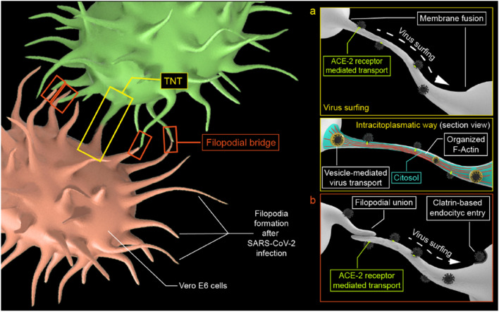
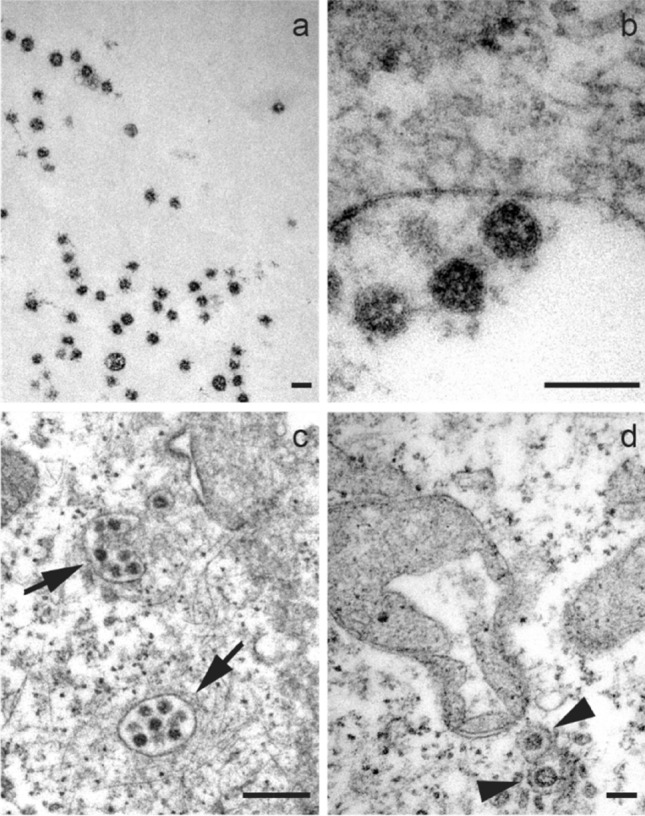
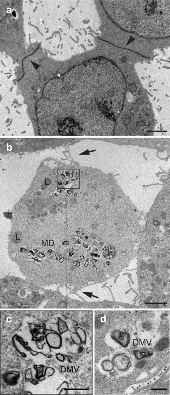
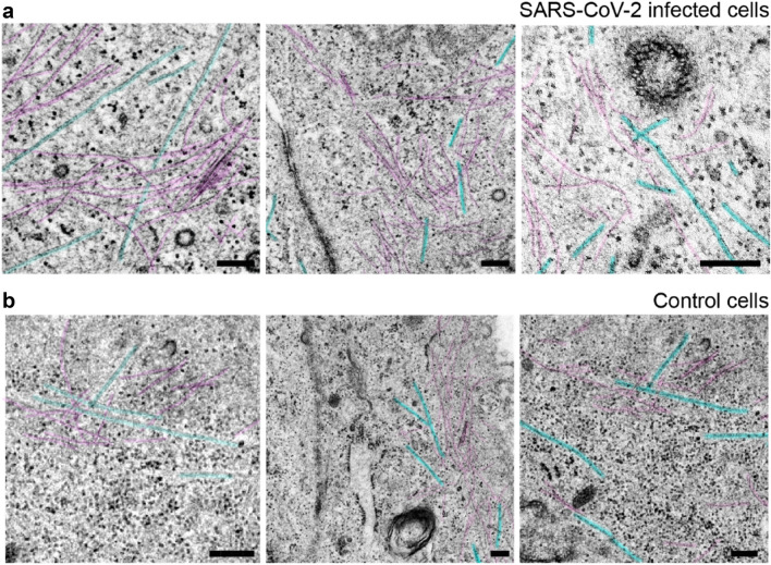
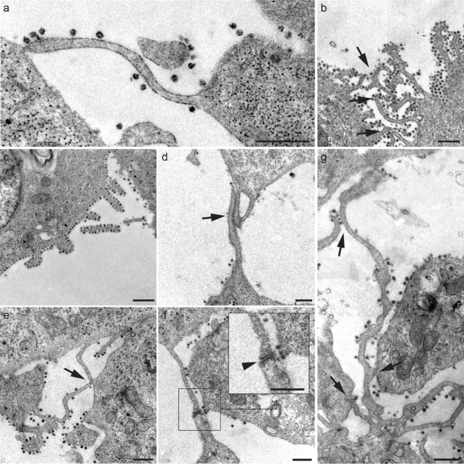
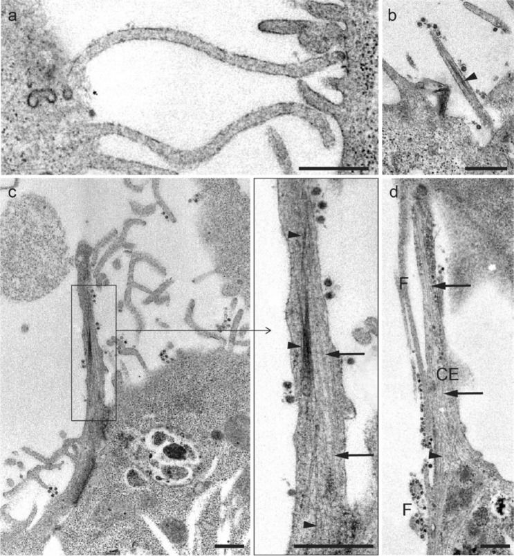
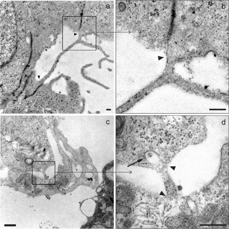
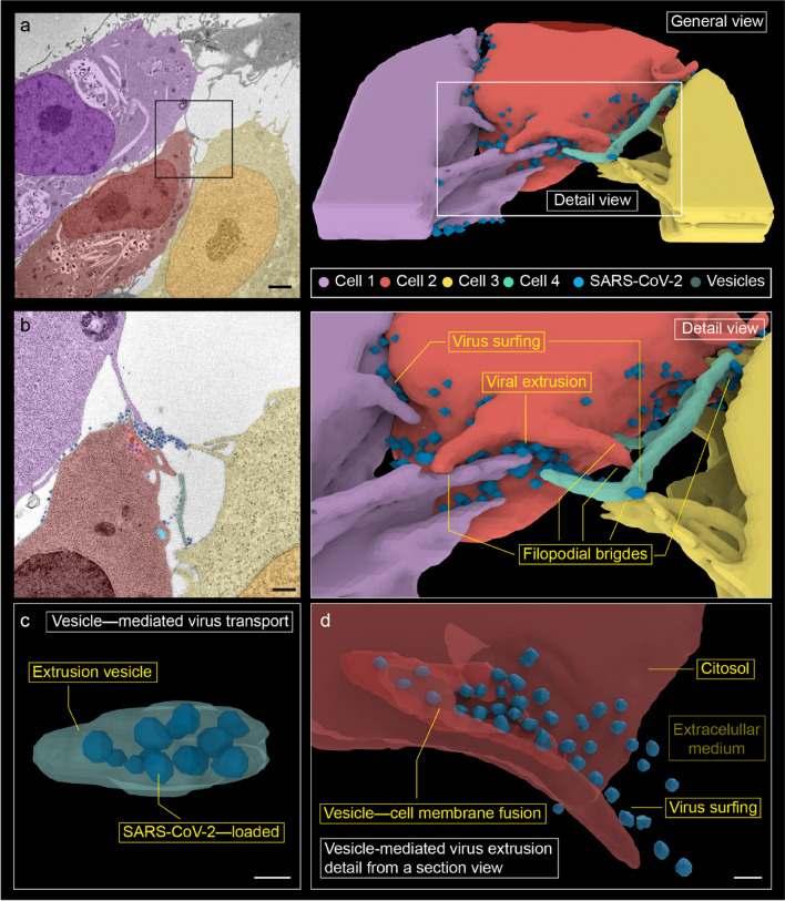
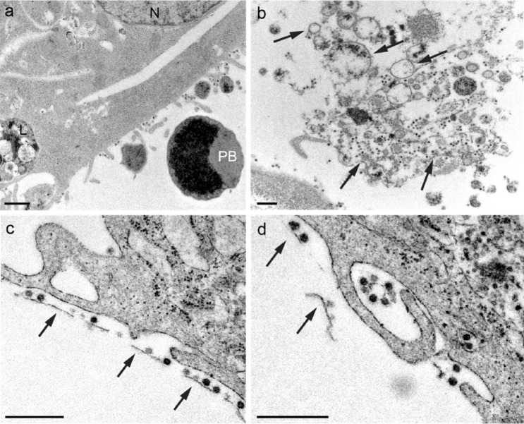
The cytoskeleton not only deals with numerous interaction and communication mechanisms at the cellular level but also has a crucial role in the viral infection cycle. Although numerous aspects of SARS-CoV-2 virus interaction at the cellular level have been widely studied, little has been reported about the structural and functional response of the cytoskeleton. This work aims to characterize, at the ultrastructural level, the modifications in the cytoskeleton of infected cells, namely, its participation in filopodia formation, the junction of these nanostructures forming bridges, the viral surfing, and the generation of tunnel effect nanotubes (TNT) as probable structures of intracellular viral dissemination. The three-dimensional reconstruction from the obtained micrographs allowed observing viral propagation events between cells in detail for the first time. More profound knowledge about these cell-cell interaction models in the viral spread mechanisms could lead to a better understanding of the clinical manifestations of COVID-19 disease and to find new therapeutic strategies.

Keywords: Actin; Cytoskeleton; Filopodia; SARS-CoV-2; TNT; Ultrastructure.


Conflict of interest statement

The authors declare no conflicts of interest.

Figures

Fig. 1
Fig. 1
Conceptual scheme of a viral surfing and the intracellular pathway in TNT. b Viral surfing along filopodial bridges
Fig. 2
Fig. 2
Micrographs show a free viral particles hoving in the extracellular medium. b Viruses adhered to the plasma membrane. c Clusters of multiple viral particles (arrows). d Individual viruses within vesicles in the cytosol (arrowhead). A mitochondrial alteration induced by SARS-CoV-2 infection is also shown in d. Scale bars represent 0.1 μm in ad
Fig. 3
Fig. 3
a Uninfected control cells establish extensive junctions between them, with a low number of filopodia and no filopodial bridges. b Filopodia formation induced by SARS-CoV-2 is observed in the infected cells, where the formation of filopodium bridges (arrows) is usually identified. c, d Detail of double membrane vesicles (DMVs). Scale bars represent 2 μm in a, b, and 0.5 μm in c, d
Fig. 4
Fig. 4
Comparative cytoskeleton micrographs from infected and control cells. a Microtubules (blue) and actin (or intermediate) filaments (purple) in the juxtamembranous region from three SARS-CoV-2-infected cells. b Cytoskeleton of three non-infected cells. It can be also appreciated nearby endocytic vesicles probably coated with clathrin and free ribosomal units. Scale bars represent 0.2 μm in a, b
Fig. 5
Fig. 5
a-g Micrographs of various filopodial structures found in SARS-CoV-2-infected Vero E6 cells. a Filopodial extension whose end adheres to the plasma membrane of a neighboring cell. b-c The number of viral particles attached to the filopodia was variable, although they were frequently observed in high density surrounding the filopodial surface. d Filopodial bridges were constituted through wide intermembrane junctions. e Junctions of filopodial bridges were often found in the distal extremes. f In many cases, bridge filopodial junctions showed electron-dense reinforcements (arrowhead; detail). g Commonly, larger filopodia contact more than one neighboring cell. Scale bars represent 0.5 μm in a-g
Fig. 6
Fig. 6
a, b Longitudinal section of filopodia, characterized by the absence of microtubules. c Longitudinal section of thick cytoplasmic extensions showing SARS-CoV-2 virus-like particles in the plasma membrane, and microtubules (arrows) and filaments (arrowheads) arranged parallelly. d Comparison between thick cytoplasmic extensions (CE) and filopodium (F) structures. Scale bars represent 0.5 μm a-d
Fig. 7
Fig. 7
Micrographs of structures similar to TNT. a A nanotube > 2.5 μm in length and 70–90 nm in diameter promoting direct contact between the cytosols of two adjacent cells is observed. b Membrane fusion (arrowhead) detail of (a). c A smaller filopodium (< 0.5 μm in length and 90–100 nm in diameter), where a structure similar to a clathrin-coated vesicle (arrow) can be seen endocytosing a viral particle. d Detail of the fusion membranes from two neighboring cell bodies (arrowheads) from c. Scale bars represent 0.2 μm in ad
Fig. 8
Fig. 8
Three-dimensional reconstruction from 14 serial electron micrographs. a Overview of cell–cell interaction in viral propagation. b Detail of the membrane projections, where filopodial connections and viral surfing are observed. Numerous membrane projections from (at least) three different cells converge in a region of high viral load. We designate as 'viral extrusion' the region where the membrane of the extrusion vesicle fuses with the plasma membrane. c A vesicle with internalized viruses from cell 2 has been reconstructed. It is observed 12 viral particles that occupy practically the entire vesicle. It is probably an extrusion vesicle externalizing the virus. d A detail of viral extrusion has been reconstructed using eight micrographs. The micrographs have been selected to show how a vesicle fuses with the membrane to extrude viruses along a filopodium. This finding supports the hypothesis that filopodia are formed to promote the spread of the virus between cells. Scale bars represent 2 μm in a, 0.5 μm in b, and ~ 0.1 μm in c, d
Fig. 9
Fig. 9
Micrographs showing a fragments of a pyroptotic body in the extracellular space (PB pyroptotic body, N nucleus, L lysosome). b, c Virions adhered to a membrane fragments (arrows). Subcellular fragments in the extracellular space have been observed as a consequence of SARS-CoV-2 infection. In membrane fragments, it has been possible to identify adherent virions. d Virus adhered to remains of membranes are occasionally encovered by the filopodia (possible macropinocytosis). Scale bars represent 0.5 μm in (a-d)

References

    1. Afzelius BA. Ultrastructure of human nasal epithelium during an episode of coronavirus infection. Virchows Arch. 1994;424:295–300. doi: 10.1007/BF00194614. - DOI - PMC - PubMed
    1. Akamatsu M, Vasan R, Serwas D, et al. Principles of self-organization and load adaptation by the actin cytoskeleton during clathrin-mediated endocytosis. Elife. 2020;9:1–40. doi: 10.7554/eLife.49840. - DOI - PMC - PubMed
    1. Akilesh S, Nicosia RF, Alpers CE, et al. Characterizing viral infection by electron microscopy: lessons from the coronavirus disease 2019 pandemic. Am J Pathol. 2021;191:222–227. doi: 10.1016/j.ajpath.2020.11.003. - DOI - PMC - PubMed
    1. Barreto-Vieira DF, da Silva MAN, de Almeida ALT, et al. SARS-CoV-2: ultrastructural characterization of morphogenesis in an in vitro system. Viruses. 2022 doi: 10.3390/v14020201. - DOI - PMC - PubMed
    1. Bedi S, Ono A. Friend or foe: the role of the cytoskeleton in influenza a virus assembly. Viruses. 2019 doi: 10.3390/v11010046. - DOI - PMC - PubMed

LinkOut - more resources