

Clinicopathological Correlations in Enucleated Globes of Late-Stage Coats Disease with a Review of the Literature
- PMID: 36175755
- PMCID: PMC9723065
- DOI: 10.1007/s44197-022-00068-y
Clinicopathological Correlations in Enucleated Globes of Late-Stage Coats Disease with a Review of the Literature
Abstract
Background: Coats disease may cause diagnostic dilemma because of its variable clinical presentation that can be suspicious of retinoblastoma. Late sequelae of the disease are blinding with possible enucleation. We demonstrate the main histopathological findings of Coats enucleated eyes with literature review.
Methods: This was a retrospective study of all enucleated globes diagnosed as Coats disease over 30 years and were reviewed by two pathologists. The corresponding demographic data, clinical presentation, pre-operative clinical impression, and indication for enucleation were collected. Descriptive analysis of our own series data was performed. Our findings were then correlated to published data that were collected from 1983 to 2021 from the PUBMED database in English-written language. Shields classification was used as an inclusion criterion for the published reports to be analyzed.
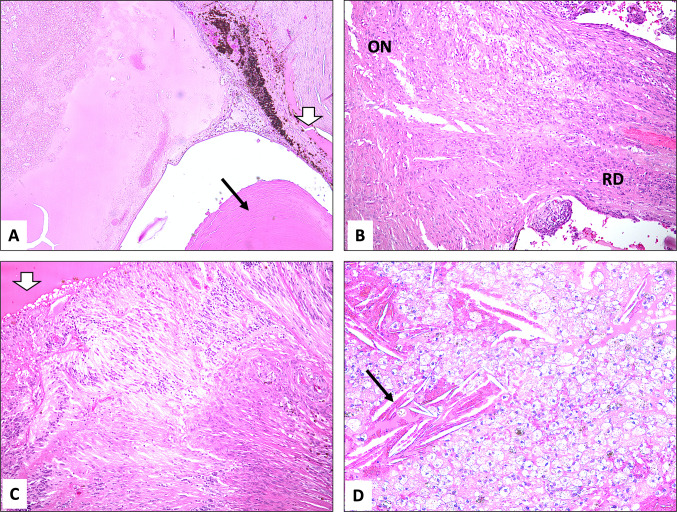
Results: We had seven enucleated globes with Coats disease. Mean age at presentation was 3.2 years (range 3 months to 9 years). Male predominance was observed in 6 and all cases were unilateral. Strabismus was the most common initial presentation (57%, n = 4), followed by leukocoria (43%, n = 3). Indication for enucleation was mostly suspected retinoblastoma (57%, n = 4). Four eyes were classified as stage 4, and 2 had advanced stage 5 changes. Histopathologically, subretinal fluid with lipid-laden macrophages was seen in all cases, the anterior chamber was shallow in 5/7 with angle neovascularization in 2/7. Telangiectatic vessels were clearly observed in 4/7.
Conclusion: Coats disease is a potentially visually disabling disease that is mostly unilateral in 95%, has male predominance of 81%, and wide age range with a mean of 17 years. In Saudi Arabia, the disease seems to present at younger age, tends to be more advanced, and may be indistinguishable from retinoblastoma at the time of diagnosis. Shields staging of Coats is highly recommended to be followed clinically to unify the pathways for treatment and to correlate the concluded outcomes.
Keywords: Coats disease; Enucleation; Globe; Leukocoria; Retinoblastoma; Subretinal exudate.
© 2022. The Author(s).
Conflict of interest statement
The authors have no conflict of interest or financial disclosures in relation to this work.
Figures

References
Publication types
MeSH terms
LinkOut - more resources
Full Text Sources
