Review
. 2022 Sep 13:10:1002909.
doi: 10.3389/fcell.2022.1002909. eCollection 2022.

HOX genes in stem cells: Maintaining cellular identity and regulation of differentiation

Affiliations
Review

HOX genes in stem cells: Maintaining cellular identity and regulation of differentiation

Jennifer Steens et al. Front Cell Dev Biol. .

Abstract

Stem cells display a unique cell type within the body that has the capacity to self-renew and differentiate into specialized cell types. Compared to pluripotent stem cells, adult stem cells (ASC) such as mesenchymal stem cells (MSCs) and hematopoietic stem cells (HSCs) exhibit restricted differentiation capabilities that are limited to cell types typically found in the tissue of origin, which implicates that there must be a certain code or priming determined by the tissue of origin. HOX genes, a subset of homeobox genes encoding transcription factors that are generally repressed in undifferentiated pluripotent stem cells, emerged here as master regulators of cell identity and cell fate during embryogenesis, and in maintaining this positional identity throughout life as well as specifying various regional properties of respective tissues. Concurrently, intricate molecular circuits regulated by diverse stem cell-typical signaling pathways, balance stem cell maintenance, proliferation and differentiation. However, it still needs to be unraveled how stem cell-related signaling pathways establish and regulate ASC-specific HOX expression pattern with different temporal-spatial topography, known as the HOX code. This comprehensive review therefore summarizes the current knowledge of specific ASC-related HOX expression patterns and how these were integrated into stem cell-related signaling pathways. Understanding the mechanism of HOX gene regulation in stem cells may provide new ways to manipulate stem cell fate and function leading to improved and new approaches in the field of regenerative medicine.

Keywords: HOX code; HSC; MSC; adult stem cell; differentiation; mesenchymal stem cell; signaling pathways.


Conflict of interest statement

The authors declare that the research was conducted in the absence of any commercial or financial relationships that could be construed as a potential conflict of interest.

Figures

FIGURE 1
FIGURE 1
Scheme summarizing the most relevant signaling networks regulating stem cell fate. Development of adult stem cells (ASCs) as well as stem cell maintenance, proliferation, differentiation and survival require complex interactions between diverse molecular signaling pathways and downstream transduction molecules. These pathways include WNT signaling, signaling by multi-functional growth factors that belong to the transforming growth factor (TGF) beta superfamily, fibroblast growth factor (FGF) signaling, sonic hedgehog (SHH), NOTCH, and retinoic acid (RA) signaling pathways. WNT ligand binding to the receptor complex consisting of FRIZZLED and low-density lipoprotein receptor-related proteins 5/6 (LPR 5/6) results in intracellular β-catenin stabilization enabling nuclear translocation. Nuclear β-catenin then elicits gene expression changes (including HOX genes) through the T-cell factor (TCF)/lymphoid enhancer factor (LEF) family of transcription factors. Signaling pathways initiated by TGFβ ligands are transduced through cell surface receptor complexes resulting in (type I; BMPR, TGFR) receptor phosphorylation and serine-threonine phosphorylations of (effector) SMAD transcription factorsmeaning activation. Following nuclear translocation (in complexes formed with SMAD4) target gene transcriptions (including HOX genes) are activated. SMADs further can interactwith β-catenin and LEF/TCF transcriptional regulators enablingWNTsignaling in a TGFβ-dependentmanner. FGF ligands, and FGF receptors (FGFR) -like other growth factor receptors including epidermal and vascular endothelial growth factor receptors- lead to autophosphorylation of the (intracellular) protein tyrosine kinase domains and activation of various effectors such as RAS/RAF/mitogen-activated protein kinase (MAPK) and phosphatidylinositol-4,5-bisphosphate 3-kinase (PI3K)/AKT/mTOR and RAS/RAF/MAPKs finally mediating NF-kB (nuclear factor “kappa-light-chain-enhancer” of activated B-cells) or FOXO (Forkhead Box O)-dependent gene expressions (including HOX genes). Other signal transducers (Phospholipase C Gamma, PLC-γ) and activators of transcription (STAT) pathways can also be activated, which intersect and synergize with other signaling pathways, e.g.,WNT, RA and TGFβ signaling (not shown). SHHsignals through a receptor complex that includes the G-protein-coupled receptor smoothened (SMO) and the (twelve-pass)membrane protein patched 1 (PTCH1). In response to SHH ligand binding, SUFU (suppressor of fused) binding, and thus cytoplasmic sequestrations of GLI (glioma-associated oncogene family members) transcription factors become inhibited, leading to GLI stabilization and nuclear translocation resulting in SHH signal transduction, namely transcriptional activation of SHH target genes (includingHOX genes). NOTCH receptor activation results in NOTCH cleavage (through a cascade of proteolytic cleavages by ADAM metalloprotease and γ-secretase) releasing the intracellular domain of the receptor (NICD). NICD translocates to the nucleus, displaces corepressor complex, and recruits coactivators finally forming a ternary complex with the DNA binding protein CSL and the transcriptional coactivator Mastermind (MAM) to activate transcription of Notch target genes (including HOX genes). NICD can also activate the NF-κB transcription factor and thus cooperatewith growth factor signaling. All-trans RA and other active retinoids generated fromvitamin A (retinol)mediate their action by binding toRAreceptors (RAR), nuclear receptors acting as transcription factors, which are bound to DNA as a heterodimer with the retinoid X receptor (RXR) in regions called retinoic acid response elements (RAREs). The multi-transmembrane protein STRA6 was shown to mediate mediates vitamin A uptake from plasma retinol binding protein 4 (RBP4). DNA interaction of RA following nuclear transport by CRABP (cellular retinoic acid binding protein) induces transcription of genes encoding transcription factors and signaling proteins that further modify gene expression particularly early HOX genes, e.g., HOXA1 with sequential activation of the clustered HOX genes in an anterior-posterior order that resembles their positions in the chromosomal cluster. RA can also activate FOXOtranscription factors and thus cooperate with growth factor, particularly FGF signaling. Generally, it is assumed that anteriorHOXgenes locatedmore at the 3′ end of a chromosome are preferentially activated by RA pathways, while activation of posterior 5′ HOX genes are preferred by BMP and WNT signaling.
FIGURE 2
FIGURE 2
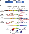
HOX gene structure and genome organization (schematic representation). (A) HOX genes are comprised of one intron separating two exons with the second exon having a 120-nucleotide sequence encoding for the 60 amino acid DNA-binding domain known as the homeobox (homeodomain). (B) The 39 human HOX genes are clustered into the four HOX families HOXA, HOXB, HOXC, and HOXD with each family consisting of nine to eleven paralogous genes (assigned by numbers based on sequence similarity and cluster position), which are responsible for the anterior-posterior specification of body segments. The position of non-coding RNAs that are interspersed within the coding HOX genes are marked (miR,microRNAs; AS, antisense RNAs). HOX gene expressions exhibit spatial and temporal collinearity: nested domains of HOX genes are generated with anteriorly HOX expressions operating earlier in development and posteriorly HOX expressions occurring later.
FIGURE 3
FIGURE 3
The HOX code of mesodermal stem cells. Stem cells derived from different tissues present patterns of HOX gene expression (“the HOX code”) that mirrors their developmental origin. According to the fact that HOX genes are not expressed before gastrulation, HOX genes were not found to be transcribed in non-differentiated, pluripotent stem cells (SC) due to active epigenetic repression of HOX genes. In cells, particularly stem cells lying at equivalent anteroposterior positions but in distinct embryonic germ layers, HOX proteins have distinct regulatory activities. (A) The reported HOX expression pattern for the mesodermal-derived adult stem cell types were listened: mesenchymal stem cell (MSC), hematopoietic stem cell (HSC) and endothelial progenitor cell (EPC). (For details: see main text.) (B) HOX expression pattern for bone marrow (BM)-, vascular wall (VW-), adipose tissue (AT)-, and fetal tissue (FT; summarizing umbilical cord, placenta and amniotic fluid MSCs)-MSCs were separately shown. Capital letters name more frequently identified HOX genes potentially representing the cell type-specific HOX code, whereas smaller letters designate individual additionally identified HOX genes above it. Same colors highlight same HOX genes that are common in different MSC types; underlined HOX genes emphasize similar HOX genes between BM-MSCs and HSCs. ESC, embryonic stem cell; iPSC, induced pluripotent stem cell; NSC neural stem cell.

References

    1. Abate-Shen C. (2002). Deregulated homeobox gene expression in cancer: Cause or consequence? Nat. Rev. Cancer 2, 777–785. 10.1038/nrc907 - DOI - PubMed
    1. Abdal Dayem A., Lee S. B., Kim K., Lim K. M., Jeon T. I., Seok J., et al. (2019). Production of mesenchymal stem cells through stem cell reprogramming. Ijms 20, 1922. 10.3390/ijms20081922 - DOI - PMC - PubMed
    1. Abdelgawad M. E., Desterke C., Uzan G., Naserian S. (2021). Single-cell transcriptomic profiling and characterization of endothelial progenitor cells: New approach for finding novel markers. Stem Cell Res. Ther. 12, 145. 10.1186/s13287-021-02185-0 - DOI - PMC - PubMed
    1. Ackema K. B., Charité J. (2008). Mesenchymal stem cells from different organs are characterized by distinct TopographicHoxCodes. Stem Cells Dev. 17, 979–992. 10.1089/scd.2007.0220 - DOI - PubMed
    1. Afzal Z., Krumlauf R. (2022). Transcriptional regulation and implications for controlling hox gene expression. Jdb 10, 4. 10.3390/jdb10010004 - DOI - PMC - PubMed