

A bioprinted complex tissue model for myotendinous junction with biochemical and biophysical cues
- PMID: 36176596
- PMCID: PMC9472009
- DOI: 10.1002/btm2.10321
A bioprinted complex tissue model for myotendinous junction with biochemical and biophysical cues
Abstract
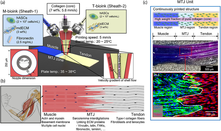
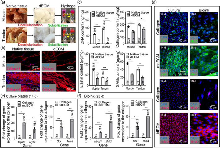
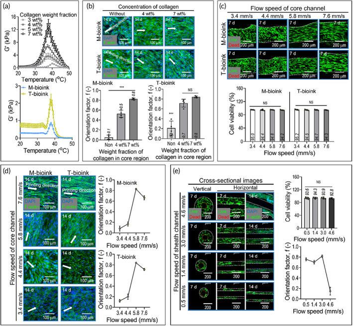
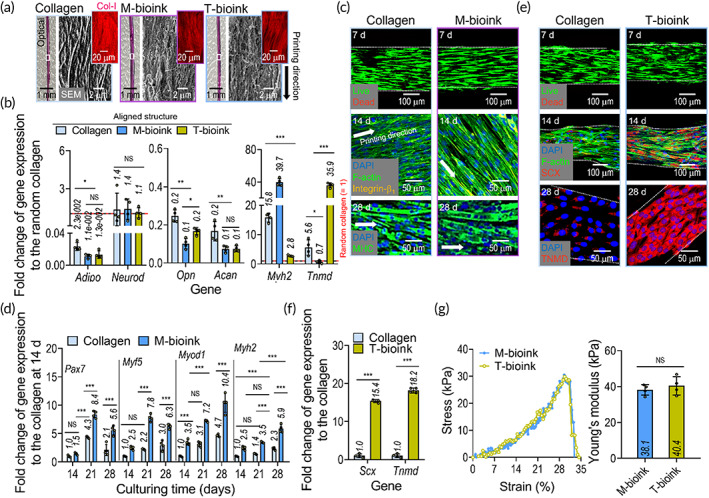
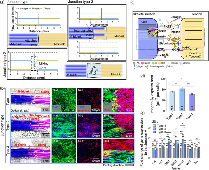
In the musculoskeletal system, the myotendinous junction (MTJ) is optimally designed from the aspect of force transmission generated from a muscle through a tendon onto the bone to induce movement. Although the MTJ is a key complex tissue in force transmission, the realistic fabrication, and formation of complex tissues can be limited. To obtain the MTJ construct, we prepared two bioinks, muscle- and tendon-derived decellularized extracellular matrix (dECM), which can induce myogenic and tenogenic differentiation of human adipose-derived stem cells (hASCs). By using a modified bioprinting process supplemented with a nozzle consisting of a single-core channel and double-sheath channels, we can achieve three different types of MTJ units, composed of muscle, tendon, and interface zones. Our results indicated that the bioprinted dECM-based constructs induced hASCs to myogenic and tenogenic differentiation. In addition, a significantly higher MTJ-associated gene expression was detected at the MTJ interface with a cell-mixing zone than in the other interface models. Based on the results, the bioprinted MTJ model can be a potential platform for understanding the interaction between muscle and tendon cells, and even the bioprinting method can be extensively applied to obtain complex tissues.
Keywords: 3D bioprinting; bioink; hASCs; myotendinous junction; tissue engineering.
© 2022 The Authors. Bioengineering & Translational Medicine published by Wiley Periodicals LLC on behalf of American Institute of Chemical Engineers.
Figures
Similar articles
-
In vitro development of a muscle-tendon junction construct using decellularised extracellular matrix: Effect of cyclic tensile loading.Biomater Adv. 2024 Jul;161:213873. doi: 10.1016/j.bioadv.2024.213873. Epub 2024 Apr 27. Biomater Adv. 2024. PMID: 38692180
-
Bioprinted hASC-laden cell constructs with mechanically stable and cell alignment cue for tenogenic differentiation.Biofabrication. 2023 Jul 25;15(4). doi: 10.1088/1758-5090/ace740. Biofabrication. 2023. PMID: 37442127
-
An engineered in vitro model of the human myotendinous junction.Acta Biomater. 2024 May;180:279-294. doi: 10.1016/j.actbio.2024.04.007. Epub 2024 Apr 10. Acta Biomater. 2024. PMID: 38604466 Free PMC article.
-
Designing Decellularized Extracellular Matrix-Based Bioinks for 3D Bioprinting.Adv Healthc Mater. 2020 Dec;9(24):e2000734. doi: 10.1002/adhm.202000734. Epub 2020 Jul 21. Adv Healthc Mater. 2020. PMID: 32691980 Review.
-
Recent Advances in Decellularized Extracellular Matrix-Based Bioinks for 3D Bioprinting in Tissue Engineering.Materials (Basel). 2023 Apr 18;16(8):3197. doi: 10.3390/ma16083197. Materials (Basel). 2023. PMID: 37110034 Free PMC article. Review.
Cited by
-
How Mechanical and Physicochemical Material Characteristics Influence Adipose-Derived Stem Cell Fate.Int J Mol Sci. 2023 Feb 10;24(4):3551. doi: 10.3390/ijms24043551. Int J Mol Sci. 2023. PMID: 36834966 Free PMC article. Review.
-
Engineering interfacial tissues: The myotendinous junction.APL Bioeng. 2024 Jun 3;8(2):021505. doi: 10.1063/5.0189221. eCollection 2024 Jun. APL Bioeng. 2024. PMID: 38841690 Free PMC article. Review.
-
Hybrid cell constructs consisting of bioprinted cell-spheroids.Bioeng Transl Med. 2022 Aug 31;8(2):e10397. doi: 10.1002/btm2.10397. eCollection 2023 Mar. Bioeng Transl Med. 2022. PMID: 36925682 Free PMC article.
-
Automated Microfluidics-Assisted Hydrogel-Based Wet-Spinning for the Biofabrication of Biomimetic Engineered Myotendinous Junction.Adv Healthc Mater. 2024 Dec;13(32):e2402075. doi: 10.1002/adhm.202402075. Epub 2024 Sep 23. Adv Healthc Mater. 2024. PMID: 39313990 Free PMC article.
-
Advances focusing on the application of decellularization methods in tendon-bone healing.J Adv Res. 2025 Jan;67:361-372. doi: 10.1016/j.jare.2024.01.020. Epub 2024 Jan 17. J Adv Res. 2025. PMID: 38237768 Free PMC article. Review.
References
-
- Mahjoub M, Berenbaum F, Houard X. Why subchondral bone in osteoarthritis? The importance of the cartilage bone interface in osteoarthritis. Osteoporos Int. 2012;23:841‐846. - PubMed
-
- Baldino L, Cardea S, Maffulli N, Reverchon E. Regeneration techniques for bone‐to‐tendon and muscle‐to‐tendon interfaces reconstruction. Br Med Bull. 2016;117:25‐37. - PubMed
-
- Tellado SF, Balmayor ER, Van Griensven M. Strategies to engineer tendon/ligament‐to‐bone interface: biomaterials, cells and growth factors. Adv Drug Deliv Rev. 2015;94:126‐140. - PubMed
-
- Bayrak E, Yilgor HP. Engineering musculoskeletal tissue interfaces. Front Mater. 2018;5:24.
LinkOut - more resources
Full Text Sources
