

Bile Leak Due to Luschka Duct Injury After Laparoscopic Cholecystectomy: A Case Report
- PMID: 36176857
- PMCID: PMC9512329
- DOI: 10.7759/cureus.28427
Bile Leak Due to Luschka Duct Injury After Laparoscopic Cholecystectomy: A Case Report
Abstract
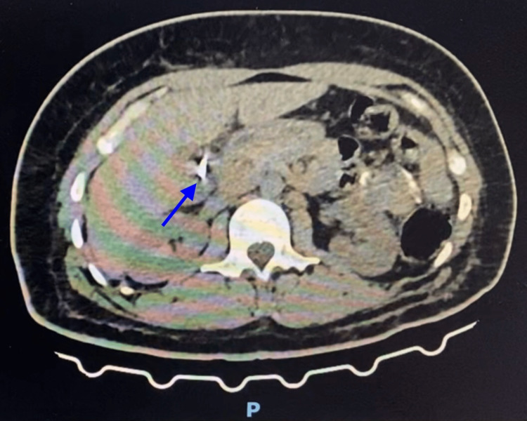
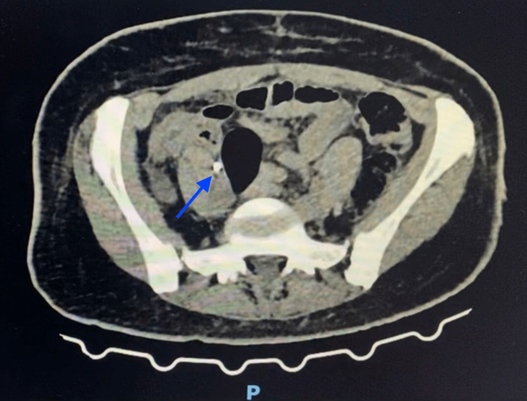
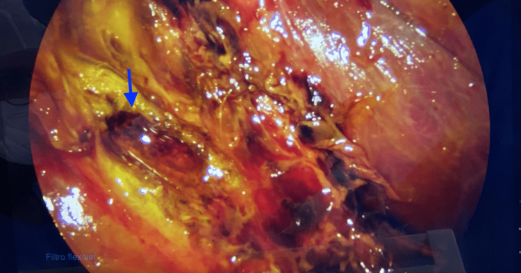
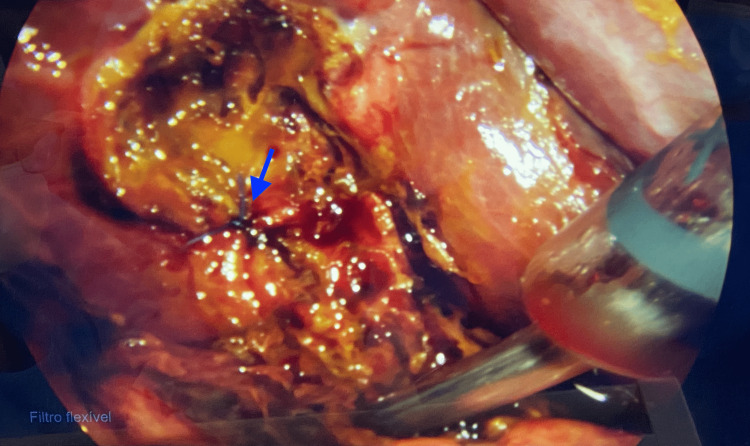
Bile leak is a common complication after laparoscopic cholecystectomy. Anatomical variations in the biliary tree can go unnoticed by the surgical team and cause complications such as this. This case report presents a patient admitted to the emergency department a week after a laparoscopic cholecystectomy due to abdominal pain and nausea. After a computed tomography, the patient was brought to the operating room for an exploratory laparoscopy, where an injured Luschka duct was found. The biliary tree has many variations that the surgeon should be aware of to minimize the risk of complications of this nature after laparoscopic cholecystectomy. There are imagining techniques with various grades of effectiveness, but in the end, the surgeon's expertise and experience are the main factors in avoiding these complications.
Keywords: bile leak; biliary tree; complications; laparoscopic cholecystectomy; luschka duct; luschka duct injury.
Copyright © 2022, Garcia et al.
Conflict of interest statement
The authors have declared that no competing interests exist.
Figures
References
-
- Acute cholecystitis: a review. Gallaher JR, Charles A. JAMA. 2022;327:965–975. - PubMed
-
- Biliary leak after laparoscopic cholecystectomy: incidence and management. Hassan AM, Mohammed AQ. https://journals.lww.com/ejos/Fulltext/2021/40020/Biliary_leak_after_lap... Egypt J Surg. 2021;40:685–694.
-
- Luschka ducts of the gallbladder in adults: case series report and review of the medical literature. Handra-Luca A, Ben Romdhane HM, Hong SM. Int J Surg Pathol. 2020;28:482–489. - PubMed
-
- Anatomy variations of biliary tree. Surgical implicance. Tolino MJ, Tartaglione AS, Sturletti CD, García MI. Int J Morphol. 2010;28:1235–1240.
Publication types
LinkOut - more resources
Full Text Sources