Review
. 2022 Sep;12(9):3548-3566.
doi: 10.1016/j.apsb.2022.03.019. Epub 2022 Mar 31.

Molecular glues modulate protein functions by inducing protein aggregation: A promising therapeutic strategy of small molecules for disease treatment

Affiliations
Review

Molecular glues modulate protein functions by inducing protein aggregation: A promising therapeutic strategy of small molecules for disease treatment

Hongyu Wu et al. Acta Pharm Sin B. 2022 Sep.

Abstract

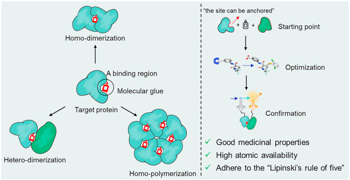
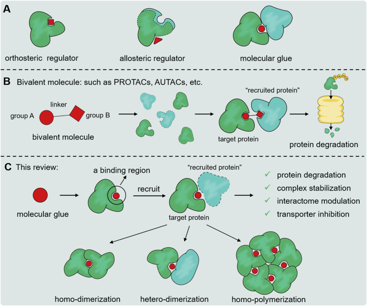
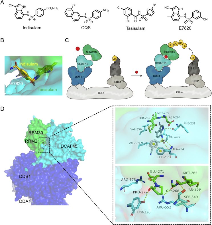
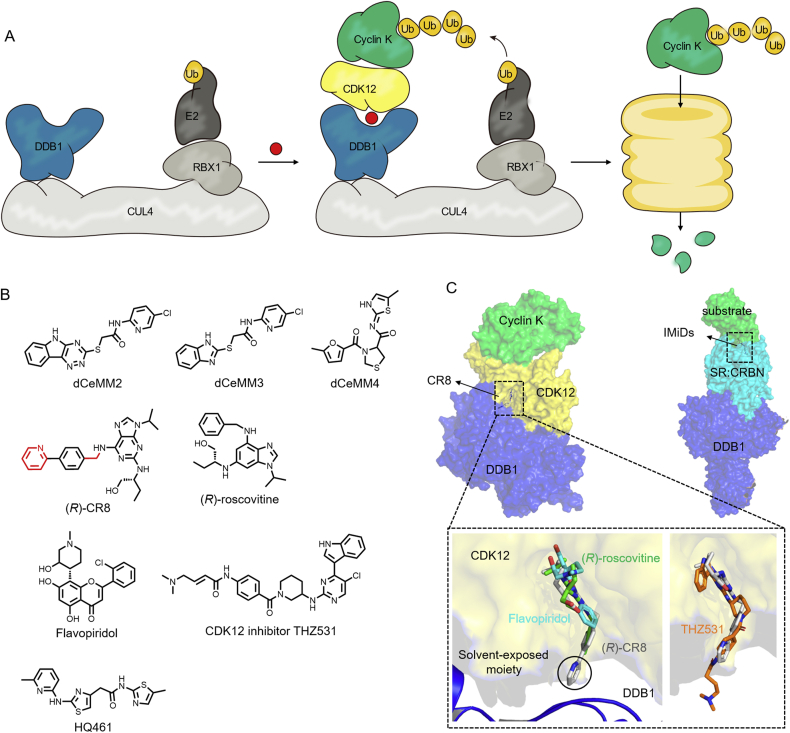
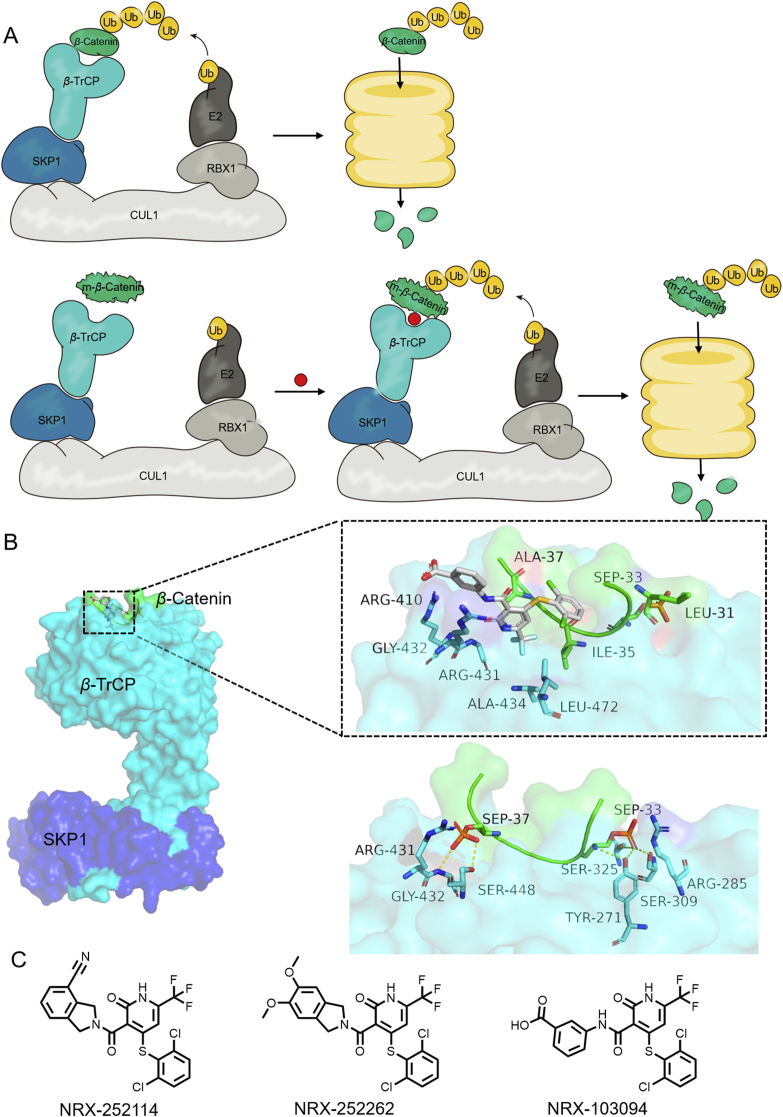
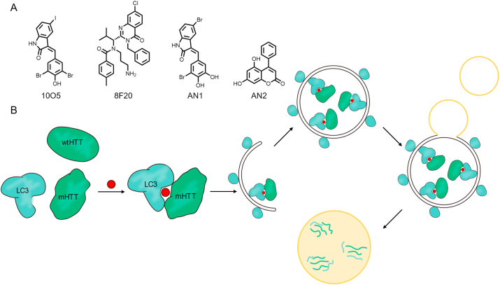
Molecular glues can specifically induce aggregation between two or more proteins to modulate biological functions. In recent years, molecular glues have been widely used as protein degraders. In addition, however, molecular glues play a variety of vital roles, such as complex stabilization, interactome modulation and transporter inhibition, enabling challenging therapeutic targets to be druggable and offering an exciting novel approach for drug discovery. Since most molecular glues are identified serendipitously, exploration of their systematic discovery and rational design are important. In this review, representative examples of molecular glues with various physiological functions are divided into those mediating homo-dimerization, homo-polymerization and hetero-dimerization according to their aggregation modes, and we attempt to elucidate their mechanisms of action. In particular, we aim to highlight some biochemical techniques typically exploited within these representative studies and classify them in terms of three stages of molecular glue development: starting point, optimization and identification.

Keywords: Dimerization; Molecular glue; Polymerization; Protein–protein interaction; Small molecule.


Figures

Image 1
Graphical abstract
Figure 1
Figure 1
(A) Difference of orthosteric regulator, allosteric regulator and molecular glue. (B) Schematic diagram of divalent molecular interaction mode. (C) Schematic diagram of molecular interaction mode and classification of protein polymerization patterns induced by molecular glues.
Figure 2
Figure 2
(A) The molecular glue BMS-202 mediates the homo-dimerization of PD-L1 (monomer a: green, monomer b: cyan, PDB:5J89). (B) Compounds 15 were the best compounds reported by Muszak, Liu, Ouyang, Wang and Song, respectively.
Figure 3
Figure 3
(A) REV1 CTD–REV7 complex (REV7: gray, REV1: green, PDB:4FJO). (B) The molecular glue JH-RE-06 mediates homo-dimerization of cREV1 CTD (monomer a: green, monomer b: cyan, PDB:6C8C).
Figure 4
Figure 4
(A) Structures of the reported molecular glues mediating MDMX dimerization. (B) The molecular glue RO-2443 mediates the homo-dimerization of MDMX (monomer a: green, monomer b: cyan, PDB:3U15).
Figure 5
Figure 5
(A) Structures of molecular glues BI-3802, CCT369260 and their corresponding inhibitors. (B) BI-3802 mediates the homo-polymerization of BCL6 (monomer a: green, monomer b: cyan, monomer c: palegreen, PDB: 6XMX).
Figure 6
Figure 6
(A) The conservative hydrophobic interactions of CoV N-NTD dimers are shown on the upper left and hydrogen bonds on the lower left (monomer a: green, monomer b: cyan, PDB:4UD1). P3 mediates the homo-polymerization of MERS-CoV N-NTD (right, PDB:6KL6). (B) Diagram of molecular glue P3-mediated polymerization of viral proteins.
Figure 7
Figure 7
(A) Structures of CRBN-binding drugs. (B) Diagram of molecular glues mediated hetero-dimerization of DDB1CRBN and substrate. (C) CC-885 mediates the hetero-dimerization of CRBN‒GSPT1 (DDB1 colored in blue, CRBN colored in cyan, GSPT1 colored in green; PDB: 5HXB). Hydrogen bonds are shown as yellow dotted lines.
Figure 8
Figure 8
(A) Chemical structures of aryl-sulfonamides molecular glues. (B) A combination of E7820 and indisulam on the surface of DCAF15 and RBM39 (indisulam PDB:6Q0W). (C) Schematic diagram of the action mechanism of aryl sulfonamides molecular glues. (D) DDB1-DCAF15-E7820-RBM39 co-crystal structure (DDB1 colored in blue, DCAF15 colored in cyan, RBM39 colored in green, DDA1 colored in lightblue; PDB:6Q0R). Hydrogen bonds are shown as yellow dotted lines.
Figure 9
Figure 9
(A) Schematic diagram of the action mechanism of molecular glue CR8. (B) Structures of three types of DDB1 and CDK12/cyclinK molecular glues. (C) Comparison of protein complex structures of CR8 (PDB:6TD3) and IMiDs molecular glues. A partial enlargement of the CR8 solvent exposure area compared with (R)-roscovitine (PDB:2A4L), flavopiridol (PDB:3BLR) and THZ531 (PDB:5ACB).
Figure 10
Figure 10
(A) Schematic diagram of the action mode of molecular glue NRX-103094. (B) NRX-103094 mediate the hetero-dimerization of β-catenin‒β-TrCP (up, PDB:6M91), and comparison with wild-type (down, PDB:1P22). (C) Structures of NRX series as molecular glues.
Figure 11
Figure 11
(A) Structures of ATTEC. (B) Schematic diagram of the action mode of ATTEC.
Figure 12
Figure 12
(A) Structure of LSN3160440. (B) GLP-1(9–36)–LSN3140660–GLP-1R complex (right, GLP-1R: cyan, GLP-1: green; PDB:6VCB), and comparison with GLP-1(7–36)–GLP-1R complex (left, PDB:5VAI).
Figure 13
Figure 13
(A) Crystallographic overlay of 14-3-3(C42)-ERα (PDB:6HMT); 14-3-3-ERRγ(C180) (PDB:6Y3W) and the structures of fragments they screened out. (B) Crystallographic overlay of 14-3-3/P65 with TCF521 (PDB:6YOW) and 14-3-3/P65 with TCF521-123 (PDB:6YPY).
Figure 14
Figure 14
Structural optimization strategy of NRX-103094. β-TrCP‒NRX-1933‒β-catenin complex (PDB:6M93), other proteins PDB have been mentioned above.
Figure 15
Figure 15
Schematic illustration of TR-FRET assay and the structures of the resulting molecular glues Ro-31-8220 and Go-6983.

Similar articles

Cited by

References

    1. Roberts N.A., Martin J.A., Kinchington D., Broadhurst A.V., Craig J.C., Duncan I.B., et al. Rational design of peptide-based HIV proteinase inhibitors. Science. 1990;248:358–361. - PubMed
    1. Erickson J., Neidhart D.J., VanDrie J., Kempf D.J., Wang X.C., Norbeck D.W., et al. Design, activity, and 2.8 Å crystal structure of a C2 symmetric inhibitor complexed to HIV-1 protease. Science. 1990;249:527–533. - PubMed
    1. Dorsey B.D., Levin R.B., McDaniel S.L., Vacca J.P., Guare J.P., Darke P.L., et al. L-735,524: the design of a potent and orally bioavailable HIV protease inhibitor. J Med Chem. 1994;37:3443–3451. - PubMed
    1. Anderson A.C. The process of structure-based drug design. Chem Biol. 2003;10:787–797. - PubMed
    1. Wenthur C.J., Gentry P.R., Mathews T.P., Lindsley C.W. Drugs for allosteric sites on receptors. Annu Rev Pharmacol Toxicol. 2014;54:165–184. - PMC - PubMed

LinkOut - more resources