

Fiber-based hand-held RCM-OCT probe for noninvasive assessment of skin lesions and therapy guidance
- PMID: 36176918
- PMCID: PMC9514145
- DOI: 10.1002/tbio.202200002
Fiber-based hand-held RCM-OCT probe for noninvasive assessment of skin lesions and therapy guidance
Abstract
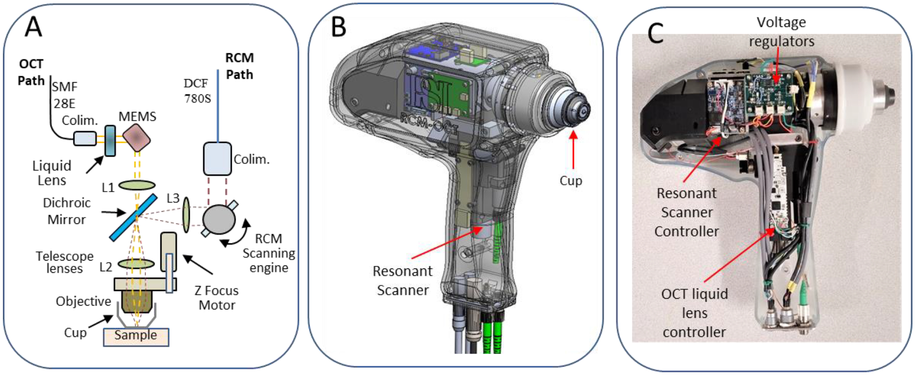
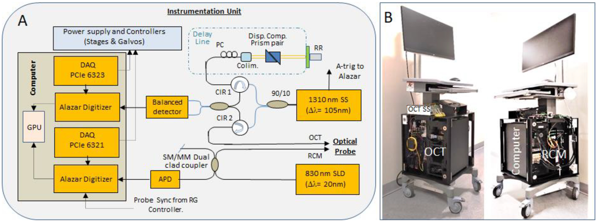
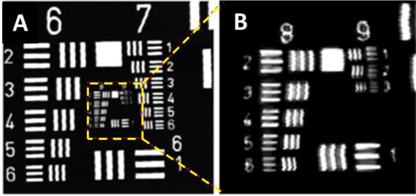
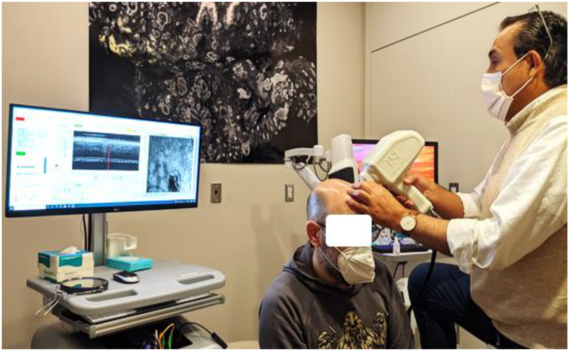
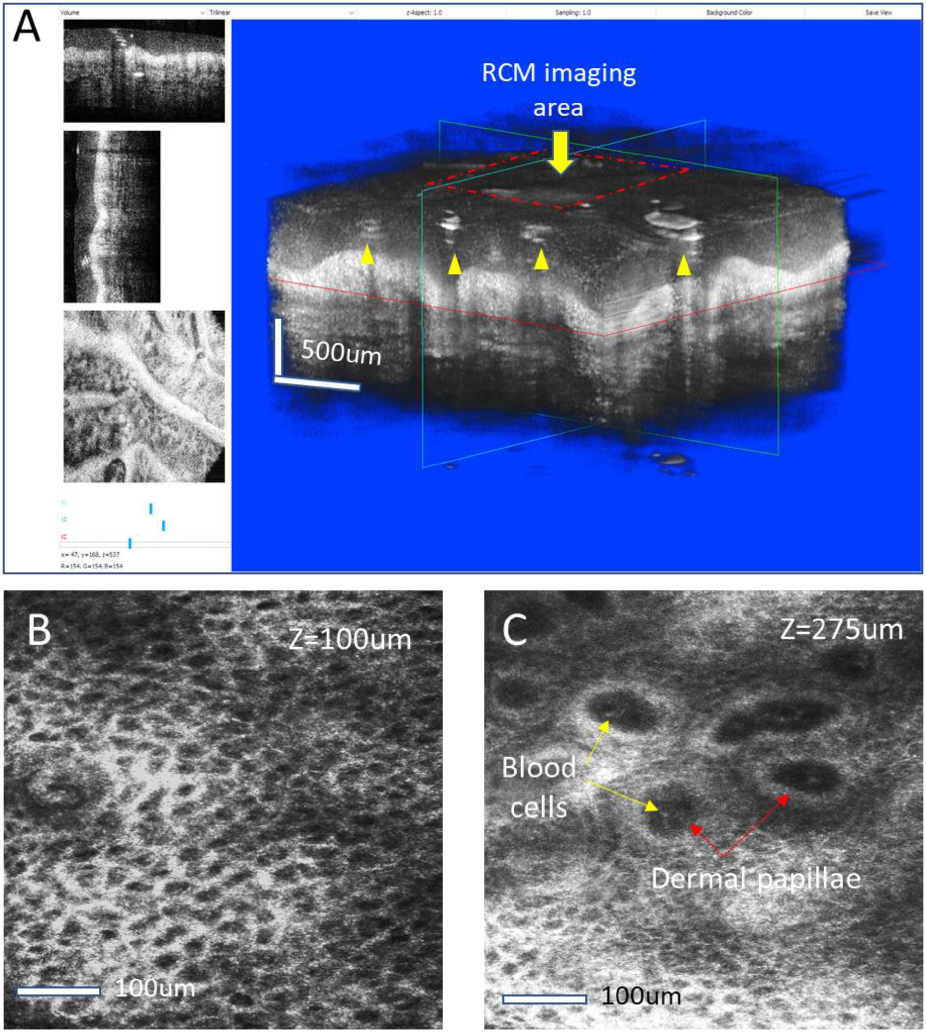
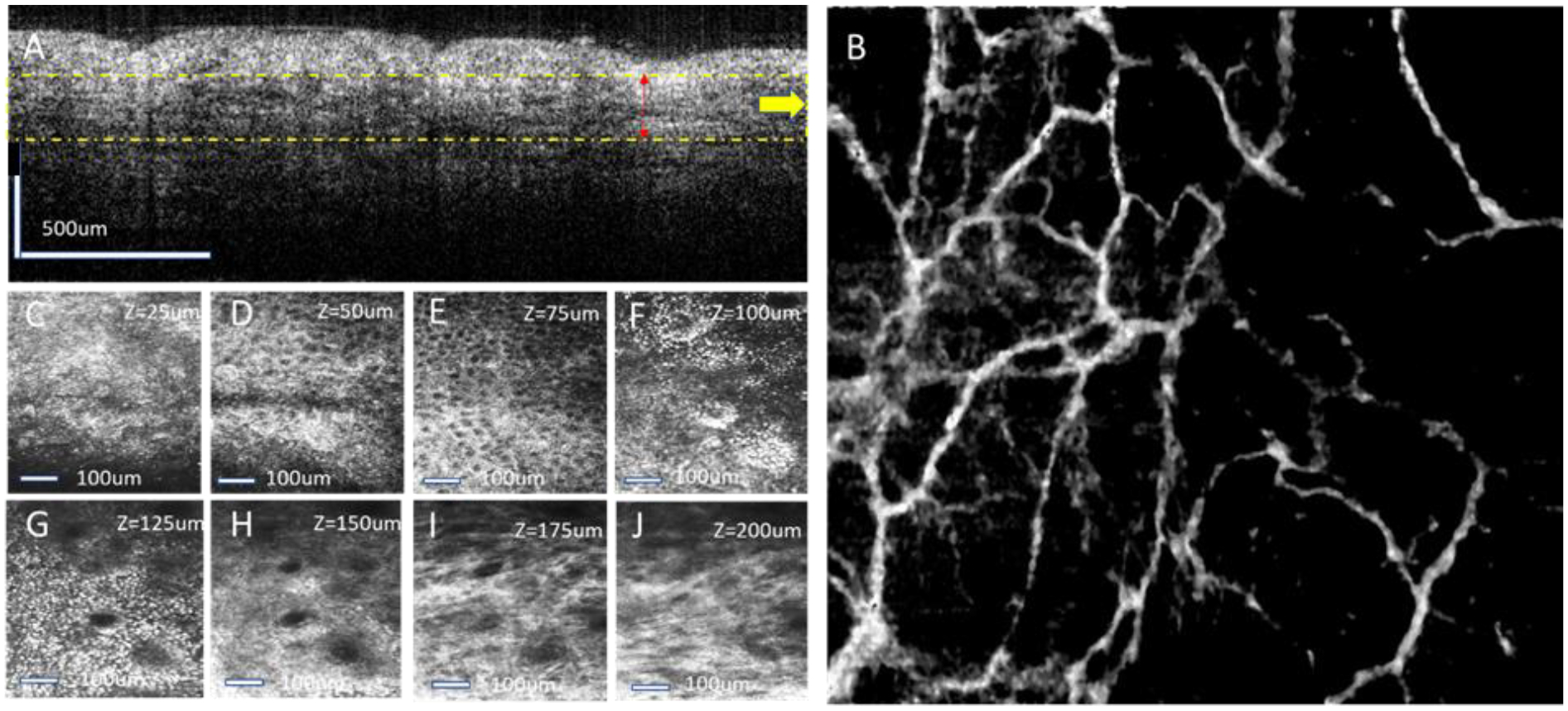
Noninvasive assessment of skin lesions, especially of basal cell carcinoma (BCC), has benefited more recently from the use of optical imaging techniques such as optical coherence tomography (OCT) and reflectance confocal microscopy (RCM). While RCM provides submicron scale resolution and thus enables identification of skin morphological changes of the skin, with the downside of limited penetration depth, OCT imaging of the same lesion brings the benefit of better resolving its depth of invasion. OCT and RCM can be used either individually or combined within the same instrument for the noninvasive diagnosis of nonmelanoma skin cancers (NMSCs). Their combined use has shown to provide certain benefits such as better characterization of the lesion's margins, both in depth and laterally, as well as improved sensitivity and specificity, as previously demonstrated by our team. In this paper we report a new "fiber-based" implementation of the second-generation RCM-OCT hand-held probe. The fiber-based implementation of both imaging modalities enabled the construction of a smaller footprint/lower weight hand-held probe. Its preliminary evaluation on the skin of healthy volunteers is reported here, demonstrating improved capabilities for resolving sub-cellular structures and image skin morphology with micron-scale resolution to a higher depth than in the previous implementation, while also enabling the construction of angiography maps showing vascular remodeling.
Keywords: noninvasive evaluation; optical coherence tomography; reflectance confocal microscopy; skin lesions.
Conflict of interest statement
CONFLICT OF INTEREST PSI has a patent on RCM-OCT: US 9,655,521
Figures
Similar articles
-
Evaluation of a Combined Reflectance Confocal Microscopy-Optical Coherence Tomography Device for Detection and Depth Assessment of Basal Cell Carcinoma.JAMA Dermatol. 2018 Oct 1;154(10):1175-1183. doi: 10.1001/jamadermatol.2018.2446. JAMA Dermatol. 2018. PMID: 30140851 Free PMC article.
-
Handheld optical coherence tomography-reflectance confocal microscopy probe for detection of basal cell carcinoma and delineation of margins.J Biomed Opt. 2017 Jul 1;22(7):76006. doi: 10.1117/1.JBO.22.7.076006. J Biomed Opt. 2017. PMID: 28697233 Free PMC article.
-
Combining Reflectance Confocal Microscopy, Optical Coherence Tomography and Ex-Vivo Fluorescence Confocal Microscopy for Margin Assessment in Basal Cell Carcinoma Excision.Dermatol Pract Concept. 2024 Apr 1;14(2):e2024090. doi: 10.5826/dpc.1402a90. Dermatol Pract Concept. 2024. PMID: 38810079 Free PMC article.
-
Role of In Vivo Reflectance Confocal Microscopy in the Analysis of Melanocytic Lesions.Acta Dermatovenerol Croat. 2018 Apr;26(1):64-67. Acta Dermatovenerol Croat. 2018. PMID: 29782304 Review.
-
A Review of Noninvasive Techniques for Skin Cancer Detection in Dermatology.Am J Clin Dermatol. 2020 Aug;21(4):513-524. doi: 10.1007/s40257-020-00517-z. Am J Clin Dermatol. 2020. PMID: 32383142 Review.
Cited by
-
Recent Advances in Melanoma Diagnosis and Prognosis Using Machine Learning Methods.Curr Oncol Rep. 2023 Jun;25(6):635-645. doi: 10.1007/s11912-023-01407-3. Epub 2023 Mar 31. Curr Oncol Rep. 2023. PMID: 37000340 Free PMC article. Review.
-
Line-field confocal optical coherence tomography based on tandem interferometry with a focus-tunable lens.Biomed Opt Express. 2024 Aug 19;15(9):5384-5399. doi: 10.1364/BOE.530717. eCollection 2024 Sep 1. Biomed Opt Express. 2024. PMID: 39296418 Free PMC article.
References
-
- Drexler W and Fujimoto JG, “Optical Coherence Tomography -Technology and Applications. 2 ed,” Switzerland: Springer International Publishing; 2015.
-
- Guitera P, Pellacani G, Longo C, Seidenari S, Avramidis M, Menzies SW. J. Invest. Dermatol 2009, 129,131–138. - PubMed
-
- Guitera P, Menzies SW, Longo C, et al. J Invest Dermatol. 2012, 132(10), 2386–2394. - PubMed
-
- Hinz T, K Ehler L, Hornung T, Voth H, Fortmeier I, Maier T, Höller T, Schmid-Wendtner MH Acta Derm Venereol. 2012; 92, 132–137. - PubMed
Grants and funding
LinkOut - more resources
Full Text Sources