. 2022 Sep 13:15:947490.
doi: 10.3389/fnmol.2022.947490. eCollection 2022.

Selective transduction of cerebellar Purkinje and granule neurons using delivery of AAV-PHP.eB and AAVrh10 vectors at axonal terminal locations

Affiliations

Selective transduction of cerebellar Purkinje and granule neurons using delivery of AAV-PHP.eB and AAVrh10 vectors at axonal terminal locations

Magdalena Surdyka et al. Front Mol Neurosci. .

Abstract

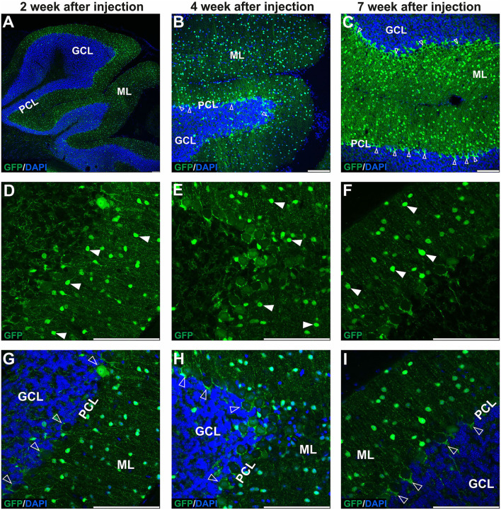
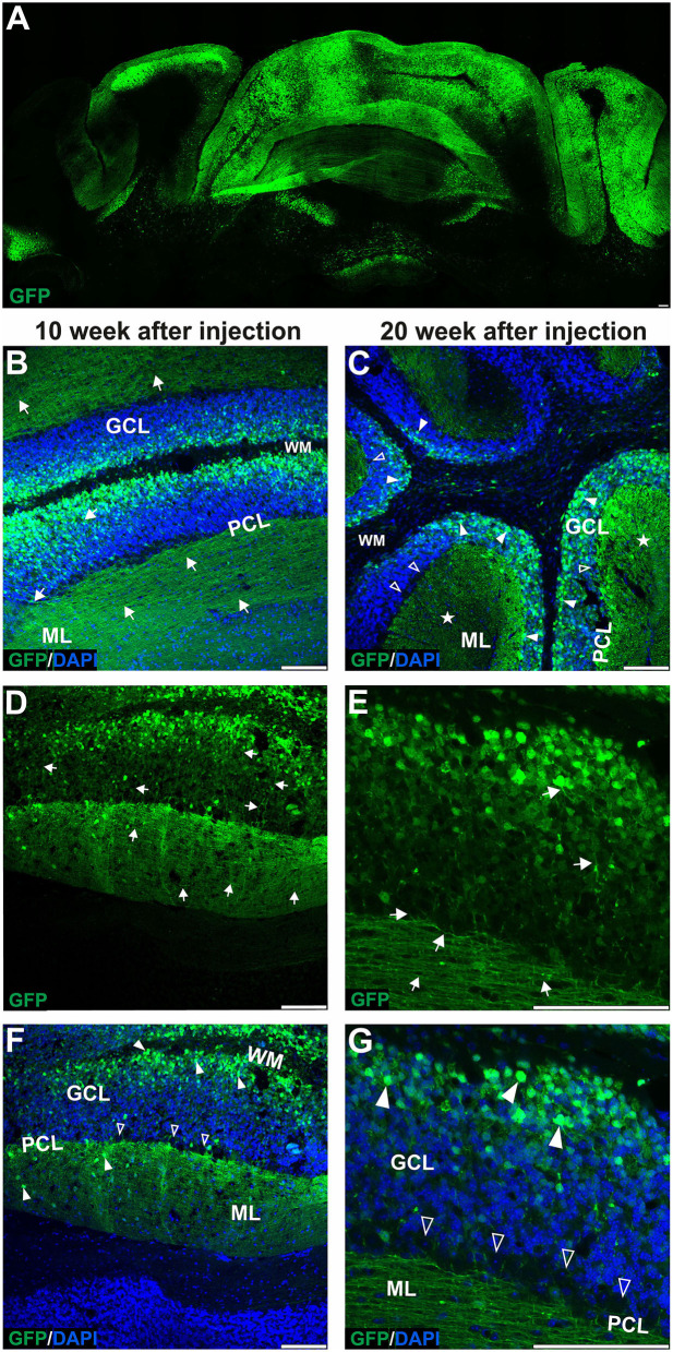
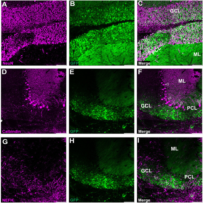
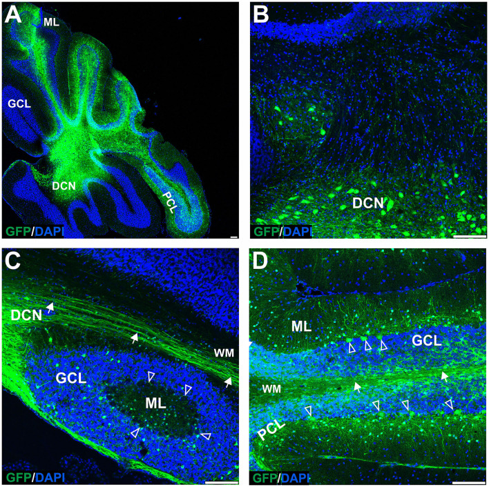
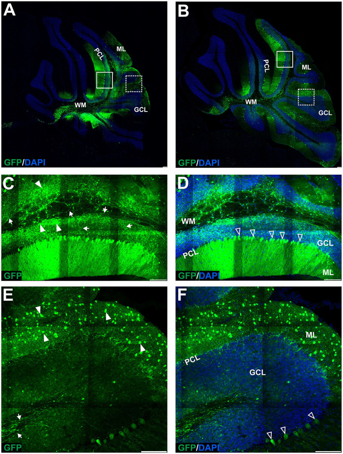
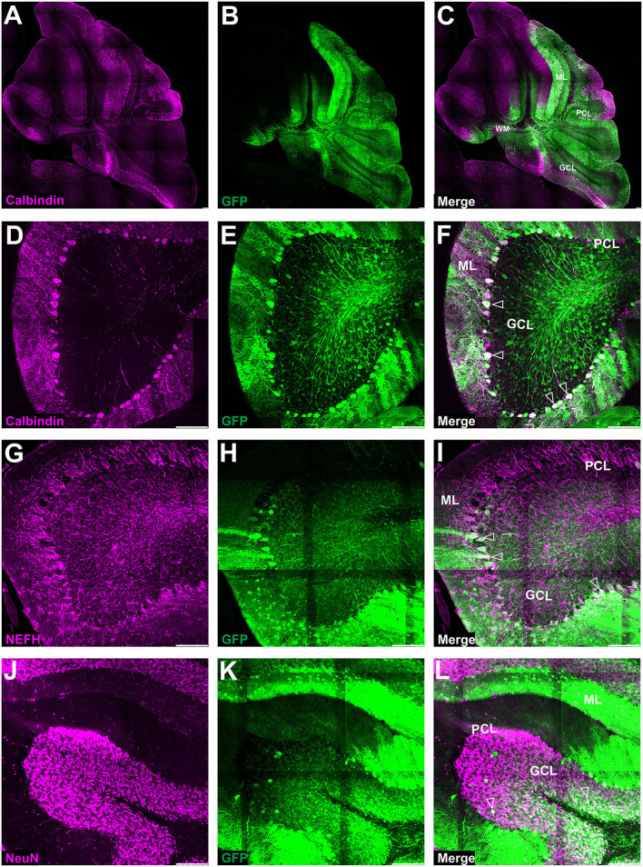
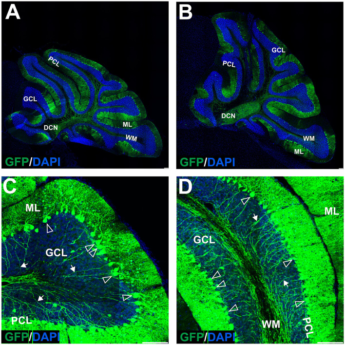
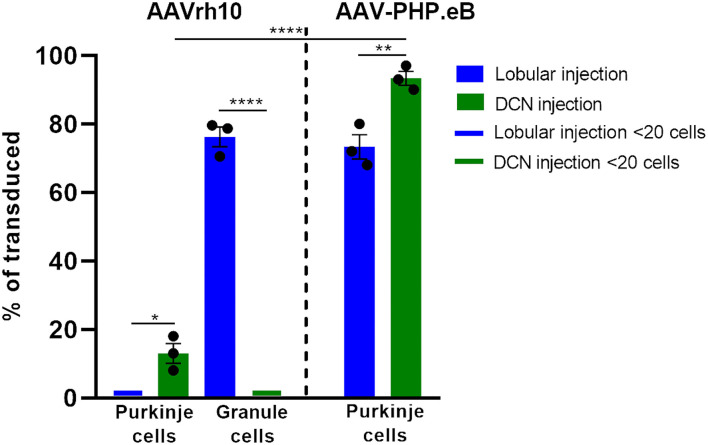
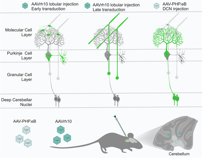
Adeno-associated virus (AAV)-based brain gene therapies require precision without off-targeting of unaffected neurons to avoid side effects. The cerebellum and its cell populations, including granule and Purkinje cells, are vulnerable to neurodegeneration; hence, conditions to deliver the therapy to specific cell populations selectively remain challenging. We have investigated a system consisting of the AAV serotypes, targeted injections, and transduction modes (direct or retrograde) for targeted delivery of AAV to cerebellar cell populations. We selected the AAV-PHP.eB and AAVrh10 serotypes valued for their retrograde features, and we thoroughly examined their cerebellar transduction pattern when injected into lobules and deep cerebellar nuclei. We found that AAVrh10 is suitable for the transduction of neurons in the mode highly dependent on placing the virus at axonal terminals. The strategy secures selective transduction for granule cells. The AAV-PHP.eB can transduce Purkinje cells and is very selective for the cell type when injected into the DCN at axonal PC terminals. Therefore, both serotypes can be used in a retrograde mode for selective transduction of major neuronal types in the cerebellum. Moreover, our in vivo transduction strategies are suitable for pre-clinical protocol development for gene delivery to granule cells by AAVrh10 and Purkinje cells by AAV-PHPeB.

Keywords: AAV-PHP.eB; AAVrh10; Purkinje cells; adeno-associated virus (AAV); cerebellum; deep cerebellar nuclei; granule cells; retrograde.


Conflict of interest statement

The authors declare that the research was conducted in the absence of any commercial or financial relationships that could be construed as a potential conflict of interest.

Figures

Figure 1
Figure 1
The early transduction phase of the mouse cerebellum after injection of the eGFP-encoding AAVrh10 vector into cerebellar lobules. The transduction pattern was detected by imaging eGFP+ cells at 2 (A,D,G), 4 (B,E,H), and 7 weeks post-injection (C,F,I). The eGFP+ cells were observed in the molecular layer (arrowhead, D–F), which demonstrated small cell bodies and characteristic locations reminiscent of stellate cells. Importantly, no eGFP expression was observed in Purkinje cells. In addition, a strong eGFP+ signal was observed in characteristic structures called pinceau and wrapped around eGFP-negative Purkinje cell bodies (unfilled arrowhead, B,C,G–I). Such characteristic structures are long known exclusively as parts of the processes of basket cells. In summary, the transduction phase demonstrated transduction of the molecular cell layer and lack of transduction in Purkinje cell and granule cell layers. (A–C,G–I) eGFP green staining was merged with DAPI blue nuclear staining. Arrowheads: eGFP+ cells in ML. Unfilled arrowheads: pericellular baskets and pinceau; GCL, granule cell layer; ML, molecular layer; PCL, Purkinje cell layer. Coronal sections. Bars: 100 μm in the lower right corner of images. Purkinje cell and granule cell transduction: <1%; N = 3 animals, see Figure 8.
Figure 2
Figure 2
The late transduction phase of the mouse cerebellum after injection of the eGFP-encoding AAVrh10 vector into cerebellar lobules. The imaging of eGFP+ cells was performed after 10 (B,D,F) and 20 weeks after injection (A,C,E,G) and demonstrated densely packed round transduced cell bodies in the granule cell layer (arrowheads, B,C,F,G). Both 10 and 20 weeks after injection, there were characteristic eGFP-positive axons of granule cells running in the granule cell layer (arrows, D-E) and further eGFP-positive parallel fibers in ML (arrow, B,D,E, asterisks C). Similar to early transfection, the eGFP signal was not observed in the Purkinje cells (unfilled arrowheads indicate unstained PC bodies, C,F,G); however, few cells in the ML were still showing some eGFP signal. Two panels (E,G) show bodies and axons of granule cells under higher magnification. In summary, the late transduction phase using the AAVrh10 serotype demonstrated that eGFP+ signal shift results in almost exclusive transduction of granule cells and labeling of their proximal axons and distant parallel fibers. Unfilled arrowheads: Purkinje cell bodies. Arrows: eGFP expression in parallel fibers or axons of granule cells. Asterisks: parallel fibers on cross-section. GCL, granule cell layer; ML, molecular layer; PCL, Purkinje cell layer; WM, white matter. (A,B,D–G): Coronal sections, (C): Sagittal sections. Bars: 100 μm in the lower right corner of images. Granule cell transduction efficiency after lobular injections of AAVrh10 (late stage): 76.3% (N = 3 animals; see Figure 8 for histogram).
Figure 3
Figure 3
Fluorescent coimmunostaining of mouse cerebellar cryosections between 10 and 20 weeks after lobular AAVrh10 injections. eGFP green signal (B,E,H) was imaged together with NeuN (magenta) or calbindin 1 (magenta) or NEFH neurofilament (magenta). The granule cell marker, NeuN (A), co-localized with green cells (B), showing an overlayed white signal (C). Immunostaining for Purkinje cell marker calbindin 1 (D) did not reveal any co-localization with eGFP signal (F). The NEFH marker, which is known for staining Purkinje and basket cell processes in the cerebellum (Wiatr et al., 2021) did not co-localize with eGFP (G). Unfilled arrowheads: co-localized in granule cells (C); or Purkinje cell bodies (F); or neurofilaments (I). GCL, granule cell layer; ML, molecular layer; PCL, Purkinje cell layer. Coronal section. Bars: 100 μm in the lower right corner of images. A slight misalignment of the composite images in (A–C) panels is due to the optical properties of Opera Phenix. Images acquired from N = 3 animals.
Figure 4
Figure 4
The transduction of the mouse cerebellum after injection of the eGFP-encoding AAVrh10 vector into deep cerebellar nuclei. Green eGFP+ signal was observed in deep cerebellar nuclei (A,B) and white matter (A–D), in DCN neurons (B), Purkinje cells, and cells of the molecular and granule cell layers (D). Despite the selection of relatively prolonged time of observation (10 weeks post-injection) and the occurrence of intensive staining of the white matter fibers (likely Purkinje cell axons), the eGFP+ Purkinje cell bodies and dendrites did not occur very frequently. Unfilled arrowheads: Purkinje cell bodies. Arrows: eGFP expression in PC axons. DCN, deep cerebellar nuclei; GCL, granule cell layer; ML, molecular layer; PCL, Purkinje cell layer; WM, white matter. Sagittal sections. Bars: 100 μm in the lower right corner of images. Purkinje cell transduction efficiency at DCN injection with AAVrh10: 13% (N = 3 animals, see Figure 8).
Figure 5
Figure 5
The transduction of the mouse cerebellum after injection of the eGFP-encoding AAV-PHP.eB vector into the cerebellar lobules. Green signal in eGFP+ cells on the sagittal sections was evident at 4 (A,C–F) and 7 (B) weeks after injection. Two transduction patterns were observed: pattern 1 indicated by the square (A,B), and pattern 2 by dashed line square (A,B) and presented, respectively, in (C–F) (4 weeks). Both patterns demonstrated various types and counts of transduced cells in ML, PCL, and GCL. Unfilled arrowheads: eGFP-positive Purkinje cells. Arrowheads: eGFP-positive cells; Arrows: eGFP+ axons of PCs. GCL, granule cell layer; ML, molecular layer; PCL, Purkinje cell layer; WM, white matter. Sagittal sections. Bars: 100μ m in the lower right corner of images. Images acquired from N = 3 animals.
Figure 6
Figure 6
Fluorescent coimmunostaining of mouse cerebellar cryosections after lobular injection of eGFP-encoding AAV-PHP.eB. Green eGFP signal (B,E,H,K) was imaged together with calbindin 1 [magenta, (C,F)] or NEFH [magenta, (I)], or NeuN [magenta, (L)]. The co-localization signal (white) is present for all types of cells in the cerebellum, indicating the transduction of Purkinje cells [calbindin 1; (A,D)], basket cells [NEFH visible in Purkinje pericellular baskets; (G)] and granule cells [NeuN in GCL; (J)]. Unfilled arrowheads: Purkinje cell bodies (C) or pericellular baskets (F) or granule cells (I). GCL, granule cell layer; ML, molecular layer; PCL, Purkinje cell layer. Micrographs containing only the eGFP channel were separately demonstrated for distribution evaluation. Sagittal section. Bars: 100 μm in the lower right corner of images. Images acquired from N = 3 animals.
Figure 7
Figure 7
The transduction of the mouse cerebellum after injection of the eGFP-encoding AAV-PHP.eB into the deep cerebellar nuclei. A robust green signal has been observed throughout the cerebellar ML and was localized almost exclusively in Purkinje cells on the sagittal sections at 4 (A,C,D) and 7 (B) weeks after injection. Higher magnification of the transduction pattern was shown in (C,D), 4 weeks after injection. eGFP signal was observed in all compartments of the PC, including bodies and dendritic trees (unfilled arrowheads), and axons (arrows). DCN, deep cerebellar nuclei; GCL, granule cell layer; ML, molecular layer; PCL, Purkinje cell layer; WM, white matter. Sagittal section. Bars: 100 μm in the lower right corner of images. Purkinje cell transduction efficiency at DCN injection: 93% (N = 3 animals, see Figure 8).
Figure 8
Figure 8
Histogram summarizing the transduction efficiency of Purkinje cells and granule cells after lobular and DCN injection of AAVrh10-eGFP and AAV-PHP.eB-eGFP. Purkinje cells transduction efficiency after AAVrh10 lobular vs. DCN injections was close to 1% vs. 13%, respectively (N = 3; difference between means 12% 1 2.887 SEM; p < 0.0142, *). Granule cell transduction efficiency after AAVrh10 lobular vs. DCN injections was 76% (late stage) vs. close to 1%, respectively (N = 3; difference between means 75% ± 2.895 SEM; t-test p < 0.0001, ****). Purkinje cells transduction efficiency after AAV-PHP.eB lobular vs. DCN injections was 73% (Pattern 1; note Figure 5) vs. 93%, respectively, AAV-PHP.eB (N = 3; difference between means 20% ± 4.069 SEM; t-test p < 0.008, **). Pattern 2 demonstrated hardly any transduction of Purkinje cells (see Figure 5). In addition, Purkinje cells transduction efficiency after injections in DCN with AAVrh10 vs. AAV-PHP.eB vector showed 13% vs. 93% (N = 3; difference between means 80% ± 3.528 SEM; t-test p < 0.0001, ****).
Figure 9
Figure 9
The cerebellum has several layers and structures, with many cell types, including Purkinje cells, granule neurons, stellate neurons, and basket neurons. All cell types are tightly interconnected by their axonal and dendritic processes, forming a perfect framework for retrograde delivery of AAV-based therapies to cell types of interest. We demonstrated that the AAVrh10 and AAV-PHP.eB viral particles placed in the vicinity of the axonal terminals by targeted brain injections into lobules, and deep cerebellar nuclei (DCN) can selectively transduce neuronal types of cerebellar networks, such as Purkinje cells and granule cells. AAVrh10 injected into lobules produces early transduction in stellate and basket cells. Subsequently, AAVrh10 retrogradely transduces granule cells. AAV-PHP.eB injected into DCN at the axonal terminals of Purkinje cells produces their selective transduction. Therefore, both serotypes can be used in a retrograde mode for selective transduction of major neuronal types in the cerebellum for effective and selective gene delivery. DCN, deep cerebellar nuclei.

Similar articles

Cited by

References

    1. Albright B. H., Storey C. M., Murlidharan G., Castellanos Rivera R. M., Berry G. E., Madigan V. J., et al. . (2018). Mapping the structural determinants required for AAVrh.10 transport across the blood-brain barrier. Mol. Ther. 26, 510–523. 10.1016/j.ymthe.2017.10.017 - DOI - PMC - PubMed
    1. Bauer S., Maier S. K. G., Neyses L., Maass A. H. (2005). Optimization of gene transfer into neonatal rat cardiomyocytes and unmasking of cytomegalovirus promoter silencing. DNA Cell Biol. 24, 381–387. 10.1089/dna.2005.24.381 - DOI - PubMed
    1. Bosch M. K., Nerbonne J. M., Ornitz D. M. (2014). Dual transgene expression in murine cerebellar purkinje neurons by viral transduction in vivo. PLoS ONE 9, e104062. 10.1371/journal.pone.0104062 - DOI - PMC - PubMed
    1. Boussicault L., Alves S., Lamazière A., Planques A., Heck N., Moumné L., et al. . (2016). CYP46A1, the rate-limiting enzyme for cholesterol degradation, is neuroprotective in Huntington's disease. Brain 139, 953–970. 10.1093/brain/awv384 - DOI - PMC - PubMed
    1. Bravo-Hernandez M., Tadokoro T., Navarro M. R., Platoshyn O., Kobayashi Y., Marsala S., et al. . (2020). Spinal subpial delivery of AAV9 enables widespread gene silencing and blocks motoneuron degeneration in ALS. Nat. Med. 26, 118–130. 10.1038/s41591-019-0674-1 - DOI - PMC - PubMed