

Case Reports
doi: 10.1259/bjrcr.20210207.
eCollection 2022 Mar 10.
Diverse imaging features of adolescent glioblastoma
Affiliations
- PMID: 36177265
- PMCID: PMC9499436
- DOI: 10.1259/bjrcr.20210207
Item in Clipboard
Case Reports
Diverse imaging features of adolescent glioblastoma
BJR Case Rep.
.
Abstract
We highlight an unusual case of multifocal glioblastoma in an adolescent patient, manifesting as four discrete brain lesions, each distinct in appearance. Familiarity with the diverse imaging features of glioblastoma can reduce misdiagnosis and avoid treatment delays.
© 2022 The Authors. Published by the British Institute of Radiology.
Figures
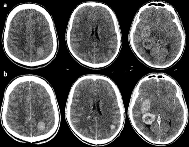
Pre- (a) and post-contrast (b) CT imaging performed on admission demonstrating bilateral cerebral tumours.
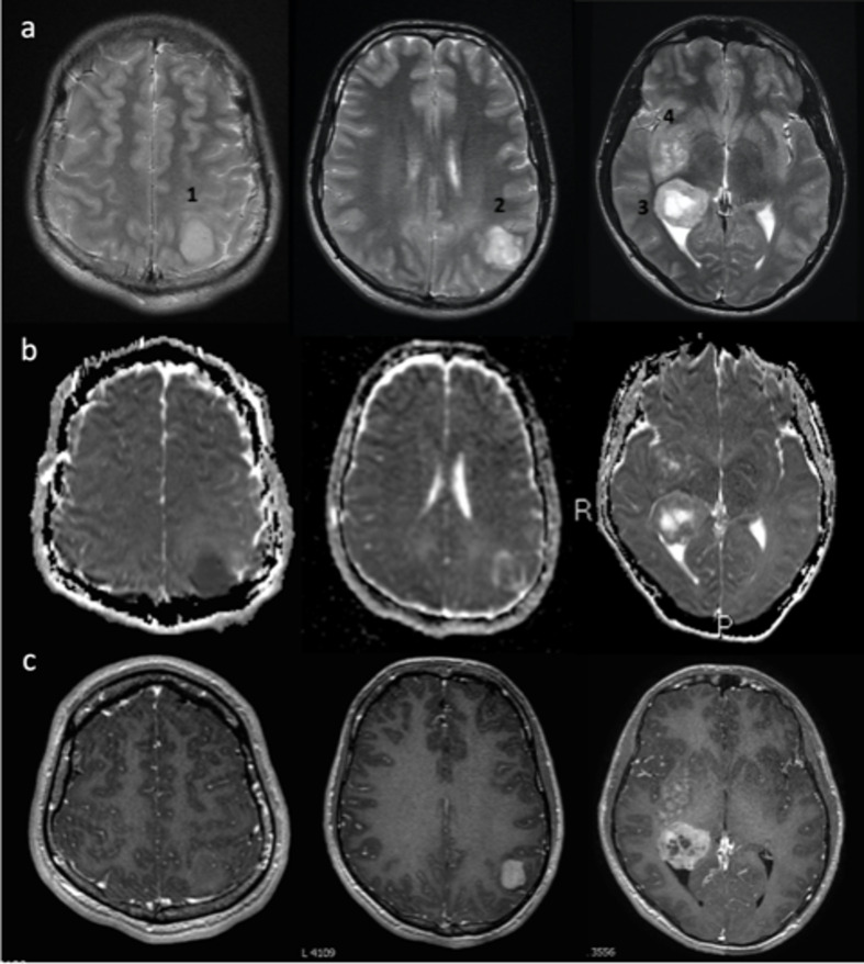
T2 (a), ADC (b), T1+Gad (c) images showing multiple masses: Lesion 1 is circumscript with marked diffusion restriction and no significant Gadolinium enhancement. Lesion 2 is heterogenous featuring a rim of oedema/infiltration, ADC values similar to surrounding brain and avid enhancement. Lesion 3 exhibits rim enhancement and central necrosis. Lesion 4 is poorly marginated on all sequences demonstrating mild patchy Gadolinium uptake. ADC, apparent diffusion coefficient.
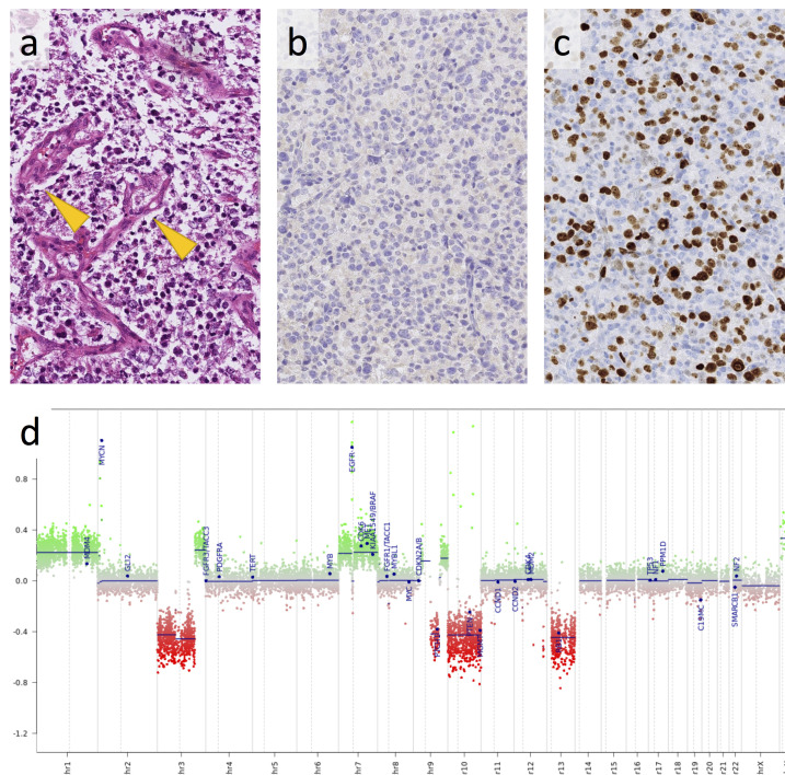
Histology of the tumour shows a monomorphic population of neoplastic glial glial cells with frequent microvascular proliferations (arrow) (a). Immunoreactivity for mutant IDH1 (R132H) is negative (b) and ki67 immunostain for proliferating tumour cells shows a very high labelling index (c). The copy number profile, derived from the Illumina 450 K methylation array, shows the characteristic chromosome seven gain, 10q loss, and amplification of MYC and EGFR (d). The scale bar corresponds to 200 µm in (a)–(c). EGFR, epidermal growth factor receptor.
References
Publication types
LinkOut - more resources
Full Text Sources
