

White matter microstructure shows sex differences in late childhood: Evidence from 6797 children
- PMID: 36177528
- PMCID: PMC9842921
- DOI: 10.1002/hbm.26079
White matter microstructure shows sex differences in late childhood: Evidence from 6797 children
Abstract
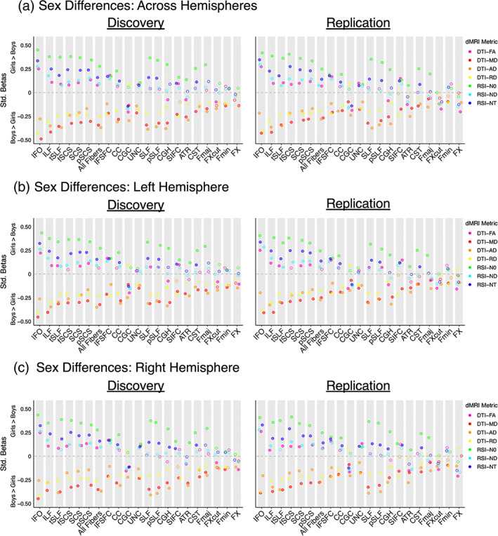
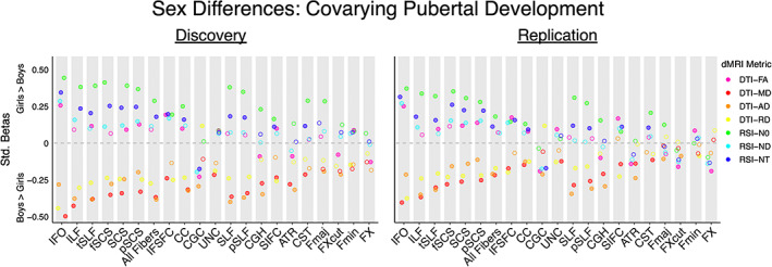
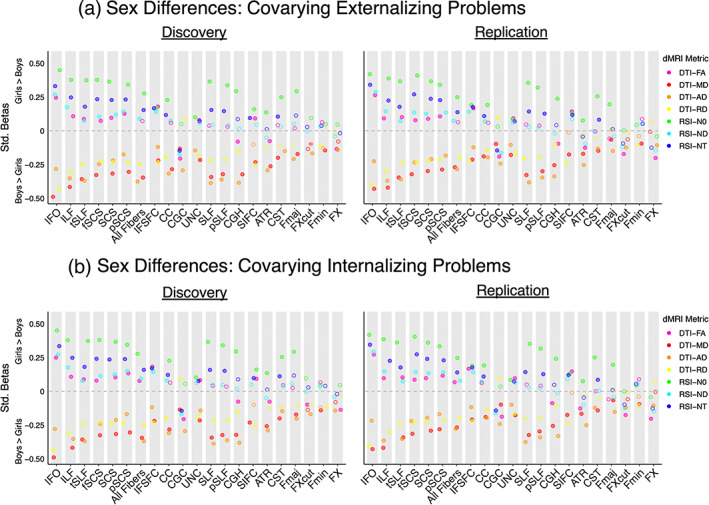
Sex differences in white matter microstructure have been robustly demonstrated in the adult brain using both conventional and advanced diffusion-weighted magnetic resonance imaging approaches. However, sex differences in white matter microstructure prior to adulthood remain poorly understood; previous developmental work focused on conventional microstructure metrics and yielded mixed results. Here, we rigorously characterized sex differences in white matter microstructure among over 6000 children from the Adolescent Brain Cognitive Development study who were between 9 and 10 years old. Microstructure was quantified using both the conventional model-diffusion tensor imaging (DTI)-and an advanced model, restriction spectrum imaging (RSI). DTI metrics included fractional anisotropy (FA) and mean, axial, and radial diffusivity (MD, AD, RD). RSI metrics included normalized isotropic, directional, and total intracellular diffusion (N0, ND, NT). We found significant and replicable sex differences in DTI or RSI microstructure metrics in every white matter region examined across the brain. Sex differences in FA were regionally specific. Across white matter regions, boys exhibited greater MD, AD, and RD than girls, on average. Girls displayed increased N0, ND, and NT compared to boys, on average, suggesting greater cell and neurite density in girls. Together, these robust and replicable findings provide an important foundation for understanding sex differences in health and disease.
Keywords: development; diffusion tensor imaging; diffusion-weighted MRI; microstructure; restriction spectrum imaging; sex differences; white matter.
© 2022 The Authors. Human Brain Mapping published by Wiley Periodicals LLC.
Conflict of interest statement
P. M. T. reports a research grant from Biogen, Inc. for research unrelated to this manuscript. J. T. M. reports consultant income from Roche, TRIS Pharmaceuticals, Octapharma, and GW Pharmaceuticals, expert witness income from Lannett, and research contracts with Roche, Octapharma, and GW Pharmaceuticals for research unrelated to this manuscript. The other authors declare no conflicts of interest.
Figures
Similar articles
-
White matter microstructure across the adult lifespan: A mixed longitudinal and cross-sectional study using advanced diffusion models and brain-age prediction.Neuroimage. 2021 Jan 1;224:117441. doi: 10.1016/j.neuroimage.2020.117441. Epub 2020 Oct 9. Neuroimage. 2021. PMID: 33039618
-
A diffusion MRI study of brain white matter microstructure in adolescents and adults with a Fontan circulation: Investigating associations with resting and peak exercise oxygen saturations and cognition.Neuroimage Clin. 2022;36:103151. doi: 10.1016/j.nicl.2022.103151. Epub 2022 Aug 12. Neuroimage Clin. 2022. PMID: 35994923 Free PMC article.
-
Associations between white matter microstructure and amyloid burden in preclinical Alzheimer's disease: A multimodal imaging investigation.Neuroimage Clin. 2014 Feb 19;4:604-14. doi: 10.1016/j.nicl.2014.02.001. eCollection 2014. Neuroimage Clin. 2014. PMID: 24936411 Free PMC article.
-
Harmonizing DTI measurements across scanners to examine the development of white matter microstructure in 803 adolescents of the NCANDA study.Neuroimage. 2016 Apr 15;130:194-213. doi: 10.1016/j.neuroimage.2016.01.061. Epub 2016 Feb 10. Neuroimage. 2016. PMID: 26872408 Free PMC article.
-
Systematic Review of the Clinical and Experimental Research Assessing the Effects of Craniosynostosis on the Brain.J Craniofac Surg. 2023 Jun 1;34(4):1160-1164. doi: 10.1097/SCS.0000000000009060. Epub 2022 Oct 3. J Craniofac Surg. 2023. PMID: 36184763
Cited by
-
Sex Differences in White Matter Diffusivity in Children with Developmental Dyslexia.Children (Basel). 2024 Jun 13;11(6):721. doi: 10.3390/children11060721. Children (Basel). 2024. PMID: 38929300 Free PMC article.
-
Microstructural Characterization of Short Association Fibers Related to Long-Range White Matter Tracts in Normative Development.Hum Brain Mapp. 2025 Jun 1;46(8):e70255. doi: 10.1002/hbm.70255. Hum Brain Mapp. 2025. PMID: 40490429 Free PMC article.
-
Exposure to multiple ambient air pollutants changes white matter microstructure during early adolescence with sex-specific differences.Commun Med (Lond). 2024 Aug 1;4(1):155. doi: 10.1038/s43856-024-00576-x. Commun Med (Lond). 2024. PMID: 39090375 Free PMC article.
-
Multivariate Links Between the Developmental Timing of Adversity Exposure and White Matter Tract Connectivity in Adulthood.Biol Psychiatry Cogn Neurosci Neuroimaging. 2025 Feb 18:S2451-9022(25)00060-6. doi: 10.1016/j.bpsc.2025.02.003. Online ahead of print. Biol Psychiatry Cogn Neurosci Neuroimaging. 2025. PMID: 39978462
-
Subiculum-BNST structural connectivity in humans and macaques.Neuroimage. 2022 Jun;253:119096. doi: 10.1016/j.neuroimage.2022.119096. Epub 2022 Mar 15. Neuroimage. 2022. PMID: 35304264 Free PMC article.
References
-
- Achenbach, T. M. (2009). The Achenbach system of empirically based assessment (ASEBA): Development, findings, theory and applications. University of Vermont Research Center for Children, Youth, and Families.
-
- Barch, D. M. , Albaugh, M. D. , Avenevoli, S. , Chang, L. , Clark, D. B. , Glantz, M. D. , … Sher, K. J. (2018). Demographic, physical and mental health assessments in the adolescent brain and cognitive development study: Rationale and description. Developmental Cognitive Neuroscience, 32, 55–66. 10.1016/j.dcn.2017.10.010 - DOI - PMC - PubMed
Publication types
MeSH terms
Grants and funding
LinkOut - more resources
Full Text Sources
Research Materials
Miscellaneous
