Review
. 2023 Jan 2;62(1):e202204745.
doi: 10.1002/anie.202204745. Epub 2022 Nov 17.

Photochemical Mechanisms of Fluorophores Employed in Single-Molecule Localization Microscopy

Affiliations
Review

Photochemical Mechanisms of Fluorophores Employed in Single-Molecule Localization Microscopy

Kai Kikuchi et al. Angew Chem Int Ed Engl. .

Abstract

Decoding cellular processes requires visualization of the spatial distribution and dynamic interactions of biomolecules. It is therefore not surprising that innovations in imaging technologies have facilitated advances in biomedical research. The advent of super-resolution imaging technologies has empowered biomedical researchers with the ability to answer long-standing questions about cellular processes at an entirely new level. Fluorescent probes greatly enhance the specificity and resolution of super-resolution imaging experiments. Here, we introduce key super-resolution imaging technologies, with a brief discussion on single-molecule localization microscopy (SMLM). We evaluate the chemistry and photochemical mechanisms of fluorescent probes employed in SMLM. This Review provides guidance on the identification and adoption of fluorescent probes in single molecule localization microscopy to inspire the design of next-generation fluorescent probes amenable to single-molecule imaging.

Keywords: Fluorescence; Photochemistry; Photoswitching; Sensors; Super-Resolution.


Conflict of interest statement

The authors declare no conflict of interest.

Figures
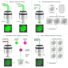
Figure 1
Figure 1
Principles of conventional and super‐resolution microscopy techniques: a) widefield and confocal microscopy; b) structured illumination microscopy; c) stimulated emission depletion microscopy; d) single molecule localization microscopy.
Figure 2
Figure 2
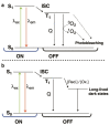
Simplified Jablonski diagram representing processes to be considered for SMLM. For simplicity, neither vibrational levels nor possible additional dark states have been indicated. a) Schematic representation showing electronic transitions during normal fluorescence. In the ON‐state, fluorophores are excited from the ground state (S0) to the excited singlet state (S1). From S1, they may relax radiatively to S0 or undergo intersystem crossing to the dark triplet state (T1). The T1 state can be returned to S0 by triplet–triplet energy transfer with 3O2 to generate 1O2, thereby resulting in photobleaching. Triplet quenchers (Q) such as COT or Ni2+ prevent the generation of 1O2. b) Redox‐active buffers can be used to depopulate the T1 state to generate long‐lived dark states and ground state. Fluorophores then return to S0 through inverse redox reactions or UV light.
Figure 3
Figure 3
The effects of fluorophore properties on SMLM image reconstruction. a) The effect of photon count (PC) on the point spread function (PSF) and thus localization error. A low photon count increases the width of the PSF, while a high PC decreases the width. b) The effect of duty cycle (DC) on fluorophore localization and final image. A low duty cycle increases the number of localizations in high density areas, which gives a more accurate representation of the target once the image is reconstructed.
Figure 4
Figure 4
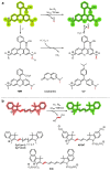
The photoconversion mechanisms of fluorophores. a) Fluorophores containing an N‐alkyl substituent such as rhodamines (tetramethylrhodamine (TMR)), coumarins, and triarylmethanes (crystal violet (CV)) can undergo photocatalyzed dealkylation. b) Cyanines such as Cy5, Cy7, AlexaFluor 647 (AF647), and indocyanine green (ICG) can be photochemically truncated by the action of singlet oxygen.
Figure 5
Figure 5
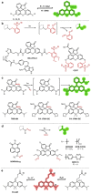
Common caging groups and examples of fluorophores containing them. a) The spirolactonization equilibrium of fluoresceins (X=O) and rhodamines (X=N) gives the off (closed) and on (open) states of these fluorophores, which can be manipulated by the addition of caging groups. b) O‐Nitrobenzyl cages can be cleaved by UV light (ca. 300–360 nm) with the loss of O‐nitrosobenzaldehydes via a bicyclic intermediate. c) Rhodamines can be caged by replacement of the lactone/lactam with diazo groups, which are uncaged through a Wolff rearrangement on exposure to UV light. d) Nitrosamines readily undergo photolysis of the N−N bond with single photon excitation by UV light (365 nm). e) Photoactivation of PA‐SiR by protonation.
Figure 6
Figure 6
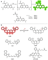
Cage‐free photoactivatable fluorophores. a) Cage‐free PaX dyes undergo 6‐endo cyclizations via the triplet diradical to give an emissive product. b) Reductive caging of dyes allows photoactivation with UV light, and has been demonstrated with both cyanine‐ and rhodamine‐based dyes.
Figure 7
Figure 7
Schematic representation of photoswitching mechanisms: a) Redox photoswitching of a discrete fluorophore. This requires the use of a reductant (red.) and irradiation () or an oxidant (ox.). b) A photochromic fluorophore can be reversibly switched between an emissive and non‐emissive state by irradiation. c) A spontaneously blinking fluorophore that stochastically switches between a non‐emissive and emissive state. This does not require additives. d) An activator and reporter fluorophore dyad. Excitation switches the reporter to a dark state. Illumination with light within the absorbance of the activator restores the emissive state of the reporter. e) A photochromic quencher and reporter dyad. The quencher is reversibly switched between an activated quenching and non‐quenching form.
Figure 8
Figure 8
The photoswitching mechanism of cyanines, exemplified with the Cy5 derivative AF647 and its non‐emissive adduct formed by reaction with a reductant.
Figure 9
Figure 9
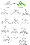
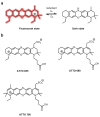
a) The reduction of rhodamine to form a long‐lived non‐emissive radical. b) Examples of rhodamine dyes that photoswitch in this manner.
Figure 10
Figure 10
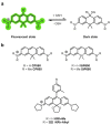
a) Photochromic photoswitching rhodamine‐amides. b) Examples of photochromic rhodamine‐amides.
Figure 11
Figure 11
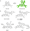
a) A spontaneously blinking rhodamine with an intramolecular nucleophile (Y) that switches between the stochastically temporarily formed ring‐open emissive form and the non‐emissive ring‐closed spirocycle. b) Examples of spontaneously blinking rhodamines used in super‐resolution imaging.
Figure 12
Figure 12
a) A pyronine dye that spontaneously blinks as a result of the reversible intermolecular nucleophilic addition of GSH (or biological thiols) to form a nonfluorescent adduct. b) Examples of spontaneously blinking pyronine dyes used for super‐resolution imaging.
Figure 13
Figure 13
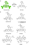
Oxazine photoswitching and examples of fluorophores a) Schematic of photoswitching mechanism of oxazines. ATTO 655. The dark state is the fully reduced, protonated species which can be oxidized by molecular oxygen back to the fluorescent on‐state. b) Examples of oxazine dyes used in super‐resolution imaging.
Figure 14
Figure 14
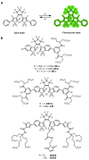
a) The photochromic photoswitching mechanism of diarylethenes. The ring‐open isomer is converted into the ring‐closed isomer by irradiation and vice versa. Both isomers are thermally stable. b) Examples of diarylethene dyes used in super‐resolution imaging.
Figure 15
Figure 15
Schematic representation of an activator–reporter dyad consisting of Cy3 (activator) and Cy5 (reporter) used in the seminal report of STORM imaging.
Figure 16
Figure 16
a) Photochromic diarylethene quencher dyads D‐A and D‐P. The fluorescence of the anthracene and perylene fluorophores is quenched when the diarylethene groups are switched to the closed isomer. b) The spiropyran photochromic dye OA‐4 containing a conjugated coumarin. c) Spirooxazine‐rhodamine dyad SpiroDyad‐2. Upon opening of the closed spirocycle to give the merocyanine, the fluorescence of the rhodamine group is quenched. d) Spiropyran‐naphthalimide‐based dye NpG. Upon reaction with β‐galactosidase, the photochromic switching of the spiropyran is restored.

Similar articles

Cited by

References

    1. Combs C. A., Curr. Protoc. Neurosci. 2010, 50. - PMC - PubMed
    1. Valli J., Garcia-Burgos A., Rooney L. M., Vale de Melo e Oliveira B., Duncan R. R., Rickman C., J. Biol. Chem. 2021, 297, 100791. - PMC - PubMed
    1. None
    1. Miller D. R., Jarrett J. W., Hassan A. M., Dunn A. K., Curr. Opin. Biomed. Eng. 2017, 4, 32–39; - PMC - PubMed
    1. Williams R. M., Zipfel W. R., Webb W. W., Curr. Opin. Chem. Biol. 2001, 5, 603–608; - PubMed

Publication types

MeSH terms

Substances

LinkOut - more resources