Review
. 2022 Oct 1;149(19):dev200373.
doi: 10.1242/dev.200373. Epub 2022 Sep 30.

The emerging significance of splicing in vertebrate development

Affiliations
Review

The emerging significance of splicing in vertebrate development

Anouk M Olthof et al. Development. .

Abstract

Splicing is a crucial regulatory node of gene expression that has been leveraged to expand the proteome from a limited number of genes. Indeed, the vast increase in intron number that accompanied vertebrate emergence might have aided the evolution of developmental and organismal complexity. Here, we review how animal models for core spliceosome components have provided insights into the role of splicing in vertebrate development, with a specific focus on neuronal, neural crest and skeletal development. To this end, we also discuss relevant spliceosomopathies, which are developmental disorders linked to mutations in spliceosome subunits. Finally, we discuss potential mechanisms that could underlie the tissue-specific phenotypes often observed upon spliceosome inhibition and identify gaps in our knowledge that, we hope, will inspire further research.

Keywords: Introns; Spliceosome; Spliceosomopathies; Splicing; Tissue-specific phenotypes; Vertebrate development.


Conflict of interest statement

Competing interests The authors declare no competing or financial interests.

Figures

Fig. 1.
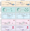
Fig. 1.
Splicing in cell cycle regulation and lineage specification. (A) Spliceosome activity can be regulated during the cell cycle via the phosphorylation of spliceosome components by cyclin-dependent kinases (CDKs). (B) Phosphorylation of the splicing factor SRSF10 regulates alternative splicing of the pro-apoptotic gene Bclx. (C) The role of alternative splicing in cell differentiation and lineage specification. Examples are shown for the genes FOXP1, BIN1 and PAX6.
Fig. 2.
Fig. 2.
Knockout of core spliceosome components results in early embryonic lethality. (A,B) Stages of early embryonic development with timeline for mouse (A) and zebrafish (B). Knockout of spliceosome components listed in black results in lethality at the time point indicated on the timeline. For spliceosome components listed in gray, the exact time point of embryonic lethality was not established. In zebrafish, lethality is not observed until around 2 dpf. E, embryonic day; hpf, hours post-fertilization. More details can be found in Table S2.
Fig. 3.
Fig. 3.
Tissues affected in spliceosomopathies. Colouring (red to blue gradient) represents the percentage of spliceosomopathies that are characterized by defects in the different organ systems. Phenotype data for spliceosomopathies was obtained from Human Phenotype Ontology database.
Fig. 4.
Fig. 4.
The role of core spliceosome components in neuroectoderm development. Schematics showing the expression patterns of the spliceosome components Pqbp1, Rnu11 and Eftud2 in mouse embryos. Conditional knockout of these factors results in aberrant splicing and cellular defects affecting neuronal and neural crest cell development. This ultimately leads to gross phenotypes, such as microcephaly or craniofacial malformations.
Fig. 5.
Fig. 5.
Potential mechanisms underlying cell type- and tissue-specific phenotypes observed upon ubiquitous spliceosome inhibition. Explanations for cell type-restricted phenotypes observed in animal models and spliceosomopathies may include cell-intrinsic differences, such as transcription and/or splicing kinetics, or differences in proliferation rates. Other causes could include differential expression of genes highly dependent on spliceosome function or differential expression of factors that can compensate for the loss of function of some spliceosome components. Finally, the differential activity of downstream pathways frequently affected by aberrant splicing, such as p53 signaling, could contribute to differences in cellular defects.

Similar articles

Cited by

References

    1. Alam, S. S., Kumar, S., Beauchamp, M.-C., Bareke, E., Boucher, A., Nzirorera, N., Dong, Y., Padilla, R., Zhang, S. J., Majewski, J. et al. (2022). Snrpb is required in murine neural crest cells for proper splicing and craniofacial morphogenesis. Dis. Models Mech. 15, dmm049544. 10.1242/dmm.049544 - DOI - PMC - PubMed
    1. Allende-Vega, N., Dayal, S., Agarwala, U., Sparks, A., Bourdon, J.-C. and Saville, M. K. (2013). p53 is activated in response to disruption of the pre-mRNA splicing machinery. Oncogene 32, 1-14. 10.1038/onc.2012.38 - DOI - PubMed
    1. Amsterdam, A., Nissen, R. M., Sun, Z., Swindell, E. C., Farrington, S. and Hopkins, N. (2004). Identification of 315 genes essential for early zebrafish development. Proc. Natl. Acad. Sci. USA 101, 12792-12797. 10.1073/pnas.0403929101 - DOI - PMC - PubMed
    1. An, M. and Henion, P. D. (2012). The zebrafish sf3b1b460 mutant reveals differential requirements for the sf3b1 pre-mRNA processing gene during neural crest development. Int. J. Dev. Biol. 56, 223-237. 10.1387/ijdb.113383ma - DOI - PMC - PubMed
    1. Balestra, D., Giorgio, D., Bizzotto, M., Fazzari, M., Ben Zeev, B., Pinotti, M., Landsberger, N. and Frasca, A. (2019). Splicing mutations impairing CDKL5 expression and activity can be efficiently rescued by U1snRNA-based therapy. Int. J. Mol. Sci. 20, E4130. 10.3390/ijms20174130 - DOI - PMC - PubMed

Publication types