. 2022 Sep 30;29(10):379-389.
doi: 10.1101/lm.053558.121. Print 2022 Oct.

Category-specific memory encoding in the medial temporal lobe and beyond: the role of reward

Affiliations

Category-specific memory encoding in the medial temporal lobe and beyond: the role of reward

Heidrun Schultz et al. Learn Mem. .

Abstract

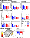
The medial temporal lobe (MTL), including the hippocampus (HC), perirhinal cortex (PRC), and parahippocampal cortex (PHC), is central to memory formation. Reward enhances memory through interplay between the HC and substantia nigra/ventral tegmental area (SNVTA). While the SNVTA also innervates the MTL cortex and amygdala (AMY), their role in reward-enhanced memory is unclear. Prior research suggests category specificity in the MTL cortex, with the PRC and PHC processing object and scene memory, respectively. It is unknown, however, whether reward modulates category-specific memory processes. Furthermore, no study has demonstrated clear category specificity in the MTL for encoding processes contributing to subsequent recognition memory. To address these questions, we had 39 healthy volunteers (27 for all memory-based analyses) undergo functional magnetic resonance imaging while performing an incidental encoding task pairing objects or scenes with high or low reward, followed by a next-day recognition test. Behaviorally, high reward preferably enhanced object memory. Neural activity in the PRC and PHC reflected successful encoding of objects and scenes, respectively. Importantly, AMY encoding effects were selective for high-reward objects, with a similar pattern in the PRC. The SNVTA and HC showed no clear evidence of successful encoding. This behavioral and neural asymmetry may be conveyed through an anterior-temporal memory system, including the AMY and PRC, potentially in interplay with the ventromedial prefrontal cortex.


Figures

Figure 1.
Figure 1.
Results overview. (A) Recognition memory results in the memory sample (n = 27). Gray boxes indicate results from individual two-way repeated measures ANOVAs with the factors category (C) and reward (R). (CHR) Corrected hit rate. (B) Average beta values for each condition of model 1 (memory sample, n = 27) in each of the five ROIs. Gray boxes indicate results from individual three-way repeated measures ANOVAs with the factors category (C), reward (R), and subsequent memory (M). Paired t-tests (hits vs. misses) were computed for all ROIs and conditions for visualization. (C) Voxel-wise activity for the high > low reward contrast in model 2 (full sample, n = 39). The statistical map is projected onto the normalized, averaged T1 (display threshold P < 0.001 uncorrected, k ≥ 20 voxels). Black outline indicates vmPFC cluster surviving at PFWE < 0.05. Bar plot depicts average beta values within the FWE-corrected vmPFC cluster. The gray box indicates results from a post-hoc two-way repeated measures ANOVA with the factors category (C) and reward (R). (*) P < 0.05, [(*)] P < 0.1. Error bars indicate SEM.
Figure 2.
Figure 2.
Experimental paradigm. (A) Day 1: incidental encoding task. (B) Day 2: recognition task. Note that text options (e.g., remember—know—guess) were arranged horizontally in the experiment. See the text for details. (OB) Objects, (SC) scenes, (HI) high reward, (LO) low reward. Example stimuli have been replaced with similar photographs due to copyright.
Figure 3.
Figure 3.
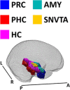
ROIs. Single-participant regions of interest were MNI-normalized, averaged over the full sample (n = 39), and thresholded at 0.5. Here they are visualized within the standard SPM12 brain mask (mask_ICV.nii). (PRC) Perirhinal cortex, (PHC) parahippocampal cortex, (HC) hippocampus, (AMY) amygdala, (SNVTA) substantia nigra/ventral tegmental area, (A) anterior, (P) posterior, (R) right, (L) left.

References

    1. Adcock RA, Thangavel A, Whitfield-Gabrieli S, Knutson B, Gabrieli JDE. 2006. Reward-motivated learning: mesolimbic activation precedes memory formation. Neuron 50: 507–517. 10.1016/j.neuron.2006.03.036 - DOI - PubMed
    1. Amaral DG, Insausti R. 1992. Retrograde transport of D-[3H]-aspartate injected into the monkey amygdaloid complex. Exp Brain Res 88: 375–388. 10.1007/BF02259113 - DOI - PubMed
    1. Awipi T, Davachi L. 2008. Content-specific source encoding in the human medial temporal lobe. J Exp Psychol Learn Mem Cogn 34: 769–779. 10.1037/0278-7393.34.4.769 - DOI - PMC - PubMed
    1. Beckstead RM, Domesick VB, Nauta WJ. 1979. Efferent connections of the substantia nigra and ventral tegmental area in the rat. Brain Res 175: 191–217. 10.1016/0006-8993(79)91001-1 - DOI - PubMed
    1. Berron D, Neumann K, Maass A, Schütze H, Fliessbach K, Kiven V, Jessen F, Sauvage M, Kumaran D, Düzel E. 2018. Age-related functional changes in domain-specific medial temporal lobe pathways. Neurobiol Aging 65: 86–97. 10.1016/j.neurobiolaging.2017.12.030 - DOI - PubMed

LinkOut - more resources