Review
. 2022 Dec;23(12):711-724.
doi: 10.1038/s41583-022-00631-3. Epub 2022 Sep 30.

How mechanisms of stem cell polarity shape the human cerebral cortex

Affiliations
Review

How mechanisms of stem cell polarity shape the human cerebral cortex

Madeline G Andrews et al. Nat Rev Neurosci. 2022 Dec.

Abstract

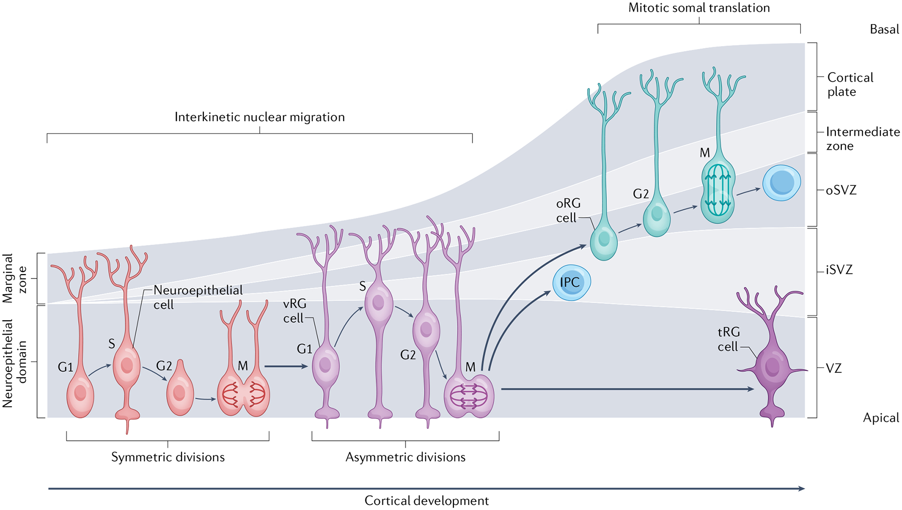
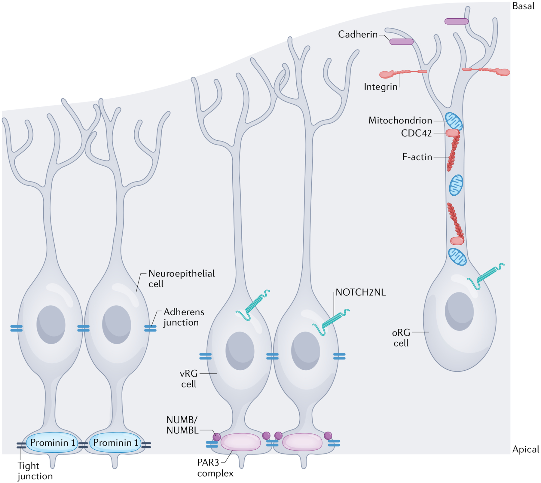
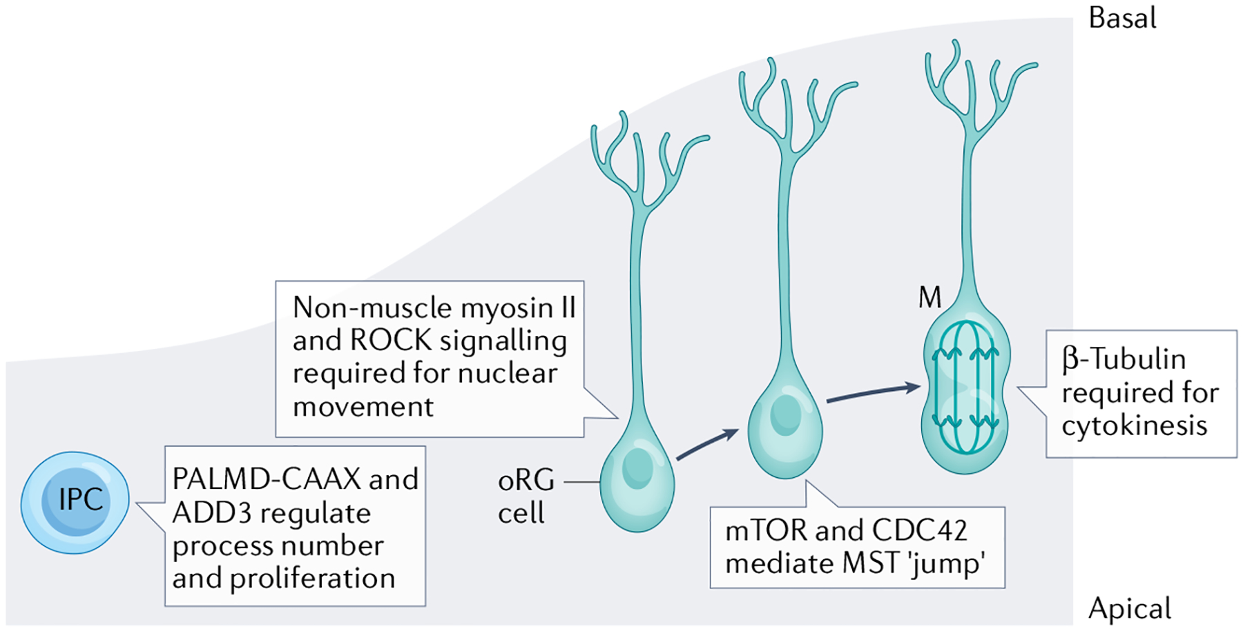
Apical-basal progenitor cell polarity establishes key features of the radial and laminar architecture of the developing human cortex. The unique diversity of cortical stem cell populations and an expansion of progenitor population size in the human cortex have been mirrored by an increase in the complexity of cellular processes that regulate stem cell morphology and behaviour, including their polarity. The study of human cells in primary tissue samples and human stem cell-derived model systems (such as cortical organoids) has provided insight into these processes, revealing that protein complexes regulate progenitor polarity by controlling cell membrane adherence within appropriate cortical niches and are themselves regulated by cytoskeletal proteins, signalling molecules and receptors, and cellular organelles. Studies exploring how cortical stem cell polarity is established and maintained are key for understanding the features of human brain development and have implications for neurological dysfunction.


Conflict of interest statement

Competing interests

A.R.K. is a co-founder, consultant and member of the Board of Neurona Therapeutics. The other authors declare no competing interests.

Figures

Fig. 1 |
Fig. 1 |. Progenitor types and division programmes in the developing human cortex.
During early human forebrain development a pseudostratified layer of neuroepithelial cells populate the developing neural tube. Neuroepithelial cells divide symmetrically at the ventricular surface in self-renewing divisions. From the end of the first trimester, neuroepithelial cells differentiate into multiple subtypes of radial glial cells, including apically localized, early-born ventricular radial glial (vRG) cells and later-born outer radial glial (oRG) cells,,. vRG cells divide in a process known as interkinetic nuclear migration, in which the nucleus moves in a basal direction during DNA synthesis (S phase) and then moves in an apical direction to undergo mitosis at the ventricular surface. oRG cells divide in a process known as mitotic somal translocation, in which the cell body ‘jumps’ (moves quickly) in a basal direction before dividing horizontally by mitosis. Both vRG cells and oRG cells differentiate into neurogenic intermediate progenitor cells (IPCs) or directly into neurons (not shown),,–. Later in neurogenesis, the apically localized radial glial cells become truncated radial glial (tRG) cells. The cortex is divided into distinct zones, in which each of these cellular populations resides. Early in development, the cortex comprises the neuroepithelial domain and the marginal zone, whereas later — as the complexity of cell types and cortical size expands — the progenitor zone is divided into the ventricular zone (VZ), inner subventricular zone (iSVZ) and outer subventricular zone (oSVZ), and the neuronal domains are divided into the cortical plate, containing layers II–IV of excitatory neurons, and the marginal zone (which contains Cajal–Retzius cells), in layer I above the cortical plate. As progenitor cells differentiate into neurons, they move basally along the radial glial scaffold to form the intermediate zone and neurons differentiate to contribute to the growing cortical plate.
Fig. 2 |
Fig. 2 |. Subcellular structures and organelles regulating progenitor polarity.
The specific morphology and polarity of different cortical progenitor subtypes are regulated by distinct molecular mechanisms. Although the dynamic regulation of human progenitor polarity is being revealed, most mechanistic studies have been performed in rodent or other model species. This figure represents a cumulative understanding of polarity from animal and human studies. The apical membranes of neuroepithelial cells form the ventricular surface of the cortex. Neuroepithelial cells express the transmembrane glycoprotein prominin 1 at the apical membrane and tight junctions form between neuroepithelial cells to establish the ventricular surface and establish their polarity. In a more basal location within the cells, along the cell body, adherens junctions are also formed between neuroepithelial cells,,. Ventricular radial glial (vRG) cells have a similar apical–basal polarity, but their apical anchoring to the ventricular surface is regulated by a different set of proteins, including those that comprise the PAR3 complex, and the vRG cells are predominantly connected to one another by adherens junctions. The proteins NUMB and/or NUMBL help maintain these adherens junctions at the apical surface. Junctional complexes regulate receipt of NOTCH signals, which are involved in mediating proliferation and polarity. The human-specific receptor, NOTCH2NL, impacts radial glial cell proliferation,. Outer radial glial (oRG) cells retain a basal polarity, but without an apical attachment. Their connection to the basal lamina is regulated by cadherins and integrins, in the oRG cell endfeet and the integrity of their basal processes is maintained by appropriate F-actin activity (regulated by the GTPase CDC42) and mitochondrial function,.
Fig. 3 |
Fig. 3 |. Human-specific regulation of the cytoskeleton in outer radial glial cells.
In the developing human cortex, outer radial glial (oRG) cells divide via a process known as mitotic somal translocation (MST). Appropriate oRG cell basal polarity is vital for the basal direction and appropriate migration of differentiating oRG cells during MST. Each phase of MST is regulated by distinct molecular components. Nuclear movement prior to division is regulated by non-muscle myosin II and requires ROCK signalling. The distance of the MST ‘jump’ is regulated by mTOR-mediated CDC42 activity. Finally, oRG cell cytokinesis requires β-tubulin. Intermediate progenitor cell (IPC) multipolar process numbers are regulated by membrane-bound PALMD-CAAX and ADD3 (REF.).

References

    1. Geschwind DH & Rakic P Cortical evolution: judge the brain by its cover. Neuron 80, 633–647 (2013). - PMC - PubMed
    1. Chou F-S, Li R & Wang P-S Molecular components and polarity of radial glial cells during cerebral cortex development. Cell. Mol. Life Sci 75, 1027–1041 (2018). - PMC - PubMed
    1. Arai Y & Taverna E Neural progenitor cell polarity and cortical development. Front. Cell. Neurosci 11, 384 (2017). - PMC - PubMed
    1. Götz M & Huttner WB The cell biology of neurogenesis. Nat. Rev. Mol. Cell Biol 6, 777–788 (2005). - PubMed
    1. Mira H & Morante J Neurogenesis from embryo to adult — lessons from flies and mice. Front. Cell Dev. Biol 8, 533 (2020). - PMC - PubMed

Publication types