. 2022 Sep 30;12(1):16390.
doi: 10.1038/s41598-022-20831-2.

Isolation and characterization of a Vibrio owensii phage phi50-12

Affiliations

Isolation and characterization of a Vibrio owensii phage phi50-12

Ling-Chun Lin et al. Sci Rep. .

Abstract

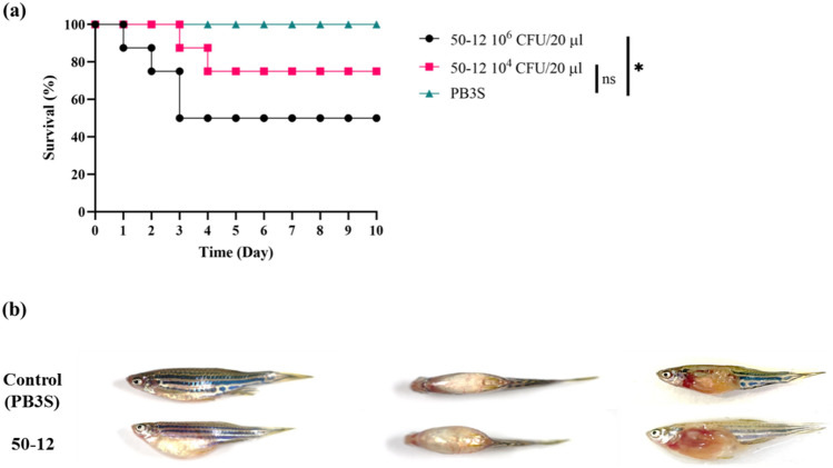
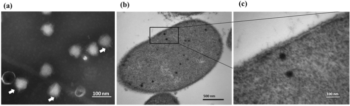
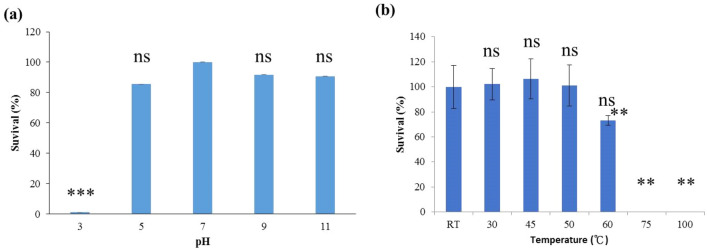
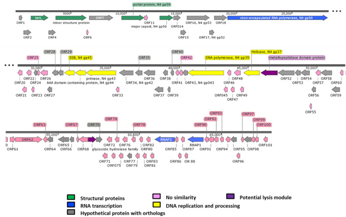
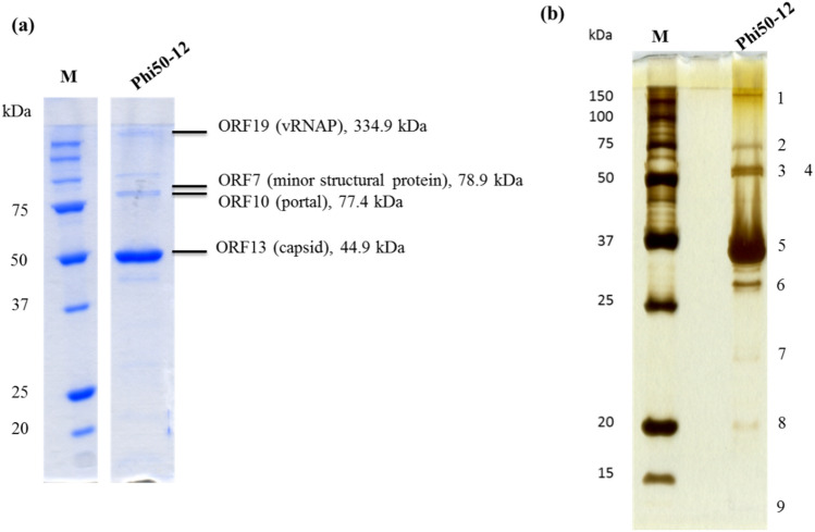
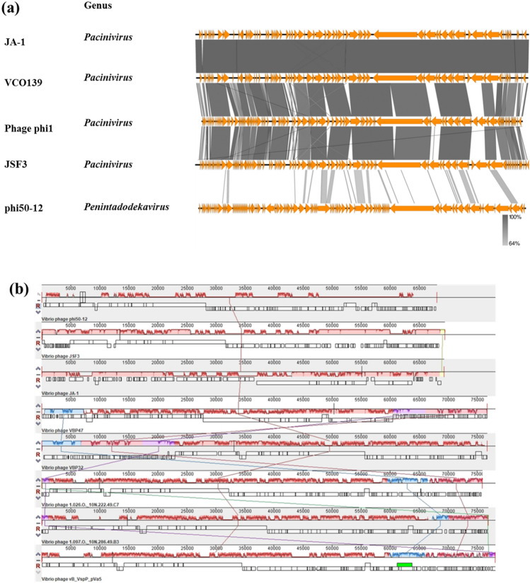
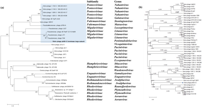
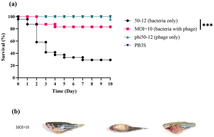
Vibrio owensii is a widely distributed marine vibrio species that causes acute hepatopancreatic necrosis in the larvae of Panulirus ornatus and Penaeus vannamei, and is also associated with Montipora white syndrome in corals. We characterized V. owensii GRA50-12 as a potent pathogen using phenotypic, biochemical, and zebrafish models. A virulent phage, vB_VowP_phi50-12 (phi50-12), belonging to the N4-like Podoviridae, was isolated from the same habitat as that of V. owensii GRA50-12 and characterized. This phage possesses a unique sequence with no similar hits in the public databases and has a short latent time (30 min), a large burst size (106 PFU/infected cell), and a wide range of pH and temperature stabilities. Moreover, phi50-12 also demonstrated a strong lysis ability against V. owensii GRA50-12. SDS-PAGE revealed at least nine structural proteins, four of which were confirmed using LC-MS/MS analysis. The size of the phi50-12 genome was 68,059 bp, with 38.5% G + C content. A total of 101 ORFs were annotated, with 17 ORFs having closely related counterparts in the N4-like vibrio phage. Genomic sequencing confirmed the absence of antibiotic resistance genes or virulence factors. Comparative studies have shown that phi50-12 has a unique genomic arrangement, except for the well-conserved core regions of the N4-like phages. Phylogenetic analysis demonstrated that it belonged to a group of smaller genomes of N4-like vibrio phages. The therapeutic effect in the zebrafish model suggests that phi50-12 could be a potential candidate for application in the treatment of V. owensii infection or as a biocontrol agent. However, further research must be carried out to confirm the efficacy of phage50-12.


Conflict of interest statement

The authors declare no competing interests.

Figures

Figure 1
Figure 1
Pathogenicity of V. owensii GRA50-12 was confirmed by using zebrafish as a model. (a) Three groups (8 fishes/group) were considered, and their survival rates were measured after intraperitoneal injection of V. owensii GRA50-12 cells (2.2 × 106 CFU/20 µl, black line; 2.2 × 104 CFU/20 µl, red line) or PB3S (PBS with 3% NaCl) as control (green line). The X-axis represents the time post-infection and the Y-axis represents the survival percentage of the zebrafish. (b) The disease symptoms in V. owensii GRA50-12 infected zebrafish. Control represents PB3S buffer injected fish; thus, no symptoms were observed. 50-12 represents zebrafish injected with GRA50-12 and these showed swollen abdomen and bleeding at body surface (left: side view; middle: top view; right: abdomen anatomy). The anatomical analysis showed swollen and bleeding intestines. The significance of the differences between groups was performed by Log-rank and Gehan–Breslow–Wilcoxon test in GraphPad Prism 9. “*” indicates p < 0.05.
Figure 2
Figure 2
Morphology of phi50-12. (a) Electron micrograph of phi50-12 demonstrates that it resembles podoviruses with short tails; arrow heads indicate the tails of phi50-12. (b) Electron micrograph of thin section reveals phi50-12 virions inside the cell and (c) the loss of cell surface integrity during the release of virions.
Figure 3
Figure 3
pH and thermal stability of phi50-12. (a) 106 PFU of phi50-12 were subjected to various pH at 30 °C for one hour and titrated by plaque assay using a double-layer method; (b) phi50-12 stability (106 PFU) under various temperatures for 1 h was measured by plaque assay. The survival rate was measured by comparison with initial phage number. The experiments were performed in triplicates and the data are shown as the mean ± SEM. Student’s t test was performed for significance. Ns no significance; **p < 0.01; ***p < 0.001.
Figure 4
Figure 4
Genomic organization of phi50-12. Direction of the arrow represents transcription orientation. Colored box represents their functionality or uniqueness.
Figure 5
Figure 5
Structural protein analysis by 12% SDS-PAGE. (a) Phage particles (5 × 1011 PFU) were boiled in protein sample buffer (80 mM Tris, pH 6.8; 2% SDS; 2-mecraptoethanol; 0.0006% Bromophenol blue) and subjected to SDS-PAGE. Parallelly, the sliced bands were subjected to LC/MS/MS analysis. Peptides matched with the annotated ORFs, and their observed molecular mass were indicated. Four of them were identified to be virion-encapsuled RNA polymerase (ORF19), minor structural protein (ORF7), portal protein (ORF10) and capsid protein (ORF13), respectively. Original gel is presented in Supplementary Fig. S9. (b) For better visualization, a silver stain procedure was also performed, nine bands were visible on the gel. Numbers indicate the protein band location. M is the molecular weight marker, phi50-12 represents the structural protein lysate.
Figure 6
Figure 6
Comparative genomic analysis of N4-like Vibrio phages. (a) TBLASTX was performed by Easyfig. All the phages belong to N4-like Vibrio phages. “Genus” represents the taxonomy to which they belong. The similarity range is indicated by the gradient scale. (b) Mauve alignment of selected N4-like Vibrio phages infecting a variety of Vibrio hosts. The hosts include V. owensii (phi50-12), V. cholera (JSF3, JA-1), V. parahaemolyticus (VBP47, VBP32), unidentified hosts (Vibrio phage 1.026.O._10N222.49.C7, 1.097.O._10N.286.49.B3), Vibrio splendidus (vB_VspP_pVa5). Each colored block represents a region of collinear sequence (LCB, local collinear block) among genomes. White boxes represent the ORFs of each genome and their arrangements.
Figure 7
Figure 7
Phylogenetic analysis of terminase large subunit and viral proteomic tree analysis. (a) The tree generated by MEGA11 based on Neighbor-joining method with 1000 bootstraps. The subfamily and genus of the phages are indicated on the right. (b) Viral proteomic tree generated by VIPTree. This analysis involved all Vibrio phages available in the reference database at present.
Figure 8
Figure 8
Therapeutic effect of phi50-12 in zebrafish. (a) Four groups were considered, each containing 24 fishes. These were subjected to four distinct conditions: injection with V. owensii GRA50-12 cells (2.8–4.5 × 106 CFU/20 µl, black line), phi50-12 treatment (MOI = 10) for 30 min followed by challenge with bacteria (red line), phi50-12 treatment only (green line) and PB3S buffer as negative control (purple line); and survival rates were measured. The X-axis represents the time post-infection and the Y-axis represents the survival percentage of the zebra fish. (b) The disease symptoms of zebrafish rescued by phi50-12 with MOI = 10. V. owensii GRA50-12 introduced first in the fish followed by administration of phi50-12 after 30 min. MOI = 10 represents the disease symptom of sick fish in the group with phi50-12 administration. Left: side view; middle: top view; right: abdomen anatomy. The anatomical picture showed swollen abdomen and bleeding intestines. The significance of the differences between groups was performed by Log-rank and Gehan–Breslow–Wilcoxon test in GraphPad Prism 9. “***” indicates p < 0.001.

References

    1. Thompson FL, Iida T, Swings J. Biodiversity of vibrios. Microbiol. Mol. Biol. Rev. 2004;68:403–431. doi: 10.1128/MMBR.68.3.403-431.2004. - DOI - PMC - PubMed
    1. Poli A, et al. Vibrio coralliirubri sp. Nov., a new species isolated from mucus of red coral (Corallium rubrum) collected at Procida island, Italy. Antonie Van Leeuwenhoek. 2018;111:1105–1115. doi: 10.1007/s10482-017-1013-5. - DOI - PubMed
    1. Romalde JL, Dieguez AL, Lasa A, Balboa S. New Vibrio species associated to molluscan microbiota: A review. Front. Microbiol. 2014;4:413. doi: 10.3389/fmicb.2013.00413. - DOI - PMC - PubMed
    1. Culot A, et al. Isolation of Harveyi clade Vibrio spp. collected in aquaculture farms: How can the identification issue be addressed? J. Microbiol. Methods. 2021;180:106106. doi: 10.1016/j.mimet.2020.106106. - DOI - PubMed
    1. Yoshizawa S, et al. Vibrio jasicida sp. Nov., a member of the Harveyi clade, isolated from marine animals (packhorse lobster, abalone and Atlantic salmon) Int. J. Syst. Evol. Microbiol. 2012;62:1864–1870. doi: 10.1099/ijs.0.025916-0. - DOI - PubMed

Publication types

Substances

Supplementary concepts