

Body Site Distribution of Acquired Melanocytic Naevi and Associated Characteristics in the General Population of Caucasian Adults: A Scoping Review
- PMID: 36180760
- PMCID: PMC9588131
- DOI: 10.1007/s13555-022-00806-x
Body Site Distribution of Acquired Melanocytic Naevi and Associated Characteristics in the General Population of Caucasian Adults: A Scoping Review
Abstract
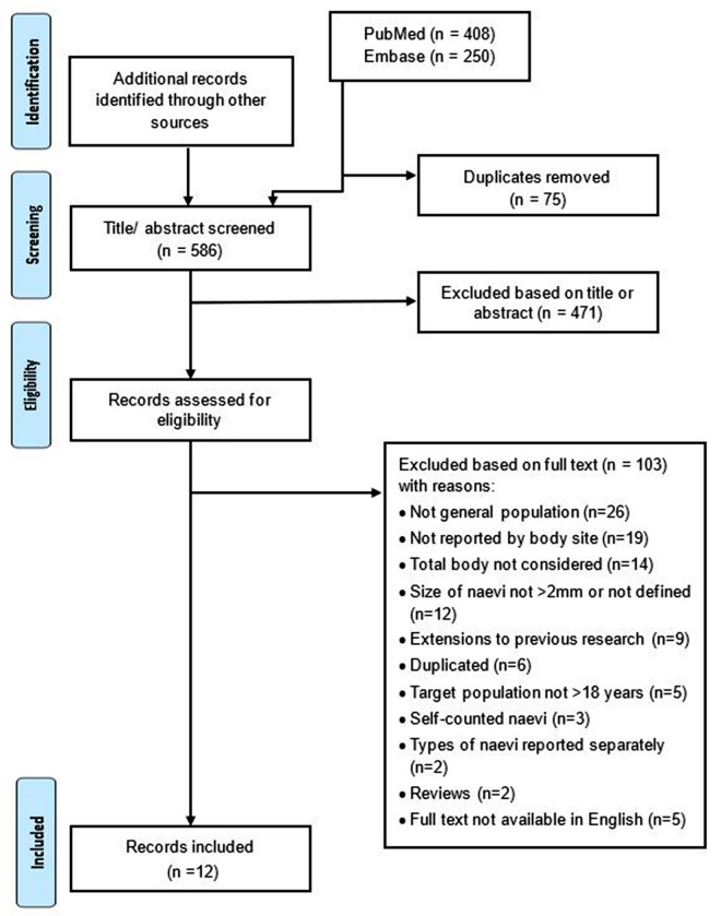
The number of melanocytic naevi is a major risk factor for melanoma. The divergent pathway hypothesis proposes that the propensity for naevus proliferation and malignant transformation may differ by body site and exposure to ultraviolet (UV) radiation. This scoping review aimed to summarise the evidence on the number and distribution of naevi (≥ 2 mm) on the body overall and by individual anatomical sites in Caucasian adults, and to assess whether studies used the International Agency for Research on Cancer (IARC) protocol to guide naevus counting processes. Systematic searches of Embase and PubMed identified 661 potentially relevant studies, and 12 remained eligible after full-text review. Studies varied widely in their counting protocols, reporting of naevus counts overall and by body sites, and used counting personnel with differing qualifications. Only one study used the IARC protocol. Studies reported that the highest number of naevi was on the trunk in males and on the arms in females. Body sites which receive intermittent exposure to UV radiation had higher density of naevi. Larger naevi (≥ 5 mm) were detected mostly on body sites intermittently exposed to UV radiation, and smaller naevi (< 5 mm) on chronically exposed sites. Studies reported that environmental and behavioural aspects related to UV radiation exposure, as well as genetic factors, all impact body site and size distribution of naevi. This review found that to overcome limitations of the current evidence, future studies should use consistent naevus counting protocols. Skin surface imaging could improve the reliability of findings. An updated IARC protocol is required that integrates these emerging standards and technologies to guide reliable and reproducible naevus counting in the future.
Keywords: Body site distribution; Melanocytic naevi; Melanoma; Melanoma risk; Moles; Population-based adults.
© 2022. The Author(s).
Figures
References
-
- Allen AC. Melanocarcinoma. In: Anderson WAD, editor. Pathology. 2. St Louis: CV Cosby Company; 1953. p. 1167.
-
- Smith JL. Malignant melanoma. In: Graeme JH, Johnson WC, Helwig EB, editors. Dermal pathology. Maryland: Harper and Row; 1972. pp. 490–491.
Publication types
Grants and funding
LinkOut - more resources
Full Text Sources
