. 2022 Dec;49(12):12253-12258.
doi: 10.1007/s11033-022-07830-2. Epub 2022 Sep 30.

The cytoprotective role of GM1 ganglioside in Huntington disease cells

Affiliations

The cytoprotective role of GM1 ganglioside in Huntington disease cells

Hannah S Hart et al. Mol Biol Rep. 2022 Dec.

Abstract

Background: Huntington disease (HD) is a neurodegenerative disease where a genetic mutation leads to excessive polyglutamine (Q) repeats in the huntingtin protein. The polyglutamine repeats create toxic plaques when the protein is cleaved, leading to neuron death. The glycolipid GM1 ganglioside (GM1) has been shown to be neuroprotective in HD models, as it prevents the cleavage of the mutant huntingtin protein by phosphorylation of serine 13 and 16. Previous studies have tested GM1 in both adult-onset and juvenile-onset HD models, but this study set out to investigate whether GM1 mediated cytoprotection is influenced by the length of polyglutamine repeats.

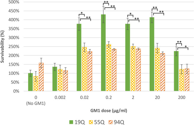
Method and result: This study utilized cell culture to analyze the effect of GM1 on cell viability, directly comparing the response between cells with adult-onset HD and juvenile-onset HD. HEK293 cells expressing either wild-type huntingtin (Htt) (19Q) exon 1, adult-onset HD mutant Htt exon 1 (55Q), or Juvenile HD mutant Htt exon 1 (94Q) were assessed for cell viability using the WST-1 assay. Our results suggested moderate doses of GM1 increased cell viability for all cell lines when compared to untreated cells. When comparing HEK293 55Q and 94Q cells, there was no difference in cell viability within each dose of GM1.

Conclusion: These data suggest cellular responses to GM1 are independent of polyglutamine repeats in HD cells and provide insight on GM1's application as a therapeutic agent for HD and other diseases.

Keywords: Cytoprotection; GM1 ganglioside; Huntington’s disease; Juvenile Huntington’s disease; Mutant huntingtin; Polyglutamine repeats.


Conflict of interest statement

The authors have no relevant financial or non-financial interests to disclose.

Figures

Fig. 1
Fig. 1
Moderate doses of GM1 increase HEK293 cell viability as evaluated by the WST-1 assay. The average viability for cells exposed to GM1 is shown for each cell line, separately. a 19Q, b 55Q, c 94Q. Drug doses increase from 0 to 200 μg/ml of GM1 left to right. Error bars represent the standard error of the group mean. Moderate dosages of GM1 significantly increased cell viability compared to untreated cells with 0 μg/ml of GM1 as indicated by ANOVA and Tukey HSD tests. A sample was defined as one well within the 96-well plate. The 19Q cell line had 12 samples within the control and 18 samples within each dose of GM1 for a total of 120 samples. The 55Q cell line had 6 samples within the control and 9 samples within each dose of GM1 for a total of 60 samples. The 94Q cell line had 6 samples within the control and 5 samples within each dose of GM1 for a total of 36 samples. *Tukey HSD tests indicate the group is significantly different compared to 0 μg/ml of GM1 where p < 0.05. **Tukey HSD tests indicates the group is significantly different compared to 0 μg/ml of GM1 where p < 0.01
Fig. 2
Fig. 2
Cell viability after treatment with GM1 is similar between adult-onset and juvenile-onset HD cells. The average viability for cells exposed to GM1 is shown for all cell lines. The data have been normalized to directly compare the response to GM1 between cell lines. To normalize, the mean survivability of untreated 19Q cells was set as 100% survival, and the averages of other cell lines and treatments were divided by the average survivability of untreated 19Q cells (0 μg/ml GM1). Drug doses increase from 0 to 200 μg/ml GM1. Error bars represent the standard error of the group mean. A sample was defined as one well within the 96-well plate. The 19Q cell line had 12 samples within the control and 18 samples within each dose of GM1 for a total of 120 samples. The 55Q cell line had 6 samples within the control and 9 samples within each dose of GM1 for a total of 60 samples. The 94Q cell line had 6 samples within the control and 5 samples within each dose of GM1 for a total of 36 samples. *HSD tests indicate there is a significant difference between the two cell lines for the specific GM1 dose where p < 0.05. **Tukey HSD tests indicate there is a significant difference between the two cell lines for the specific GM1 dose where p < 0.01.  All data were considered significant with p < 0.05

References

    1. Bates GP, Dorsey R, Gusella JF, Hayden MR, Kay C, Leavitt BR, Nance M, Ross CA, Scahill RI, Wetzel R, Wild EJ, Tabrizi SJ. Huntington disease. Nat Rev Dis Primers. 2015 doi: 10.1038/nrdp.2015.5. - DOI - PubMed
    1. Di Pardo A, Amico E, Maglione V. Impaired levels of gangliosides in the corpus callosum of Huntington disease animal models. Front Neurosci. 2016;10:457. doi: 10.3389/fnins.2016.00457. - DOI - PMC - PubMed
    1. Maglione V, Marchi P, Di Pardo A, Lingrell S, Horkey M, Tidmarsh E, Sipione S. Impaired ganglioside metabolism in Huntington's disease and neuroprotective role of GM1. J Neurosci. 2010;30:4072. doi: 10.1523/JNEUROSCI.6348-09.2010. - DOI - PMC - PubMed
    1. Posse de Chaves E, Sipione S. Sphingolipids and gangliosides of the nervous system in membrane function and dysfunction. FEBS Lett. 2010;584:1748–1759. doi: 10.1016/j.febslet.2009.12.010. - DOI - PubMed
    1. Mocchetti I. Exogenous gangliosides, neuronal plasticity and repair, and the neurotrophins. Cell Mol Life Sci. 2005;62:2283–2294. doi: 10.1007/s00018-005-5188-y. - DOI - PMC - PubMed

MeSH terms