Review
. 2022 Oct 1;15(1):34.
doi: 10.1186/s13072-022-00467-2.

A standardized nomenclature for mammalian histone genes

Affiliations
Review

A standardized nomenclature for mammalian histone genes

Ruth L Seal et al. Epigenetics Chromatin. .

Abstract

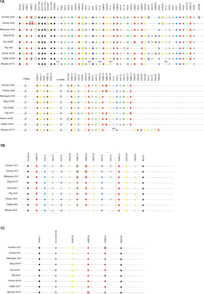
Histones have a long history of research in a wide range of species, leaving a legacy of complex nomenclature in the literature. Community-led discussions at the EMBO Workshop on Histone Variants in 2011 resulted in agreement amongst experts on a revised systematic protein nomenclature for histones, which is based on a combination of phylogenetic classification and historical symbol usage. Human and mouse histone gene symbols previously followed a genome-centric system that was not applicable across all vertebrate species and did not reflect the systematic histone protein nomenclature. This prompted a collaboration between histone experts, the Human Genome Organization (HUGO) Gene Nomenclature Committee (HGNC) and Mouse Genomic Nomenclature Committee (MGNC) to revise human and mouse histone gene nomenclature aiming, where possible, to follow the new protein nomenclature whilst conforming to the guidelines for vertebrate gene naming. The updated nomenclature has also been applied to orthologous histone genes in chimpanzee, rhesus macaque, dog, cat, pig, horse and cattle, and can serve as a framework for naming other vertebrate histone genes in the future.


Conflict of interest statement

The authors declare that they have no competing interests.

Figures

Fig. 1
Fig. 1
The three replication-dependent histone gene clusters in mammals. Gene symbols are shown across the top; species and chromosomal location of each cluster is indicated at the side. Black = non-histone genes, pink = histone H1 genes, yellow = H2A genes, red = H2B genes, blue = H3 genes, green = H4 genes. Pseudogenes are indicated by a gray box around the gene (pseudogenes have the same symbol as their protein-coding orthologs but end with -ps for mouse and P for the other species). A paler shade indicates that the gene is present but currently unannotated and unnamed; a blank space indicates that the gene is missing entirely. Mouse H1 genes contain an ‘f’ in place of the hyphen, so each mouse H1f symbol is shown above each relevant mouse gene. A: The largest replication-dependent cluster, also known as HIST1. There are two large gaps in the cluster in all species. Conservation between species is remarkable, although there are some species-specific duplications, gene losses and in situ pseudogenizations. Mouse has an expansion at the end of the cluster—these genes are shown with the mouse gene symbol format; note that all mouse symbols follow this format but for simplicity only the uppercase format used for other mammalian genes is shown for the conserved genes. B. The second largest replication-dependent cluster, also known as HIST2; each species has at least 10 genes in this cluster that contains genes for the 4 core histones but contains no histone H1 genes. The cluster contains a large inverted repeat, indicated by brackets. C. The third mammalian replication-dependent cluster, also known as HIST3. Note that H3-4 has an exceptional symbol due to the common usage of the H3.4 symbol for the protein encoded by this gene, the systematic H3C16 alias is shown in parentheses

References

    1. Marzluff WF, Wagner EJ, Duronio RJ. Metabolism and regulation of canonical histone mRNAs: life without a poly(A) tail. Nat Rev Genet. 2008;9:843–854. doi: 10.1038/nrg2438. - DOI - PMC - PubMed
    1. Lyons SM, Cunningham CH, Welch JD, Groh B, Guo AY, Wei B, et al. A subset of replication-dependent histone mRNAs are expressed as polyadenylated RNAs in terminally differentiated tissues. Nucleic Acids Res. 2016;44:9190–9205. - PMC - PubMed
    1. Kemp JP, Jr, Yang X-C, Dominski Z, Marzluff WF, Duronio RJ. Superresolution light microscopy of the Drosophila histone locus body reveals a core-shell organization associated with expression of replication-dependent histone genes. Mol Biol Cell. 2021;32:942–955. doi: 10.1091/mbc.E20-10-0645. - DOI - PMC - PubMed
    1. Kaya-Okur HS, Wu SJ, Codomo CA, Pledger ES, Bryson TD, Henikoff JG, et al. CUT&Tag for efficient epigenomic profiling of small samples and single cells. Nat Commun. 2019;10:1930. doi: 10.1038/s41467-019-09982-5. - DOI - PMC - PubMed
    1. Ye X, Wei Y, Nalepa G, Harper JW. The cyclin E/Cdk2 substrate p220(NPAT) is required for S-phase entry, histone gene expression, and cajal body maintenance in human somatic cells. Mol Cell Biol. 2003;23:8586–8600. doi: 10.1128/MCB.23.23.8586-8600.2003. - DOI - PMC - PubMed

Publication types

LinkOut - more resources