. 2023 Feb 1;44(2):612-628.
doi: 10.1002/hbm.26088. Epub 2022 Oct 1.

Multi-atlas thalamic nuclei segmentation on standard T1-weighed MRI with application to normal aging

Affiliations

Multi-atlas thalamic nuclei segmentation on standard T1-weighed MRI with application to normal aging

Adolf Pfefferbaum et al. Hum Brain Mapp. .

Abstract

Specific thalamic nuclei are implicated in healthy aging and age-related neurodegenerative diseases. However, few methods are available for robust automated segmentation of thalamic nuclei. The threefold aims of this study were to validate the use of a modified thalamic nuclei segmentation method on standard T1 MRI data, to apply this method to quantify age-related volume declines, and to test functional meaningfulness by predicting performance on motor testing. A modified version of THalamus Optimized Multi-Atlas Segmentation (THOMAS) generated 22 unilateral thalamic nuclei. For validation, we compared nuclear volumes obtained from THOMAS parcellation of white-matter-nulled (WMn) MRI data to T1 MRI data in 45 participants. To examine the effects of age/sex on thalamic nuclear volumes, T1 MRI available from a second data set of 121 men and 117 women, ages 20-86 years, were segmented using THOMAS. To test for functional ramifications, composite regions and constituent nuclei were correlated with Grooved Pegboard test scores. THOMAS on standard T1 data showed significant quantitative agreement with THOMAS from WMn data, especially for larger nuclei. Sex differences revealing larger volumes in men than women were accounted for by adjustment with supratentorial intracranial volume (sICV). Significant sICV-adjusted correlations between age and thalamic nuclear volumes were detected in 20 of the 22 unilateral nuclei and whole thalamus. Composite Posterior and Ventral regions and Ventral Anterior/Pulvinar nuclei correlated selectively with higher scores from the eye-hand coordination task. These results support the use of THOMAS for standard T1-weighted data as adequately robust for thalamic nuclear parcellation.

Keywords: T1-weighted MRI; aging; thalamic nuclei segmentation; thalamus; white matter nulled MRI.


Figures

FIGURE 1
FIGURE 1
The modified THOMAS pipeline (a) and results from THOMAS parcellation on a T1 MRI data set (b). The major difference between this pipeline and the original pipeline developed for WMn MRI data is the use of majority voting for label fusion which enables the use of WMn prior data with standard T1 input data
FIGURE 2
FIGURE 2
Left and right volumes for the 11 nuclei from the THOMAS parcellation from T1 data plotted against THOMAS parcellation from WMn data
FIGURE 3
FIGURE 3
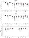
Dice similarity coefficients for the left and right whole thalamus (THAL) and 11 thalamic nuclei as well as the four composite thalamic regions for both hemispheres
FIGURE 4
FIGURE 4
Scatterplots for the left and right whole thalamus as a function of supratentorial intracranial volume (sICV) (left plot) and volume adjusted for sICV (right plot). Women are red, and men are blue. Note that women have smaller thalami and sICV, but the difference between men and women is removed when volumes are adjusted for head size. Also note the significantly smaller volumes across the age span
FIGURE 5
FIGURE 5
sICV‐adjusted volume as a function of age for the left and right whole thalamus and 11 nuclei. Women are red, and men are blue. Linear regression fits are presented for all plots plus quadratic fits for the five measures for which the quadratic was better than the linear fit
FIGURE 6
FIGURE 6
sICV‐adjusted volume as a function of time to complete the Grooved Pegboard task for the left and right whole thalamus and four combined thalamic regions: Anterior=AV; Ventral=VA+VLa+VLp+VPl; Posterior=Pul+LGN+MGN; and Medial=CM+MD+Hb

Similar articles

Cited by

References

    1. Aggleton, J. P. , Pralus, A. , Nelson, A. J. , & Hornberger, M. (2016). Thalamic pathology and memory loss in early Alzheimer's disease: moving the focus from the medial temporal lobe to Papez circuit. Brain, 139(Pt 7), 1877–1890. 10.1093/brain/aww083 - DOI - PMC - PubMed
    1. Azghadi, A. , Rajagopal, M. M. , Atkinson, K. A. , & Holloway, K. L. (2021). Utility of GPI+VIM dual‐lead deep brain stimulation for Parkinson's disease patients with significant residual tremor on medication. Journal of Neurosurgery, 136, 1364–1370. 10.3171/2021.4.JNS21502 - DOI - PubMed
    1. Bagarinao, E. , Watanabe, H. , Maesawa, S. , Kawabata, K. , Hara, K. , & Ohdake, R. (2022). Reserve and Maintenance in the Aging Brain: A Longitudinal Study of Healthy Older Adults. eNeuro, 9(1). 10.1523/ENEURO.0455-21.2022 - DOI - PMC - PubMed
    1. Bao, S. , Bermudez, C. , Huo, Y. , Parvathaneni, P. , Rodriguez, W. , & Resnick, S. M. (2019). Registration‐based image enhancement improves multi‐atlas segmentation of the thalamic nuclei and hippocampal subfields. Magnetic Resonance Imaging, 59, 143–152. 10.1016/j.mri.2019.03.014 - DOI - PMC - PubMed
    1. Bernstein, A. S. , Rapcsak, S. Z. , Hornberger, M. , Saranathan, M. , & Alzheimer's Disease Neuroimaging, I . (2021). Structural changes in thalamic nuclei across prodromal and clinical Alzheimer's disease. Journal of Alzheimer's Disease, 82(1), 361–371. 10.3233/JAD-201583 - DOI - PubMed

Publication types