. 2022 Nov;298(11):102547.
doi: 10.1016/j.jbc.2022.102547. Epub 2022 Sep 28.

Computational and functional studies of the PI(4,5)P2 binding site of the TRPM3 ion channel reveal interactions with other regulators

Affiliations

Computational and functional studies of the PI(4,5)P2 binding site of the TRPM3 ion channel reveal interactions with other regulators

Siyuan Zhao et al. J Biol Chem. 2022 Nov.

Abstract

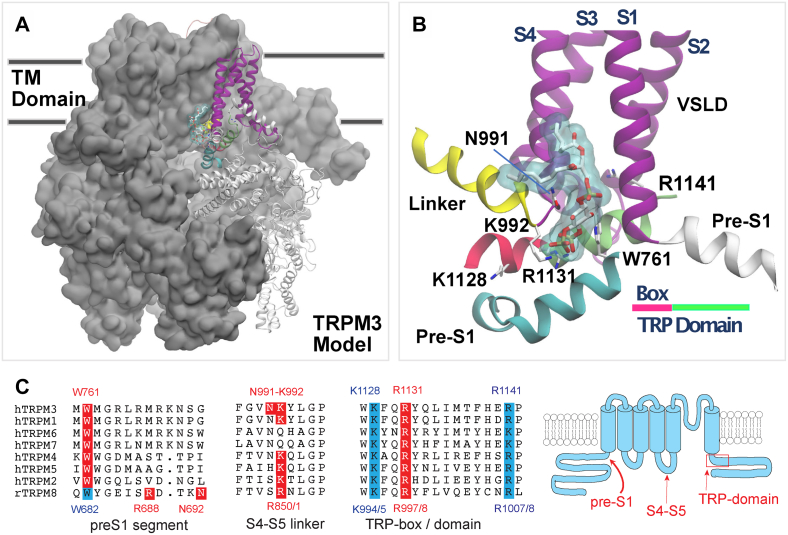
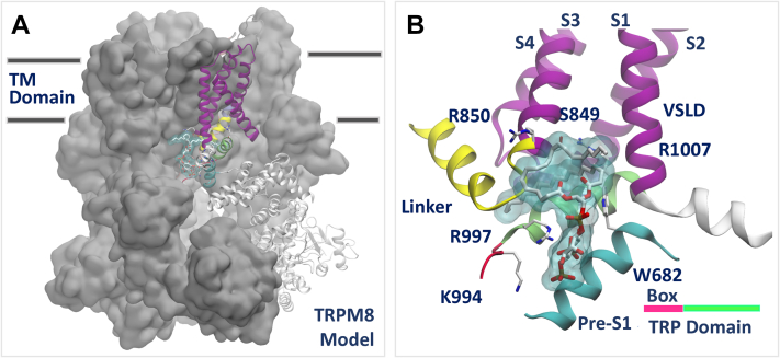
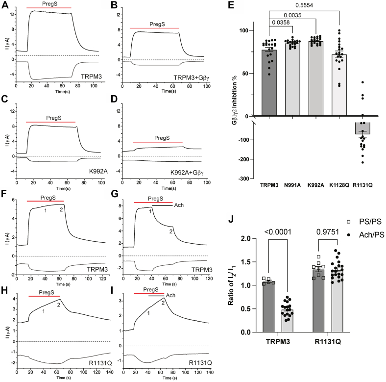
Transient receptor potential melastatin 3 (TRPM3) is a heat-activated ion channel expressed in peripheral sensory neurons and the central nervous system. TRPM3 activity depends on the membrane phospholipid phosphatidylinositol 4,5-bisphosphate (PI(4,5)P2), but the molecular mechanism of activation by PI(4,5)P2 is not known. As no experimental structure of TRPM3 is available, we built a homology model of the channel in complex with PI(4,5)P2via molecular modeling. We identified putative contact residues for PI(4,5)P2 in the pre-S1 segment, the S4-S5 linker, and the proximal C-terminal TRP domain. Mutating these residues increased sensitivity to inhibition of TRPM3 by decreasing PI(4,5)P2 levels. Changes in ligand-binding affinities via molecular mechanics/generalized Born surface area (MM/GBSA) showed reduced PI(4,5)P2 affinity for the mutants. Mutating PI(4,5)P2-interacting residues also reduced sensitivity for activation by the endogenous ligand pregnenolone sulfate, pointing to an allosteric interaction between PI(4,5)P2 and pregnenolone sulfate. Similarly, mutating residues in the PI(4,5)P2 binding site in TRPM8 resulted in increased sensitivity to PI(4,5)P2 depletion and reduced sensitivity to menthol. Mutations of most PI(4,5)P2-interacting residues in TRPM3 also increased sensitivity to inhibition by Gβγ, indicating allosteric interaction between Gβγ and PI(4,5)P2 regulation. Disease-associated gain-of-function TRPM3 mutations on the other hand resulted in no change of PI(4,5)P2 sensitivity, indicating that mutations did not increase channel activity via increasing PI(4,5)P2 interactions. Our data provide insight into the mechanism of regulation of TRPM3 by PI(4,5)P2, its relationship to endogenous activators and inhibitors, as well as identify similarities and differences between PI(4,5)P2 regulation of TRPM3 and TRPM8.

Keywords: TRP channel; TRPM3; computational modeling; ion channel; phosphoinositide.


Conflict of interest statement

Conflict of interest The authors declare that they have no conflicts of interest with the contents of this article.

Figures

Figure 1
Figure 1
Model of TRPM3 in complex with a PI(4,5)P2phospholipid. The homology model of TRPM3 was built based on the structure of TRPM4 (PDB ID: 6BCJ) (29) as described in the Results and Experimental procedures sections. For visualization purposes, only one molecule of the phospholipid is shown. A, view from the transmembrane (TM) plane of TRPM3 tetramer. Protein atoms of three of the four protomers are shown in surface representation, colored in gray. In the fourth protomer, protein atoms are shown in new cartoon representation. Atoms in the pre-S1 domain (two separate ranges), voltage sensor–like domain (VSDL), and the S4–S5 linker are shown in new cartoon representation, colored in cyan and white, magenta, and yellow, respectively. The TRP domain is colored in bright red (TRP-box) and green. The remaining structural elements in the protomer are shown in white (transparent). The atoms of the phospholipid are shown in licorice representation, with C, N, and O atoms colored in white, blue, and red, respectively. For visualization purposes, only Cα and protein side-chain atoms are shown. Phospholipid atoms are also shown in surface representation, colored in light blue (transparent). In (B), close-up view of the PI(4,5)P2 binding site in TRPM3. Protein atoms are represented as new cartoons. Phospholipid atoms are represented as in (A). C, sequence alignment and cartoon of the PI(4,5)P2-interacting regions in TRPM3 and TRPM8. Red residues are in contact with PI(4,5)P2 in the TRPM8 cryo-EM structure(s) and/or in our TRPM3 model, when conserved in other TRPM channels, they are also labeled red. Residues in cyan in the TRP domain were experimentally characterized in this study or in Ref. (36). The location of the W682 residue in TRPM8 that we experimentally characterized is also noted here in cyan. The dual numbering S4–S5 loop and in the TRP domain indicates the difference in numbering between the rTRPM8 that we use in experiments and the fcTRPM8 used in cryo-EM studies. The MHR4 region in TRPM8 that contains the K605 PI(4,5)P2 contact residue in TRPM8 is not shown, as its sequence is not conserved in TRPM3. PDB, Protein Data Bank; PI(4,5)P2, phosphatidylinositol 4,5-bisphosphate; TRPM3, transient receptor potential melastatin 3.
Figure 2
Figure 2
Refined model of TRPM8 in complex with PI(4,5)P2. PI(4,5)P2 was docked to the apo structure of the fcTRPM8 (PDB ID: 6BPQ) (30) as described in the Results and Experimental procedures sections. For visualization purposes, only one molecule of PI(4,5)P2 is shown. A, view from the transmembrane (TM) plane of TRPM8 tetramer. B, close-up view of the PI(4,5)P2 binding site in TRPM8. All representations are reproduced as for Figure 1. fcTRPM8, flycatcher apo TRPM8; PI(4,5)P2, phosphatidylinositol 4,5-bisphosphate; TRPM8, transient receptor potential melastatin 8.
Figure 3
Figure 3
Mutating putative PI(4,5)P2-interacting residues increases sensitivity of TRPM3 to inhibition by PI(4,5)P2depletion. Human TRPM31325 splice variant or its mutants were expressed in Xenopus oocytes, and two-electrode voltage-clamp (TEVC) experiments were performed to measure the activity of TRPM3 as described in the Experimental procedures section using a ramp protocol from −100 mV to +100 mV. Currents induced by 50 μM PregS were measured before and after treatment with 35 μM wortmannin to deplete PI(4,5)P2. A and B, representative traces of WT TRPM3 (A) and K992A mutant (B) before (left) and after (right) treatment of the same oocyte with 35 μM wortmannin for 2 h. Top traces show currents at +100 mV; dash lines indicate zero current; bottom traces show currents at −100 mV. Applications of 50 μM PregS are indicated by red lines. CG, data summary of the percentage inhibition of PregS-induced currents by wortmannin treatment at 100 mV for different mutants: W761F (C), N991A (D), K992A (E), R1131Q and R1141Q (F), and K1128Q (G). HL, current amplitudes of various mutants at 100 mV. Each symbol represents measurement of one oocyte from two independent preparations. Statistical significance was calculated with t test, or one-way ANOVA (F and K), p values are shown on bar graphs. For the overall ANOVA, F = 6.78, p = 0.0023 for panel F, and F = 13.01, p < 0.0001 for panel K. Individual panels show summary of measurements performed on the same experimental days. PI(4,5)P2, phosphatidylinositol 4,5-bisphosphate; PregS, pregnenolone sulfate; TRPM3, transient receptor potential melastatin 3.
Figure 4
Figure 4
Effect of mutations in the PI(4,5)P2binding site residues of TRPM3. Binding affinity changes were calculated via the MM/GBSA method, as described in the Experimental procedures section. A, change in binding affinity (ΔΔG; kcal/mol) upon mutating protein residues in silico in the PI(4,5)P2 binding site of TRPM3. In blue, mutants that bind significantly worse than the native protein, indicating loss of interaction with PI(4,5)P2 upon mutations. In gray, mutants with no significant effect on binding. BF, binding mode of PI(4,5)P2 to native and mutant TRPM3 channels. In (B), native TRPM3. In (CF), for any mutant with significant loss of interaction with the phospholipid (in blue color in A), the effect of each individual mutation is shown by visualizing the mutated residue superposed to the native counterpart; the loss of any hydrogen bond or salt bridge interactions with the phospholipid is highlighted in oval shapes. Protein atoms are shown in new cartoon representation, in gray and cyan color for native and mutant channels, respectively. For visualization purposes, hydrogens not engaged in interactions with the protein–ligand interactions are hidden. Ligand atoms are in licorice representation, with C, O, H, and P atoms colored in gray, red, white, and yellow, respectively. Hydrogen bonds and salt bridges are represented as dotted lines. MM/GBSA, molecular mechanics/generalized Born surface area; PI(4,5)P2, phosphatidylinositol 4,5-bisphosphate; TRPM3, transient receptor potential melastatin 3.
Figure 5
Figure 5
Mutating putative PI(4,5)P2-interacting residues increases sensitivity of TRPM8 to inhibition by PI(4,5)P2depletion. Rat TRPM8 or its mutants were expressed in Xenopus oocytes. TEVC experiments were performed as described in the Experimental procedures section using ramp protocol from −100 mV to +100 mV. Menthol (500 μM) was applied to activate TRPM8 channels, and 35 μM wortmannin was applied for 2 h to deplete PI(4,5)P2. AF, representative traces of TRPM8 before (A) and after wortmannin treatment (B), R851A before (C) and after wortmannin treatment (D) and W682Q before (E) and after wortmannin treatment (F). Top traces show currents at 100 mV; dash lines indicate 0 current; bottom traces show currents at −100 mV. Applications of 500 μM menthol are indicated by red lines. G, summary of inhibition evoked wortmannin treatment at 100 mV (basal plus menthol-induced current after leak subtraction) plotted for WT, R851A, and W682Q. H and I, current amplitudes of all three groups at 100 mV (H) and −100 mV (I). Each symbol represents an individual oocyte. All experiments were from two to three independent preparations. Statistical significance was calculated with one-way ANOVA. p Values for individual comparisons are shown on bar graphs. For the overall ANOVA, F = 49.08, p < 0.0001 for panel G, and F = 53.25, p < 0.0001 for panel H, and F = 122.7, p < 0.0001 for panel I. PI(4,5)P2, phosphatidylinositol 4,5-bisphosphate; TEVC, two-electrode voltage clamp; TRPM3, transient receptor potential melastatin 8.
Figure 6
Figure 6
Mutating putative PI(4,5)P2-interacting residues decreases sensitivity of TRPM8 to menthol activation. TEVC experiments were performed using a ramp protocol from −100 mV to 100 mV, as described in the Experimental procedures section. AC, representative traces of TRPM8 (A), R851A (B), and W682Q (C). Top traces show currents at +100 mV; dash lines indicate zero current; bottom traces show currents at −100 mV. Applications of various concentrations of menthol (μM) are indicated by red lines. D, Hill1 fit of the concentration dependence of menthol at 100 mV for TRPM8 and mutated channels. Currents were initially normalized to the current evoked by 500 μM menthol, then renormalized to the maximum current from the hill fits for plotting. Symbols represent mean ± SD for n = 12 to 13 measurements from two different oocyte preparations. PI(4,5)P2, phosphatidylinositol 4,5-bisphosphate; TEVC, two-electrode voltage clamp; TRPM8, transient receptor potential melastatin 8.
Figure 7
Figure 7
Mutating putative PI(4,5)P2-interacting residues decreases sensitivity of TRPM3 to PregS activation. RNAs coding for either the TRPM31325 splice variant or its mutants were injected into Xenopus oocytes. TEVC was performed to measure TRPM3 currents using a ramp protocol from −100 mV to 100 mV, as described in the Experimental procedures section. AC, representative traces of hTRPM3 WT (A), N991A (B), and K992A (C). Top traces show currents at +100 mV; dash lines indicate zero current; bottom traces show currents at −100 mV. Applications of various concentrations of PregS (micromolar) are indicated by red lines. D, Hill1 fit of the concentration dependence of PregS at 100 mV for TRPM3 and mutated channels. Currents were initially normalized to the current evoked by 100 μM PregS and then renormalized to the maximum current from the hill fits for plotting. Symbols represent mean ± SD for n = 12 to 13 measurements from two different oocyte preparations. hTRPM3, human TRPM3; PI(4,5)P2, phosphatidylinositol 4,5-bisphosphate; PregS, pregnenolone sulfate; TEVC, two-electrode voltage clamp; TRPM3, transient receptor potential melastatin 3.
Figure 8
Figure 8
Relationship between Gβγ and PI(4,5)P2regulation of TRPM3. hTRPM3 or its mutants were expressed in oocytes with or without Gβ1γ2 subunits for AE or with muscarinic hM2 receptors for FJ. TEVC was used to measure channel activity using a ramp protocol from −100 mV to 100 mV as described in the Experimental procedures section. AD, representative traces of TRPM3 (A), TRPM3 coexpressed with Gβγ subunits (B), K992A (C) and K992A coexpressed with Gβγ subunits (D). E, data summary shows the inhibition caused by Gβγ subunits in different mutant groups. Percentages of inhibition were calculated by normalizing decreased current amplitudes to the average currents induced by 50 μM PregS in control oocytes without Gβγ subunits. FJ, representative traces of TRPM3 treated with PregS alone (F), TRPM3 treated with PregS and acetylcholine (G), R1131Q treated with PregS alone (H), and TRPM3 treated with PregS and acetylcholine (I). Top traces show currents at +100 mV; dash lines indicate zero current; bottom traces show currents at −100 mV. Application of 50 μM PregS is indicated by red lines, and application of 5 μM acetylcholine is indicated by black lines. PregS-induced currents in the R1131Q mutant channels showed a continuous increase after application of PregS, which necessitated comparison to control cells where PregS was applied for the same length of time without the application of ACh (H). J, data summary shows the ratio of current amplitudes between points 2 and 1 indicated on the representative traces. Each symbol represents an individual oocyte from three (E) and two (J) independent preparations. Statistical significance was calculated with one-way ANOVA (E) (F = 9.025, p = 0.001) and two-way ANOVA (J) (F = 29.03 and p < 0.0001) for interaction between ACh and mutation. p Values for post hoc individual comparisons are shown on bar graphs. ACh, acetylcholine; hM2, human M2 muscarinic receptor; hTRPM3, human TRPM3; PI(4,5)P2, phosphatidylinositol 4,5-bisphosphate; PregS, pregnenolone sulfate; TEVC, two-electrode voltage clamp; TRPM3, transient receptor potential melastatin 3.
Figure 9
Figure 9
Disease-associated gain-of-function mutants do not change the TRPM3 sensitivity to the PI(4,5)P2depletion. hTRPM3, V990M, or P1090Q was expressed in oocytes, and 35 μM wortmannin (2 h) was used to deplete PI(4,5)P2. TEVC was performed as described in the Experimental procedures section. AF, representative traces of hTRPM3 (A), hTRPM3 after wortmannin treatment (B), V990M (C), V990M after wortmannin treatment (D), P1090Q (E) and P1090Q after wortmannin treatment (F). Top traces show currents at +100 mV; dash lines indicate zero current; bottom traces show currents at −100 mV. Application of 50 μM PregS is indicated by red lines. G, data summary of wortmannin inhibition of currents induced by 50 μM PregS. H, data summary of wortmannin inhibition of currents induced by EC50 concentrations of PregS, 17 μM, 0.6 μM, and 7 μM for hTRPM3, V990M, and P1090Q, respectively. Symbols represent individual oocytes from three (G) and two (H) different preparations. hTRPM3, human TRPM3; PI(4,5)P2, phosphatidylinositol 4,5-bisphosphate; PregS, pregnenolone sulfate; TEVC, two-electrode voltage clamp; TRPM3, transient receptor potential melastatin 3.
None

References

    1. Oberwinkler J., Philipp S.E. Trpm3. Handb Exp. Pharmacol. 2014;222:427–459. - PubMed
    1. Wagner T.F., Loch S., Lambert S., Straub I., Mannebach S., Mathar I., et al. Transient receptor potential M3 channels are ionotropic steroid receptors in pancreatic beta cells. Nat. Cell Biol. 2008;10:1421–1430. - PubMed
    1. Held K., Kichko T., De Clercq K., Klaassen H., Van Bree R., Vanherck J.C., et al. Activation of TRPM3 by a potent synthetic ligand reveals a role in peptide release. Proc. Natl. Acad. Sci. U. S. A. 2015;112:E1363–E1372. - PMC - PubMed
    1. Straub I., Krugel U., Mohr F., Teichert J., Rizun O., Konrad M., et al. Flavanones that selectively inhibit TRPM3 attenuate thermal nociception in vivo. Mol. Pharmacol. 2013;84:736–750. - PubMed
    1. Krugel U., Straub I., Beckmann H., Schaefer M. Primidone inhibits TRPM3 and attenuates thermal nociception in vivo. Pain. 2017;158:856–867. - PMC - PubMed

Publication types

MeSH terms