. 2022 Nov:185:106476.
doi: 10.1016/j.phrs.2022.106476. Epub 2022 Sep 28.

Pharmacological targeting of G protein-coupled receptor heteromers

Affiliations

Pharmacological targeting of G protein-coupled receptor heteromers

Estefanía Moreno et al. Pharmacol Res. 2022 Nov.

Abstract

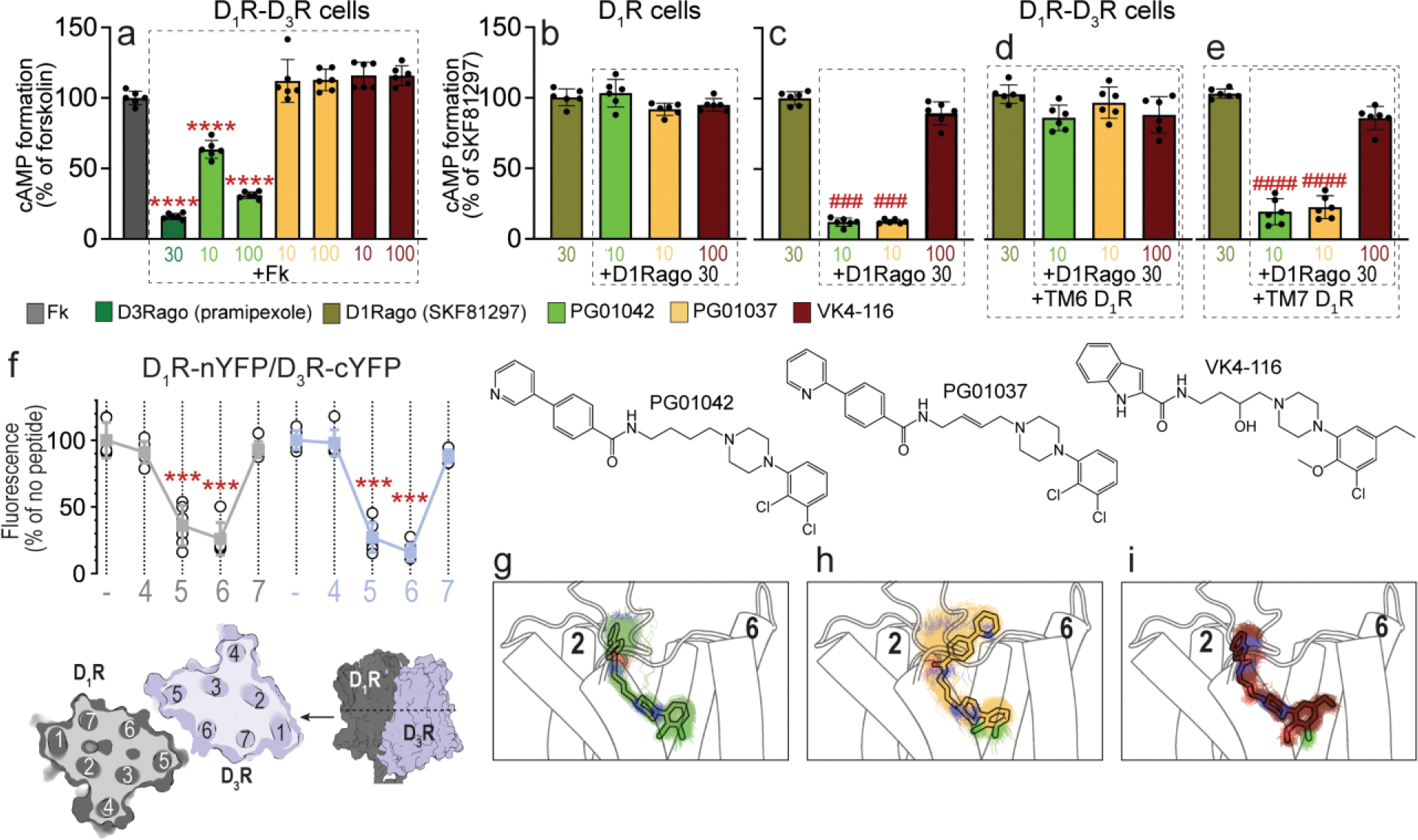
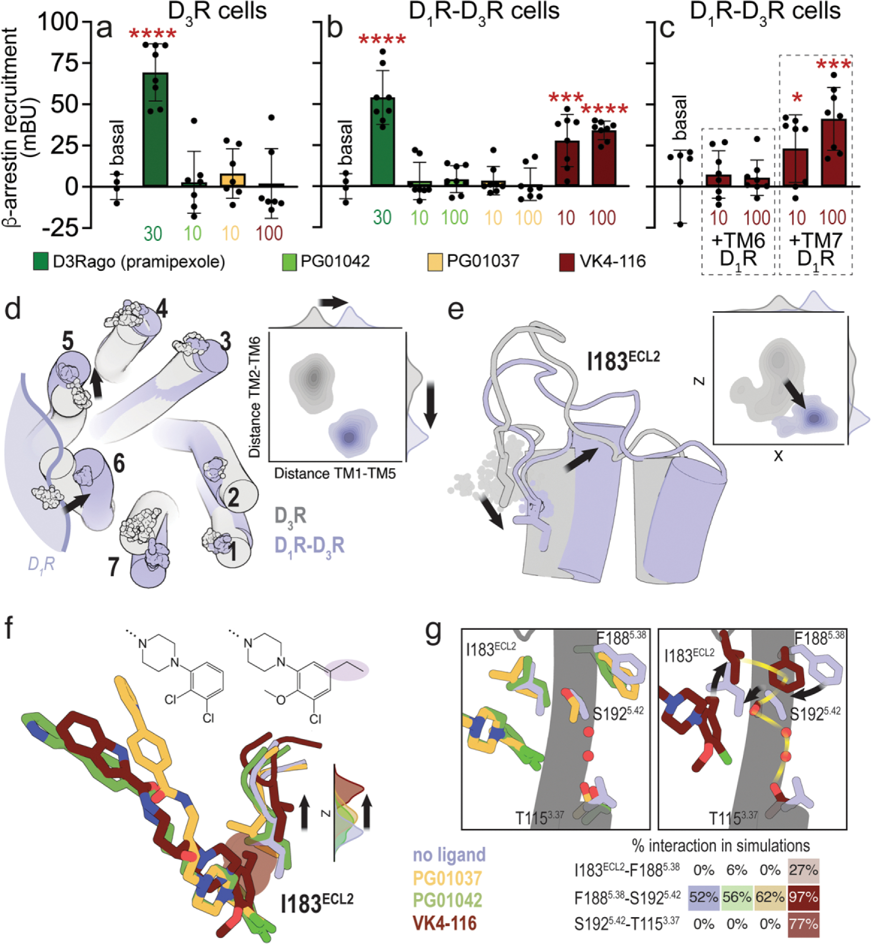
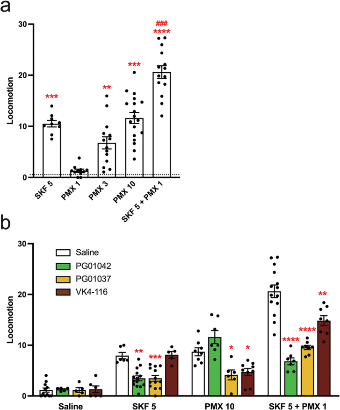
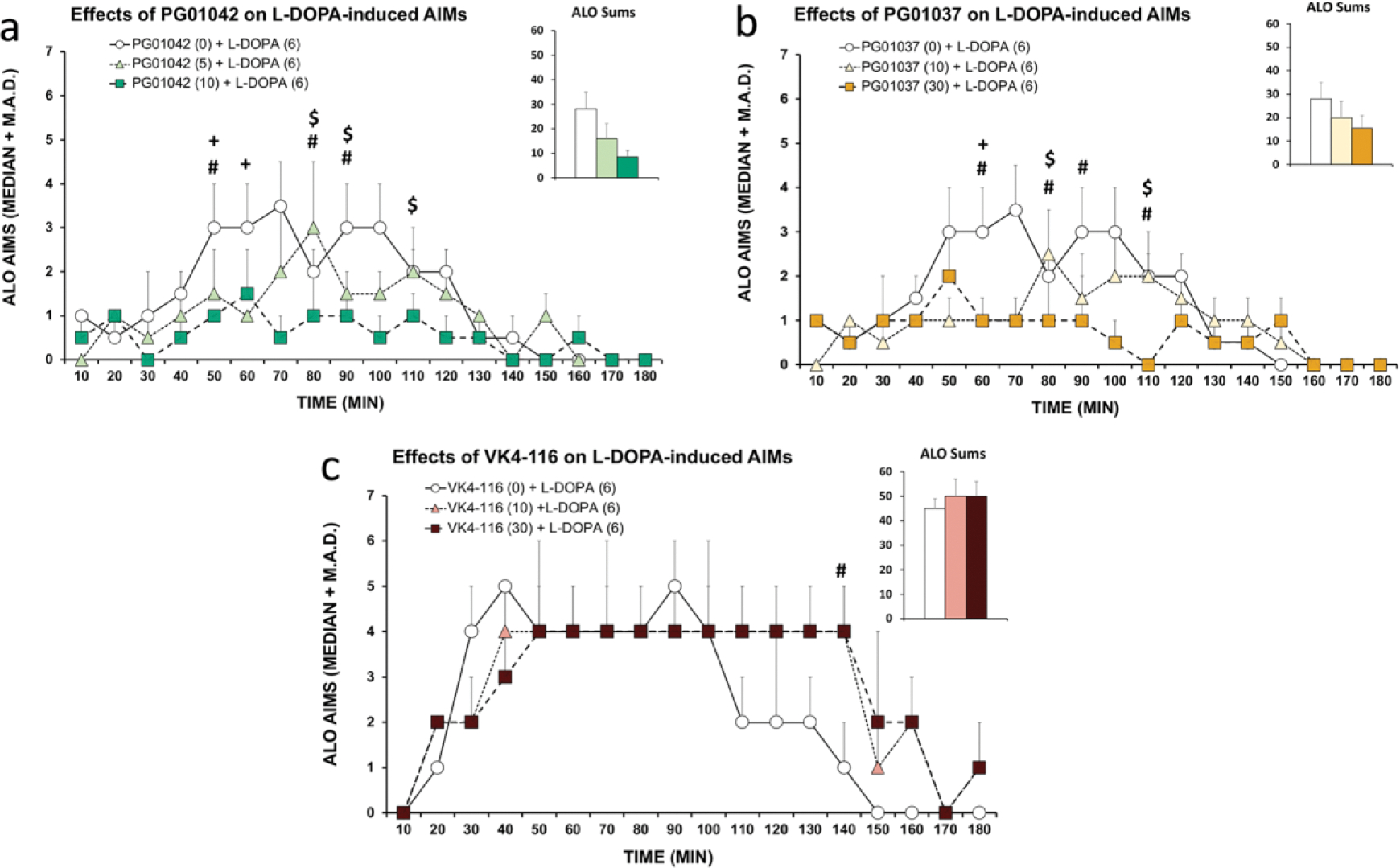
A main rationale for the role of G protein-coupled receptor (GPCR) heteromers as targets for drug development is the putative ability of selective ligands for specific GPCRs to change their pharmacological properties upon GPCR heteromerization. The present study provides a proof of concept for this rationale by demonstrating that heteromerization of dopamine D1 and D3 receptors (D1R and D3R) influences the pharmacological properties of three structurally similar selective dopamine D3R ligands, the phenylpiperazine derivatives PG01042, PG01037 and VK4-116. By using D1R-D3R heteromer-disrupting peptides, it could be demonstrated that the three D3R ligands display different D1R-D3R heteromer-dependent pharmacological properties: PG01042, acting as G protein-biased agonist, counteracted D1R-mediated signaling in the D1R-D3R heteromer; PG01037, acting as a D3R antagonist cross-antagonized D1R-mediated signaling in the D1R-D3R heteromer; and VK4-116 specifically acted as a ß-arrestin-biased agonist in the D1R-D3R heteromer. Molecular dynamics simulations predicted potential molecular mechanisms mediating these qualitatively different pharmacological properties of the selective D3R ligands that are dependent on D1R-D3R heteromerization. The results of in vitro experiments were paralleled by qualitatively different pharmacological properties of the D3R ligands in vivo. The results supported the involvement of D1R-D3R heteromers in the locomotor activation by D1R agonists in reserpinized mice and L-DOPA-induced dyskinesia in rats, highlighting the D1R-D3R heteromer as a main pharmacological target for L-DOPA-induced dyskinesia in Parkinson's disease. More generally, the present study implies that when suspecting its pathogenetic role, a GPCR heteromer, and not its individual GPCR units, should be considered as main target for drug development.

Keywords: Dopamine D(1) receptor; Dopamine D(3) receptor; G protein-coupled receptor (GPCR) heteromers; L-DOPA-induced dyskinesia; Locomotor activation; Mouse; PG01037 (PubChem CID: 11477180); PG01042 (PubChem CID: 11443078); Pramipexole (PubChem CID: 119570); Rat; SKF81297 (PubChem CID: 1218); VK4–116 (PubChem CID: 130431318).


Conflict of interest statement

Declaration of Competing Interest The authors declare no competing interests.

Figures

Figure 1.
Figure 1.. Differential effects of PG01042, PG01037, and VK4-116 on G protein-dependent signaling in HEK-293T cells transfected with D3R and D1R.
a-e. Results from cAMP formation experiments in HEK-293T cells transfected with D1R-Rluc cDNA (1 μg) with or without D3R-YFP cDNA (1.5 μg) (D1R-D3R cells and D1R cells, respectively). In a, cells are treated with the D2-like receptor agonist pramipexole (D3Rago; 30 nM for 10 min) or the D3R ligands PG01042, PG01037 and VK4-116 (all at 10 or 100 nM for 15 min) before forskolin (Fk, 0.5 μM). In b-e, cells are pre-treated or not with D1R TM6 or TM7 peptides (4 μM for 4 h) and treated with PG01042 (10 nM), PG01037 (10 nM) and VK4-116 (100 nM) for 15 min before the D1R agonist SKF81297 (30 nM; D1Rago). Values of cAMP formation are shown as mean ± S.D. (n = 6) and expressed as percentage of Fk-treated or D1Rago-treated cells in each condition (100% represents 80–100 pmols cAMP/106 cells). ****: p < 0.0001 versus FK; ### and ####: p < 0.001 and p < 0.0001 versus D1Rago, respectively (one-way ANOVA followed by Tukey’s post hoc comparisons). f. Results from BiFC experiments in HEK-293T cells co-transfected with D1R-nYFP and D3R-cYFP in the absence (−) or the presence of the indicated TM peptides (at 4 μM) from D1R (gray symbols and plots) or D3R (blue symbols and plots). Fluorescence values (in means ± S.D.) are expressed as the percentage of the fluorescence in the absence (−) of the indicated TM peptides (n = 6, with triplicates); ***: p < 0.001 versus control values (one-way ANOVA followed by Dunnett’s post hoc comparisons). The schemes illustrate extracellular and parallel to the membrane views of the computational model of the D1R-D3R heteromer built using the TM 5/6 interface (see text). g-i. Representative structures (solid sticks) and evolution (lines) of PG01042 (g, in green), PG01037 (h, in orange) and VK4-116 (i, in purple) in complex with D3R (white cylinders, only the initial structure is shown) as devised from three replicas of unbiased 1μs MD simulations. The second pharmacophore unit of PG01042 and VK4-116 remained stable at the ECD near TM 2, whereas this part of PG01037 favors its interaction with TM 6 (see Suppl. Fig. 2).
Figure 2.
Figure 2.. Differential effects of PG01042, PG01037, and VK4-116 on β-arrestin recruitment in HEK-293T cells transfected with D3R and D1R.
a-c. Results from β-arrestin recruitment experiments in HEK-293T cells transfected with β-arrestin-1-Rluc cDNA (0.5 μg), D3R-YFP cDNA (1 μg cDNA) with or without D1R cDNA (1.5 μg cDNA) (D1R-D3R cells and D3R cells, respectively). In a-b, cells are treated for 10 min with the D2-like receptor agonist pramipexole (D3Rago; 30 nM) or the D3R ligands PG01042, PG01037 and VK4-116 (10 or 100 nM). In c, cells are pre-treated or not with D1R TM6 or TM7 peptides (4 μM for 4 h) and with VK4-116 (10 or100 nM) for 15 min before the D1R agonist SKF81297 (30 nM; D1Rago). Coelenterazine H (5 μM) was added before pramipexole or the selective D3R ligands for 7 minutes and β-arrestin-1 recruitment was measured by BRET (see Material and Methods). Values are mean ± S.D. (n = 8). *, *** and ****: p < 0.05, p < 0.001 and p < 0.0001 versus basal, respectively (one-way ANOVA followed by Dunnett’s post hoc comparisons). d. Evolution of the Cα atoms (spheres) of Y321.35 in TM 1, L892.64 in TM 2, I1013.23 in TM 3, F1704.62 in TM 4, F1885.38 in TM 5, H3546.60 in TM 6, and P3627.32 in TM 7 during three replicas of unbiased 1 μs MD simulations of the D3R monomer (gray) and the D1R-D3R heteromer (blue) with no ligand bound. Contour plots of the distances between Y321.35 in TM 1 and F1885.38 in TM 5 (distance TM1-TM5) and between L892.64 in TM 2 and H3546.60 in TM 6 (distance TM2-TM6) during the MD simulations. Distributions of these distances are shown in the axes. e. Evolution of I183ECL2 (spheres), and contour plots and distributions of X,Z coordinates corresponding to the Cb atom of I183ECL2 during MD simulations. Black arrows represent the movements of D3R in the D1R-D3R heteromer (blue) relative to the D3R protomer (gray). f. Detailed view of I183ECL2 of D3R in the D1R-D3R heteromer during MD simulations with no ligand bound (blue) and PG01042 (green), PG01037 (orange) and VK4-116 (red) bound to D3R. Distributions of the Z coordinate corresponding to the Cα atom of I183ECL2 during MD simulations. The ethyl group of VK4-116 that triggers the upward movement of I183ECL2 is highlighted. g. Detailed views of I183ECL2, F1885.38, S1925.42, and T1153.37 of D3R in the D1R-D3R heteromer during MD simulations with no ligand bound (blue) and PG01042 (green), PG01037 (orange) and VK4-116 (red) bound to D3R. Frequency contacts (%) between side-chain residues, color-coded according to the ligand bound to D3R, as calculated with the GetContacts software. Black arrows represent the movements of these side chains of D3R relative to the unliganded D3R (blue). The xy plane is as defined by the Orientations of Proteins in Membranes (OPM) [73].
Figure 3.
Figure 3.. Differential effects of PG01042, PG01037, and VK4-116 on the locomotor activation induced by D1R and D2-like receptor agonists in reserpinized mice.
Mice were administered reserpine (5 mg/kg, s.c.) 20 h before administration of the D1R agonist SKF 81297 (SKF; 5 mg/kg, i.p.), the D2-like receptor agonist pramipexole (PMX; 1, 3 or 10 mg/kg, i.p.), or both, without (a) or with (b) previous administration (15 min before) of the D3R ligands PG01042 (10 mg/kg, i.p.), PG01037 (30 mg/kg, i.p.) or VK4-116 (10 mg/kg, i.p.). Values are mean ± S.E.M. (n = 5–18) and are expressed as the average of the transformed counts (squared root) obtained from the 10 min-periods recorded for 1 h. In a, **, *** and ****: p < 0.01, p < 0.001 and p < 0.0001 versus vehicle-treated animals (represented by the dotted line), respectively; ###: p < 0.001 versus SKF (one-way ANOVA followed by Tukey’s post hoc comparisons) (one-way ANOVA followed by Tukey’s post hoc comparisons). In b, *, **, *** and ****: p < 0.05, p < 0.01, p < 0.001 and p < 0.0001 versus respective saline-treated animals, respectively.
Figure 4.
Figure 4.. D1R, D2R, and D3R gene (DRD1, DRD2 and DRD3) expression in the mouse striatum.
a. RNAscope ISH results in the ventral striatum (framed area in the coronal section) showing strong expression of DRD1, DRD2 and DRD3 mRNA with a strong co-localization of DRD3 and DRD1 and limited expression of DRD3 in DRD2-expressing neurons. b. RNAscope ISH results in the dorsal striatum (framed area in the coronal section) showing a strong expression of DRD1 and DRD2 mRNA and limited expression of DRD3 mRNA, which predominates in DRD1-expressing neurons. Images were taken under 60X magnification. DRD1 in red, DRD2 in blue, DRD3 in green.
Figure 5.
Figure 5.. Differential effects of PG01042, PG01037, and VK4-116 on L-DOPA-induced dyskinesia.
In a within-subjects, counterbalanced design, hemiparkinsonian rats previously rendered dyskinetic were treated with the D3R ligands (a) PG01042 (0, 5, 10 mg/kg; i.p.), (b) PG01037 (0, 10, 30 mg/kg; i.p.), or (c) VK4-116 (VK; 0, 10, 30 mg/kg; i.p.) prior to administration of L-DOPA (6 mg/kg; s.c.). Rats were rated for Axial, Limb and Orolingual (ALO) abnormal involuntary movements (AIMs) every 10 min for 180 min post-injection. AIMs time course and ALO sums are expressed as median + median absolute deviation (M.A.D.). Data were analyzed by non-parametric Friedman ANOVAs with Wilcoxon Match Pairs post-hoc tests. + p < 0.05 for Vehicle versus low dose, # p < 0.05 for vehicle versus high dose, $ p < 0.05 for low dose versus high dose.

References

    1. Missale C, Nash SR, Robinson SW, Jaber M, Caron MG. Dopamine receptors: from structure to function. Physiol Rev. 1998;78:189–225. doi: 10.1152/physrev.1998.78.1.189 - DOI - PubMed
    1. Starr BS, Starr MS, Kilpatrick IC. Behavioural role of dopamine D1 receptors in the reserpine-treated mouse. Neuroscience. 1987;22:179–188. doi: 10.1016/0306-4522(87)90208-9 - DOI - PubMed
    1. Ferré S, Giménez-Llort L, Artigas F, Martínez E. Motor activation in short- and long-term reserpinized mice: role of N-methyl-D-aspartate, dopamine D1 and dopamine D2 receptors. Eur J Pharmacol. 1994;255:203–213. doi: 10.1016/0014-2999(94)90099-x - DOI - PubMed
    1. Schwartz JC, Diaz J, Bordet R, Griffon N, Perachon S, Pilon C, Ridray S, Sokoloff P. Functional implications of multiple dopamine receptor subtypes: the D1/D3 receptor coexistence. Brain Res Brain Res Rev. 1998;26:236–242. doi: 10.1016/s0165-0173(97)00046-5 - DOI - PubMed
    1. Fiorentini C, Busi C, Gorruso E, Gotti C, Spano P, Missale C. Reciprocal regulation of dopamine D1 and D3 receptor function and trafficking by heterodimerization. Mol Pharmacol. 2008;74:59–69. doi: 10.1124/mol.107.043885 - DOI - PubMed

Publication types

Substances