

P800SO3-PEG: a renal clearable bone-targeted fluorophore for theranostic imaging
- PMID: 36183117
- PMCID: PMC9526902
- DOI: 10.1186/s40824-022-00294-2
P800SO3-PEG: a renal clearable bone-targeted fluorophore for theranostic imaging
Abstract
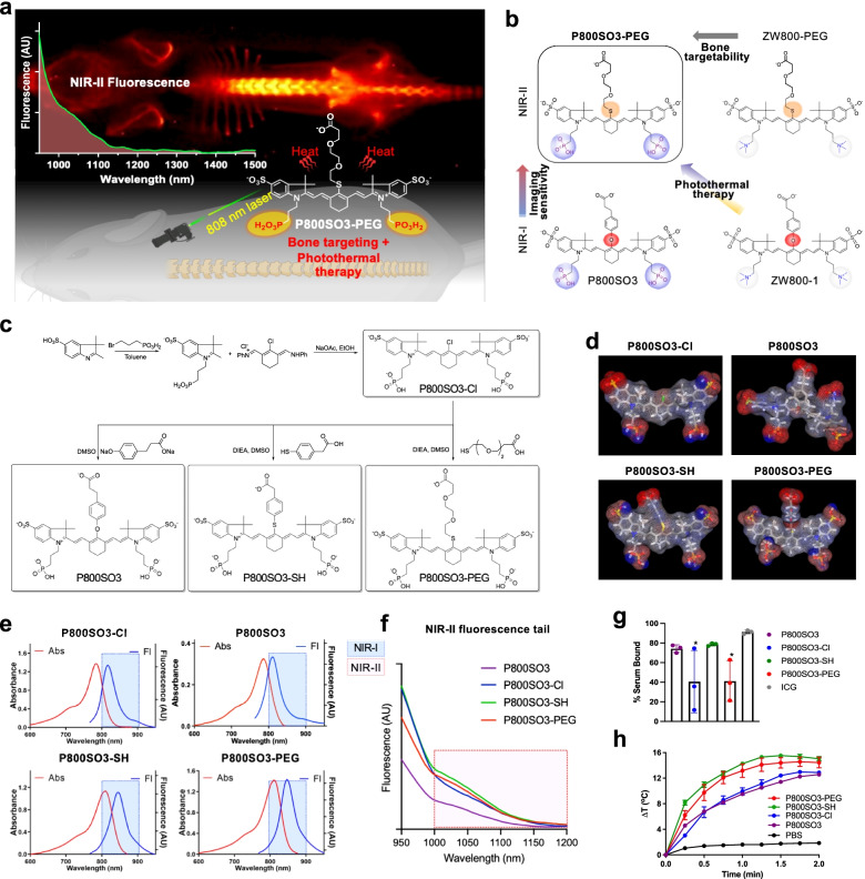
Background: Due to the deep tissue penetration and reduced scattering, NIR-II fluorescence imaging is advantageous over conventional visible and NIR-I fluorescence imaging for the detection of bone growth, metabolism, metastasis, and other bone-related diseases.
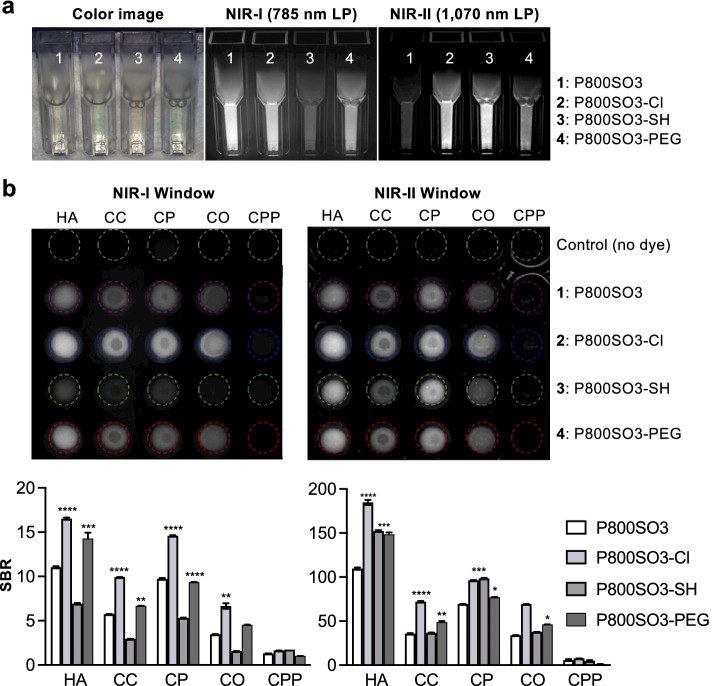
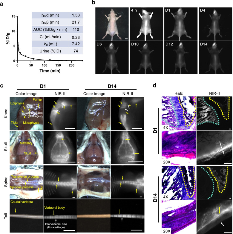
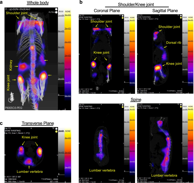
Methods: Bone-targeted heptamethine cyanine fluorophores were synthesized by substituting the meso-carbon with a sulfur atom, resulting in a bathochromic shift and increased fluorescence intensity. The physicochemical, optical, and thermal stability of newly synthesized bone-targeted NIR fluorophores was performed in aqueous solvents. Calcium binding, bone-specific targeting, biodistribution, pharmacokinetics, and 2D and 3D NIR imaging were performed in animal models.
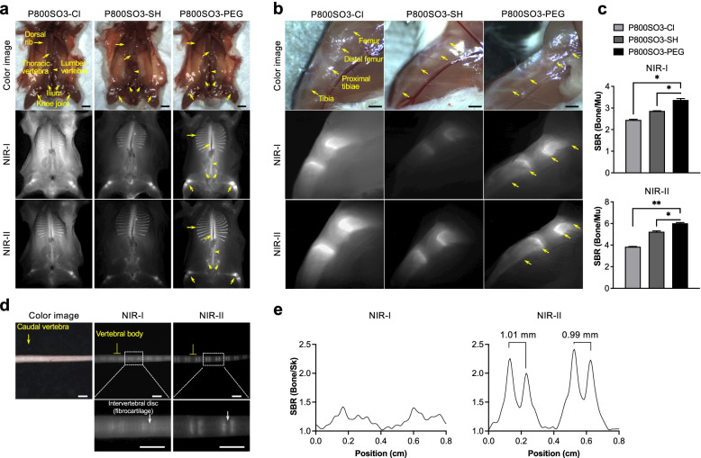
Results: The newly synthesized S-substituted heptamethine fluorophores demonstrated a high affinity for hydroxyapatite and calcium phosphate, which improved bone-specific targeting with signal-background ratios > 3.5. Particularly, P800SO3-PEG showed minimum nonspecific uptake, and most unbound molecules were excreted into the urinary bladder. Histological analyses demonstrated that P800SO3-PEG remained stable in the bone for over two weeks and was incorporated into bone matrices. Interestingly, the flexible thiol ethylene glycol linker on P800SO3-PEG induced a promising photothermal effect upon NIR laser irradiation, demonstrating potential theranostic imaging.
Conclusions: P800SO3-PEG shows a high affinity for bone tissues, deeper tissue imaging capabilities, minimum nonspecific uptake in the major organs, and photothermal effect upon laser irradiation, making it optimal for bone-targeted theranostic imaging.
Keywords: Bone targeting; NIR imaging; Renal clearance; Structure-inherent targeting; Targeted fluorophore.
© 2022. The Author(s).
Conflict of interest statement
The authors declare that they have no known competing financial interests or personal relationships that could have appeared to influence the work reported in this paper.
Figures
Similar articles
-
Near-Infrared Fluorescent Hydroxyapatite Nanoparticles for Targeted Photothermal Cancer Therapy.Pharmaceutics. 2023 Apr 29;15(5):1374. doi: 10.3390/pharmaceutics15051374. Pharmaceutics. 2023. PMID: 37242617 Free PMC article.
-
ZW800-PEG: A Renal Clearable Zwitterionic Near-Infrared Fluorophore for Potential Clinical Translation.Angew Chem Int Ed Engl. 2021 Jun 14;60(25):13847-13852. doi: 10.1002/anie.202102640. Epub 2021 May 17. Angew Chem Int Ed Engl. 2021. PMID: 33857346 Free PMC article.
-
Tumor-Targeted ZW800-1 Analog for Enhanced Tumor Imaging and Photothermal Therapy.Pharmaceutics. 2021 Oct 9;13(10):1648. doi: 10.3390/pharmaceutics13101648. Pharmaceutics. 2021. PMID: 34683940 Free PMC article.
-
Near-Infrared Contrast Agents for Bone-Targeted Imaging.Tissue Eng Regen Med. 2019 Aug 19;16(5):443-450. doi: 10.1007/s13770-019-00208-9. eCollection 2019 Oct. Tissue Eng Regen Med. 2019. PMID: 31624700 Free PMC article. Review.
-
Structure-Inherent Targeting of Near-Infrared Fluorophores for Image-Guided Surgery.Chonnam Med J. 2017 May;53(2):95-102. doi: 10.4068/cmj.2017.53.2.95. Epub 2017 May 25. Chonnam Med J. 2017. PMID: 28584787 Free PMC article. Review.
Cited by
-
Tissue-seeking dyes for in vivo applications.Smart Mol. 2024 Oct 24;2(4):e20240029. doi: 10.1002/smo.20240029. eCollection 2024 Dec. Smart Mol. 2024. PMID: 40626275 Free PMC article. Review.
-
Image-Guided Monitoring of Mitochondria and Blood-Brain Barrier Dysfunction in Amyotrophic Lateral Sclerosis Mice.Biomater Res. 2025 Mar 17;29:0162. doi: 10.34133/bmr.0162. eCollection 2025. Biomater Res. 2025. PMID: 40099231 Free PMC article.
-
A systematic study on the use of multifunctional nanodiamonds for neuritogenesis and super-resolution imaging.Biomater Res. 2023 Apr 27;27(1):37. doi: 10.1186/s40824-023-00384-9. Biomater Res. 2023. PMID: 37106432 Free PMC article.
-
A stable and biocompatible shortwave infrared nanoribbon for dual-channel in vivo imaging.Nat Commun. 2025 Jan 2;16(1):4. doi: 10.1038/s41467-024-55445-x. Nat Commun. 2025. PMID: 39747028 Free PMC article.
-
Quasi-dendritic sulfonate-based organic small molecule for high-quality NIR-II bone-targeted imaging.J Nanobiotechnology. 2023 Jul 19;21(1):230. doi: 10.1186/s12951-023-01999-9. J Nanobiotechnology. 2023. PMID: 37468990 Free PMC article.
References
Grants and funding
LinkOut - more resources
Full Text Sources
Miscellaneous
