Review
. 2022 Oct 1;26(1):50.
doi: 10.1186/s40824-022-00291-5.

Evaluating polymeric biomaterials to improve next generation wound dressing design

Affiliations
Review

Evaluating polymeric biomaterials to improve next generation wound dressing design

Jacob G Hodge et al. Biomater Res. .

Abstract

Wound healing is a dynamic series of interconnected events with the ultimate goal of promoting neotissue formation and restoration of anatomical function. Yet, the complexity of wound healing can often result in development of complex, chronic wounds, which currently results in a significant strain and burden to our healthcare system. The advancement of new and effective wound care therapies remains a critical issue, with the current therapeutic modalities often remaining inadequate. Notably, the field of tissue engineering has grown significantly in the last several years, in part, due to the diverse properties and applications of polymeric biomaterials. The interdisciplinary cohesion of the chemical, biological, physical, and material sciences is pertinent to advancing our current understanding of biomaterials and generating new wound care modalities. However, there is still room for closing the gap between the clinical and material science realms in order to more effectively develop novel wound care therapies that aid in the treatment of complex wounds. Thus, in this review, we discuss key material science principles in the context of polymeric biomaterials, provide a clinical breadth to discuss how these properties affect wound dressing design, and the role of polymeric biomaterials in the innovation and design of the next generation of wound dressings.

Keywords: Biomaterials; Polymers; Tissue Engineering; Wound Dressings; Wound Healing.


Conflict of interest statement

All authors declare that they do not have any conflict of interest except Dr. David Zamierowski. Dr. Zamierowski declares that he has sold patents to the V.A.C. to KCI/Acelity and continues to receive royalties on Prevena (KCI/Acelity, now KCI/3M). Dr. David Zamierowski is the owner and founder of Zam Research LLC.

Figures

Fig. 1
Fig. 1
Flow chart of biomaterial classifications
Fig. 2
Fig. 2
Phases of wound healing. Depiction of the phases of wound healing and comparison of acute versus chronic healing. A Progression through the physiological phases starting with uninjured skin progressing to remodeling and formation of a scab. Includes a time scale to compare temporality. B Depiction of recently injured wound in hemostatic phase of healing progressing to proper healing and scab formation. C Depiction of chronic wound not properly progressing from hemostatic phase through healing and scab formation resulting in ulcer formation and an open wound. Created using www.biorender.com software
Fig. 3
Fig. 3
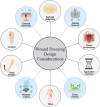
Web diagram of wound dressing design Considerations. Schematic diagram listing ten important characteristics to consider when design wound dressings. The four circles highlighted in blue represent the four design criteria listed within the texted as “key” parameters. The remaining six circles highlighted with grey are important supplementary parameters to also consider, although the degree of importance can vary depending on application. Created using www.biorender.com software
Fig. 4
Fig. 4
Polymeric hydrogel physical properties. A Depiction of a hydrogel model showing differences in mesh sizes between (left) low molecular weight polymers and (right) high molecular weight polymer hydrogels. The frequency in functional reactive sites can be seen and is depicted as teal circles at the junction point of polymer strands. B Schematic representing the function of molecular weight in the swelling of a polymeric hydrogel. C Chart of relative trends in polymeric hydrogels as a function of molecular weight. Created using www.biorender.com software
Fig. 5
Fig. 5
Material chemistry and hydrophobicity. A Depiction of the different functional groups that are commonly found in polymeric biomaterials and give rise to many of their properties. B Depicts a hydrophobicity scale with more hydrophobic (water-resistant) polymers including polymers with more hydrocarbons linkages and less hydrophobic polymers containing more reactive oxygen and nitrogen moieties. Includes different amino acids (top) and different synthetic monomers (bottom), in addition to cellulose (bottom left). Created using ChemDraw Office software
Fig. 6
Fig. 6
Polymer degradation mechanisms. A Enzymatic degradation depiction with proteolytic enzyme breaking down collagen fibril into smaller collagen peptides. B Oxidative degradation depiction with a reactive oxygen species degradation polymer with a proline derivative. C Hydrolytic degradation depiction of an ester-containing polymer reacting with water and broken down into an alcohol and carboxylic acid. Created using www.biorender.com software
Fig. 7
Fig. 7
Insertion of peptide sequences into polymeric biomaterials. Diagram to depict how different peptide sequences can be incorporated into polymeric biomaterials to modulate their properties. Shown here is the insertion of an MMP-sensitive peptide sequence (orange polygons) that is inserted into individual polymer strands (black polygons) to allow for control over degradative kinetics and release of small molecules (green), such as drugs or biologics (right). Created and adapted from www.biorender.com software
Fig. 8
Fig. 8
Bulk versus surface erosion dynamics. A Depiction of polymer structure (grey circle) that contains small molecules (red circles) within the polymeric structure. (Top) Demonstration of bulk erosion and more rapid burst release of small molecules due to the rate of solvent absorption being greater than polymer degradation, relatively. (Bottom) Demonstration of surface erosion and a more gradual controlled release of small molecules due to the rate of degradation being greater than solvent absorption, relatively. B Graphical representation of polymeric dressing properties and drug/small molecular release kinetics over time via Bulk (left) and Surface (right) erosion. Changes in polymer properties depicted in blue lines. Changes in drug release kinetics depicted by red line. C Schematic to represent the relative role of ROS compounds in wound healing. Created using www.biorender.com software
Fig. 9
Fig. 9
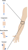
Native tissue force dynamics. Schematic representation of the common forces that skin tissue is exposed to. Created using www.biorender.com software
Fig. 10
Fig. 10
Comparing the relative permeability of dressings. Schematic representation of the different degrees of permeability a wound dressing contains. (Top, blue) Depiction of an occlusive or non-permeable dressing that is most commonly used as a superficial or outermost layer. Occlusive dressings prevent the movement of fluids, both gas and liquids, as well as cells and bacteria. (Middle, green) Depiction of a semi-permeable or semi-occlusive dressing that permits the movement of gases and water vapor (dashed black arrow) but typically limits the movement of liquids to variable degrees depending on the dressing. Semi-permeable dressings prevent the movement of cells and bacteria. (Bottom, black) Permeable or non-occlusive dressings are often depicted as foam or foam-like materials that are absorbent in nature and allow the movement of fluids, both gas and liquid, in addition to cells and bacteria. Oxygen molecules depicted as small blue circles. Carbon dioxide molecules depicted as small purple circles. Bacteria depicted as green organisms. Water is depicted as larger blue circles. Black arrows depict movement through the dressing material. Thicker arrows depict ability to evaporate into ambient environment. Red arrow accompanied by red “X” depicts lack of transport through material. Black-dashed arrow depicts that liquid water does not transport but water vapor still can. Created and adapted using www.biorender.com software
Fig. 11
Fig. 11
Example of multi-layered wound dressing system. Schematic representation of dual-layered wound dressing system, Winter’s Composite. Includes a hydrophilic, permeable base foam dressing layer (Bottom, black) covered by a hydrophobic, semi-permeable dressing layer (Top, green). Depicted in the composite dressing is the combined effects of a permeable and semi-permeable dressing, where all fluids and cells/bacteria can pass through the permeable foam base, but liquid water (and other liquids such as serous exudate) in addition to cells/bacteria get stuck within the permeable foam layer because they cannot pass through into the semi-permeable dressing on superficial surface. However, the semi-permeable layer still allows some removal of water through evaporation, where water vapor is allowed to pass but not liquid water. This combination, known as Winter’s composite, creates a permeability gradient and can aid in exudative removal in mildly exudative wounds, upon dressing changes, due to the absorptive hydrophilic foam. Oxygen molecule depicted as small blue circle. Carbon dioxide molecule depicted as small purple circle. Bacteria depicted as green organism. Water is depicted as larger blue circle. Black arrows depict movement through dressing material. Thicker arrows depict ability to evaporate into ambient environment. Red arrow accompanied by red “X” depicted lack of transport through material. Black-dashed arrow depicts that liquid water does not transport but water vapor still can. Created using www.biorender.com software
Fig. 12
Fig. 12
Fabricating a Bioengineered Skin Substitute (Graft). Schematic representation of generating a skin graft with autologous skin cells (i.e. keratinocytes and/or fibroblasts). A biopsy of a patient can be performed to remove autologous skin cells which can then be culture onto/within a polymeric scaffold in vitro. The scaffold can be fabricated a number of ways, depicted here is the methodology of 3D printing of a collagenous lattice. The skin cells are cultured on the polymeric scaffold for typically several weeks and then removed from cultured, and can be applied to a patient as a customized, autologous skin graft using their own cells. The graft is thought to work via a number of mechanisms, including coverage and protection of the wound, the embedded skin cells secrete biologics to promote wound healing within the native tissue, and the graft matrix can serve as a healthy tissue substrate for resident wound cells to grow onto/into and repopulate
Fig. 13
Fig. 13
Modifying traditional wound dressings (Graft). Schematic depictions of ways that current traditional wounds dressings have been modified to enhance their wound healing capabilities. (Film) Insertion of a plasticizing agent, such as glucose or other small molecules into a polymer network can prevent the alignment of polymer fibers and subsequently increasing the flexibility of film dressings. (Alginate) A number of ions have been investigated for wound healing capabilities, such as the use of magnesium to enhance angiogenic signaling via modulation of native endothelial cells, and silver as an antimicrobial agent that has been used for decades. (Foam) Depiction of an in situ curing/crosslinking foam that expands to fill the irregular contour of many wounds to increase surface contact area. Additionally, foams can be embedded with drugs and/or biologics that can subsequently be released into the wound bed to promote controlled wound regeneration. (Hydrocolloid) Recent investigations in hydrocolloids have shown how drugs, biologics, and platelets can be delivered into the wound bed. Platelets have been investigated as a rich source of growth factors and immunomodulatory compounds via degranulation of their intracellular cargo. Release of platelets can be controlled a number of ways, shown here is how absorption of wound exudate results in swelling of the colloidal network and subsequent release of platelets. (Smart Dressing) Smart dressings can, in theory, be incorporated into a number of different dressing types via insertion of a small, flexible electronics. Depicted here a bacterial compound sensing smart dressing that allows for real-time monitoring of wounds, such as burns, ulcers, or surgical, for bacterial infiltration. Upon detection a sensor can provide both a visual color change in the dressing, in addition to sending a signal to a phone app for outpatient monitoring, and a drug-eluting scaffold can then be triggered to release antimicrobial compounds. (Hydrogel) Schematic depiction of a hydrogel formulated to be deposited into a wound and then a secondary semi-permeable dressing can be applied superficially to protect the hydrogel. The hydrogel can be dosed with a number of bioactive compounds and cells, such as the use of angiogenic-primed stem cells. The angiogenic-primed stem cells demonstrate enhanced angiogenic activity within the wound and release compounds that promote neovascularization within the wound tissue

Similar articles

Cited by

References

    1. Langer R, Tirrell DA. Designing materials for biology and medicine. Nature. 2004;428(6982):487–492. doi: 10.1038/nature02388. - DOI - PubMed
    1. Nolan K, Millet Y, Ricordi C, Stabler CL. Tissue engineering and biomaterials in regenerative medicine. Cell Transplant. 2008;17(3):241–243. doi: 10.3727/096368908784153931. - DOI - PubMed
    1. Bhattacharya S. Wound healing through the ages. Indian J Plast Surg. 2012;45(2):177–179. doi: 10.4103/0970-0358.101255. - DOI - PMC - PubMed
    1. Hawthorne B, Simmons JK, Stuart B, Tung R, Zamierowski DS, Mellott AJ. Enhancing wound healing dressing development through interdisciplinary collaboration. J Biomed Mater Res B Appl Biomater. 2021;109(12):1967–1985. doi: 10.1002/jbm.b.34861. - DOI - PMC - PubMed
    1. Shaw TJ, Martin P. Wound repair at a glance. J Cell Sci. 2009;122(Pt 18):3209–3213. doi: 10.1242/jcs.031187. - DOI - PMC - PubMed