

Three-dimensional fracture mapping and analysis of coronal fractures in AO/OTA types 33-B3 and C3
- PMID: 36183139
- PMCID: PMC9526965
- DOI: 10.1186/s13018-022-03327-7
Three-dimensional fracture mapping and analysis of coronal fractures in AO/OTA types 33-B3 and C3
Abstract
Background: Although the relatively high incidence of coronal fractures in the supracondylar-intercondylar fractures is well established, little is currently known about the morphology of those fractures. Herein, we characterized the coronal fractures in AO/OTA type 33-C3 and assessed their differences with Busch-Hoffa fractures (33-B3).
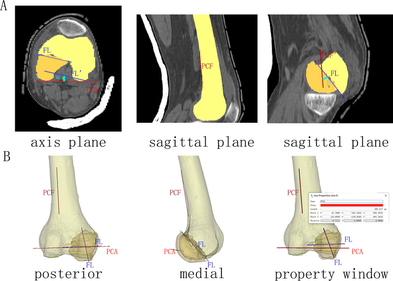
Methods: We retrospectively collected 61 cases of AO/OTA type 33-B or C fractures with coronal plane fragments and generated three-dimensional fracture maps of those with coronal fractures based on CT imaging and measured angle α (the angle between the coronal fracture and the posterior condyle axis in the axis plane) and angle β (the angle between the coronal fracture and the posterior femoral cortex in the sagittal plane).
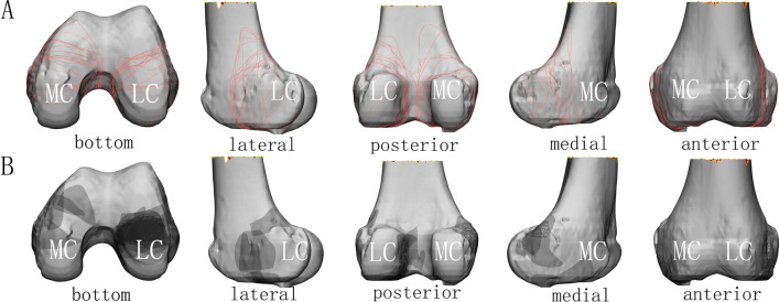
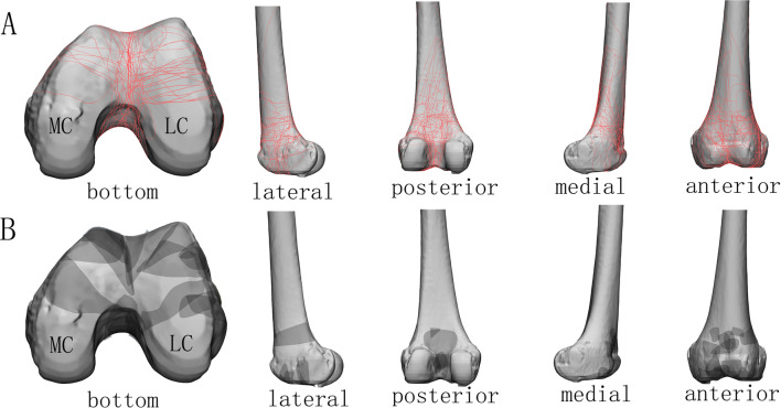
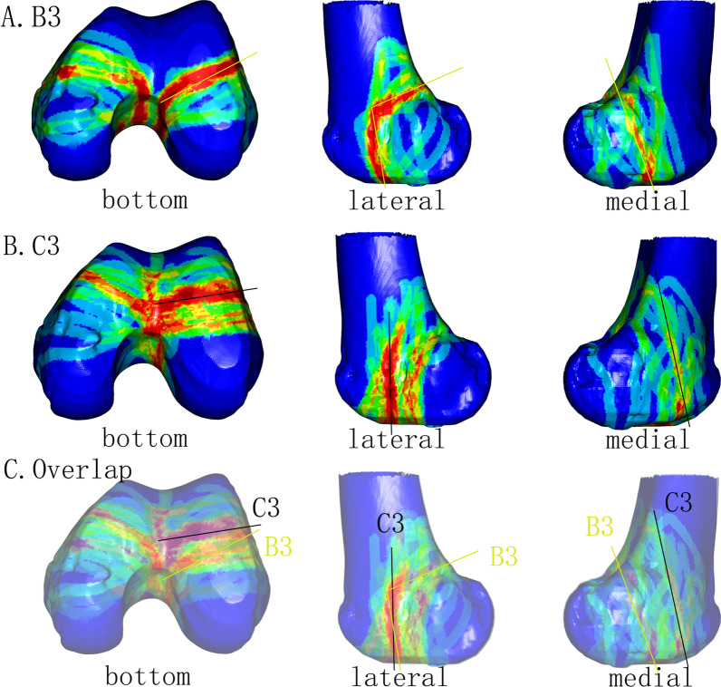
Results: Thirty-three cases (32%) of AO/OTA type 33-C fractures contained coronal fragments. Most of them were type 33-C3 fractures. Angles α and β for type 33-C3 were significantly smaller than for type B3 at the lateral condyle, while the angles at the medial condyle were not significantly different. The fracture maps showed that the coronal fractures and the articular comminution area were more anterior in type 33-C3.
Conclusions: The incidence of coronal fractures was 32% and 67% in AO/OTA types 33-C and 33-C3, respectively. Our findings suggest that coronal fractures differed between both types, emphasizing the potential need for different treatment approaches.
Keywords: Coronal fracture; Distal femur; Fracture map; Morphology; Supracondylar–intercondylar fracture.
© 2022. The Author(s).
Conflict of interest statement
There is no conflict of interest.
Figures
Similar articles
-
Two and Three-Dimensional CT Mapping of Hoffa Fractures.J Bone Joint Surg Am. 2017 Nov 1;99(21):1866-1874. doi: 10.2106/JBJS.17.00473. J Bone Joint Surg Am. 2017. PMID: 29088042
-
Fracture morphology of AO/OTA 31-A trochanteric fractures: A 3D CT study with an emphasis on coronal fragments.Injury. 2017 Feb;48(2):277-284. doi: 10.1016/j.injury.2016.12.015. Epub 2016 Dec 23. Injury. 2017. PMID: 28040260
-
Intra-articular Fracture Pattern in Intercondylar Distal Femur Fractures: An Analysis of Frequency and Major Fracture Fragments.Injury. 2021 Apr;52(4):967-970. doi: 10.1016/j.injury.2020.11.061. Epub 2020 Nov 28. Injury. 2021. PMID: 33280890
-
Busch-Hoffa fracture: A systematic review.Medicine (Baltimore). 2023 Dec 1;102(48):e36161. doi: 10.1097/MD.0000000000036161. Medicine (Baltimore). 2023. PMID: 38050206 Free PMC article.
-
Hoffa fracture of the femoral condyle: Injury mechanism, classification, diagnosis, and treatment.Medicine (Baltimore). 2019 Feb;98(8):e14633. doi: 10.1097/MD.0000000000014633. Medicine (Baltimore). 2019. PMID: 30813201 Free PMC article.
Cited by
-
Morphology and novel classification of proximal humeral fractures.Front Bioeng Biotechnol. 2024 Jul 19;12:1366089. doi: 10.3389/fbioe.2024.1366089. eCollection 2024. Front Bioeng Biotechnol. 2024. PMID: 39100622 Free PMC article.
-
Progress of fracture mapping technology based on CT three-dimensional reconstruction.Front Bioeng Biotechnol. 2024 Nov 6;12:1471470. doi: 10.3389/fbioe.2024.1471470. eCollection 2024. Front Bioeng Biotechnol. 2024. PMID: 39569162 Free PMC article. Review.
References
-
- Lundin N, Huttunen TT, Enocson A, Marcano AI, Felländer-Tsai L, Berg HE. Epidemiology and mortality of pelvic and femur fractures-a nationwide register study of 417,840 fractures in Sweden across 16 years: diverging trends for potentially lethal fractures. Acta Orthop. 2021;92(3):323–328. doi: 10.1080/17453674.2021.1878329. - DOI - PMC - PubMed
MeSH terms
LinkOut - more resources
Full Text Sources
Medical
Miscellaneous
